Original
Article
This work is licensed under a Creative Commons Attribution-NonCommercial- NoDerivatives International License. ©2021 The Editorial Committee of Annals of Thoracic and Cardiovascular Surgery
Multivariate Analysis of Risk Factor for Mortality
and Feasibility of Extracorporeal Membrane
Oxygenation in High-Risk Thoracic Surgery
Do Hyung Kim, MD,1 Jong Myung Park, MD,2 Joohyung Son, MD,1 and Sung Kwang Lee, MD1
Introduction
Extracorporeal membrane oxygenation (ECMO) is widely used in patients with respiratory or cardiac fail-ure who do not respond to conventional life support therapy. The feasibility of ECMO as intraoperative car-diorespiratory support during lung transplantation is well known; however, there were rare reports of ECMO used for thoracic surgery.1–7) These reports have ana-lyzed only 10–20 cases and simply confirmed the surgi-cal results. In many cases, it is difficult to achieve clinical significance beyond the expansion of ECMO indications, and there are no reports of efficacy in spe-cific areas.3,8–11)
Background: Extracorporeal membrane oxygenation (ECMO) as intraoperative cardio-respiratory support during lung transplantation is well known, but use for other types of surgery are limited. To assess risk factor for mortality after high-risk thoracic surgery and feasibility of ECMO, we reviewed.
Methods: This study was an observational study. Between January 2011 and October 2018, 63 patients underwent thoracic surgery with ECMO for severe airway disease, pul-monary insufficiency requiring lung surgery, and other conditions.
Results: In all, 46 patients remained alive at 30 days after surgery. The mean patient age was 50.38 ± 16.16 years. ECMO was most commonly used to prevent a lethal event (34
[73.9%]) in the Survival (S) group and rescue intervention (13 [76.5%]) in the Non-sur-vival (N) group. In all, 11 patients experienced arrest during surgery (S vs N: 2 [4.3%] vs 9 [52.9%], p ≤0.001). The multivariate analysis revealed that arrest during surgery (odds
ratio [OR], 24.44; 95% confidence interval [CI], 1.82–327.60; p = 0.016) and age (OR,
7.47; 95% CI, 1.17–47.85; p = 0.034) were independently associated with mortality.
Conclusions: ECMO provides a safe environment during thoracic surgery, and its compli-cation rate is acceptable except for extracorporeal cardiopulmonary resuscitation (ECPR). Keywords: thoracic surgery, extracorporeal membrane oxygenation, cardiopulmonary
resuscitation
1Department of Thoracic and Cardiovascular Surgery, Pusan
National University Yangsan Hospital, Yangsan, Korea
2Department of Thoracic and Cardiovascular Surgery, Busan
Medical Center, Yeonje-Gu, Busan, Korea
Received: July 17, 2020; Accepted: September 14, 2020
Corresponding author: Sung Kwang Lee, MD. Department of Thoracic and Cardiovascular Surgery, Pusan National Univer-sity Yangsan Hospital, Pusan National UniverUniver-sity College of Medicine, 20 Geumo-ro, Mulgeum-eup, Yangsan-si, Gyeong-sangnam-do, 50612, Korea
Email: drlsk@naver.com
doi: 10.5761/atcs.oa.20-00224 atcs
Annals of Thoracic and Cardiovascular Surgery
1341-1098 2186-1005
The Editorial Committee of Annals of Thoracic and Cardiovascular Surgery
atcs.oa.20-00224 10.5761/atcs.oa.20-00224 XX XX XX XX 17July2020 2020 XX2020
We have experienced various advantages and prob-lems since the introduction of ECMO in thoracic surgery in 2006. Here, we present our experience and analyze the clinical outcomes of ECMO support in thoracic surgery to report tips on its use, factors influencing surgical out-comes, and indications for ideal ECMO support. The purpose of this study is to be used as basic data for risk factors related to mortality after high-risk thoracic sur-gery and ECMO indications. Please indicate at the end of the Introduction section: We present the following article in accordance with the Strengthening the Report-ing of Observational Studies in Epidemiology (STROBE) reporting checklist.
Materials and Methods
Patient selection
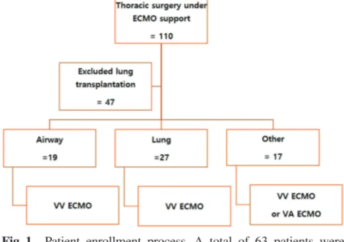
Between January 2011 and October 2018, 110 patients underwent thoracic surgery under ECMO support. Of them, 47 who underwent lung transplantation were excluded; thus, a total of 63 patients supported on ECMO were analyzed in this report. The indications for ECMO were needed for surgical correction to solve problems related to clinical disease as follows: severe airway dis-ease such as trachea stenosis, pulmonary insufficiency requiring lung surgery such as hemoptysis due to the destroyed lung, pneumonia, sepsis, ventilator-induced lung injury (VILI), and other conditions that can cause cardiac arrest during surgery such as advanced huge tho-racic cancer and major vessel injury leading to hypovo-lemic shock or tamponade (Fig. 1). We did not randomly remove all patients who met the selection criteria to reduce selection bias.
Preoperative characteristics
The medical records of these 63 patients were reviewed. The following routine clinical baseline data were collected: age, sex, body mass index (BMI), diag-nosis, purpose for ECMO, type of ECMO support, and preoperative clinical condition (such as major multiple trauma, advanced thoracic malignancy, infectious dis-ease, airway obstruction, bleeding before surgery, diabe-tes, hypertension, renal failure, and coagulopathy).
ECMO and anticoagulation protocol
ECMO was administered based on guidelines from the Extracorporeal Life Support Organization. The ECMO system consisted of an EBS (Terumo, Tokyo, Japan) or PLS (Maquet, Mahwah, NJ, USA) machine, centrifugal
pumps (Maquet, Rastatt, Germany), and simplified Bio-line-coated circuits (Maquet). All patients were cannulated peripherally through the femoral vein and artery or inter-nal jugular vein using the Seldinger technique under ultrasound guidance. Venovenous (VV) ECMO was used for respiratory support. In the majority of cases involving VV ECMO, the right femoral vein was used for venous drainage and the internal jugular vein for venous return; if additional heart support was necessary during VV ECMO, a return cannula was added to the femoral artery and VVA ECMO was implemented. If hemodynamic support was required rather than respiratory support, venoarterial (VA) ECMO was performed. Cannulation mainly used the configuration of femoral vein drain and femoral artery return. If cardiac arrest occurred, VA ECMO was performed regardless of the cause of arrest.
Preoperative extracorporeal cardiopulmonary resus-citation (ECPR) or preoperative elective ECMO was performed once with a 50–70 IU/kg heparin bolus imme-diately before insertion of the cannula for ECMO; no additional heparin was injected until the end of the oper-ation. Patients with ongoing bleeding did not start anti-coagulation therapy. After the end of the operation, if
Fig. 1 Patient enrollment process. A total of 63 patients were
included. Between January 2011 and October 2018, 110 patients underwent thoracic surgery under ECMO support. Of them, 47 who underwent lung transplantation were excluded. We divided the patients into three groups by con-dition. In all, 19 patients underwent ECMO due to severe airway disease such as trachea stenosis. In all, 27 patients underwent ECMO for pulmonary insufficiency requiring lung surgery such as hemoptysis due to a destroyed lung, pneumonia, sepsis, and VILI. In all, 17 patients underwent ECMO for other conditions that can cause cardiac arrest during surgery such as advanced huge thoracic cancer and major vessel injury leading to hypovolemic shock or tam-ponade. ECMO: extracorporeal membrane oxygenation; VILI: ventilator-induced lung injury
support of ECMO was needed, anticoagulation was ini-tiated after 24 hours. Anticoagulation was monitored by activated clotting time (ACT) and activated partial thromboplastin time (aPTT). ACT was maintained for approximately 160–190 s.
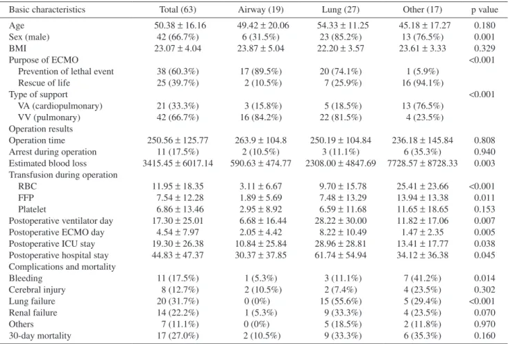
Because most surgeries are accompanied by massive bleeding, we applied autologous transfusion using a cell saver to prevent hypovolemic shock in trauma patients. Generally, cardiopulmonary bypass (CPB) with sucker could be used for direct infusion of blood loss, but to operate CPB which is an open circuit, sufficient dose of heparin and ACT prolong must be needed, which causes more bleeding. We decided to create a configuration of autologous transfusion circuit using ECMO which is a closed circuit. The circuit was configured as follows to quickly replenish the blood loss. After the blood was suctioned into the reservoir, it was sent from the reser-voir to the cell saver and recycled. Then, to reduce the amount of transfusion and fluid, the collected blood in the cell saver bag was directly connected to the Rapid Infusion System to infusion directly through the patient's central line (Fig. 2). After surgery, obstruction of femo-ral artery by the cannula was suspected, a small addi-tional cannula was inserted for distal leg perfusion.
Statistical analysis
Continuous variables are expressed as mean ± stan-dard deviation, and differences between continuous
variables were analyzed using the unpaired Student’s t-test. Categorical variables are expressed as numbers and their percentages were compared by the Pearson’s chi-squared test or Fisher’s exact test as appropriate. According to the latest European Association of Car-dio-Thoracic Surgery statistical and data reporting guidelines, we entered the threshold value of dichoto-mous variables with univariable p values <0.20 into a multivariable binary logistic-regression model to iden-tify the independent risk factors for 30-day mortality. To provide concise and informative factors for the predic-tion of 30-day mortality, the continuous variables in the multivariate logistic regression model were dichoto-mized according to the clinically meaningful cutoffs widely accepted for risk stratification in routine clinical practice, including geriatric state, age >65 years; and overweight/obese state, BMI >25 kg/m2. Results are expressed as odds ratios (ORs) with 95% confidence intervals (CIs), and two-tailed p values <0.05 were con-sidered significant. We used SPSS for Windows (version 21.0, IBM Corp., Armonk, NY, USA) for the statistical analysis.
Results
During the study period, 63 patients underwent tho-racic surgery under ECMO support for hemodynamic or respiratory support. Their baseline characteristics are
Fig. 2 Configuration of autologous transfusion using a cell saver with a rapid infusion system. We applied an
autolo-gous transfusion to prevent hypovolemic shock in trauma patients. After the blood was suctioned into the reser-voir, it was sent from the reservoir to the cell saver and recycled. The collected blood in the cell saver bag was directly connected to the Rapid Infusion System for infusion directly through the patient’s central line.
presented in Table 1. To assess risk factors affecting mortality, we divided the patients into two groups according to mortality status: 46 patients survived, while 17 died by 30 days after surgery.
Patient characteristics of survival patients
Our study consisted of 42 (66.7%) male patients (Sur-vival group (S) vs Non-sur(Sur-vival group (N): 28 [60.9%] vs 14 [82.4%], p = 0.139) with a mean age of 50.38 ± 16.16 years (S vs N: 47.4 ± 15.8 vs 58.35 ± 14.7, p = 0.016). ECMO was often applied to prevent a lethal event (34 [73.9%]) in the Survival group and to rescue life (13 [76.5%]) in the Non-survival group (p <0.001). There were significant intergroup differences. ECMO support types included VV (only respiration support) ECMO (37 [80.4%]) in the Survival group and VA (car-diopulmonary support) ECMO (12 [70.6%]) in the Non-survival group (p <0.001). There were significant intergroup differences. Surgery types and preoperative clinical conditions did not differ significantly between the two groups.
Postoperative results of survival patients
In all, 11 patients experienced arrest during surgery (S vs N: 2 [4.3%] vs 9 [52.9%], p <0.001). The mean oper-ation time was 250.56 ± 125.77 min (S vs N: 249.0 ± 115.6 vs 254.7 ± 153.9, p = 0.875). Surgery-related com-plications did not differ significantly between the two groups. There were no significant intergroup differences.
Multivariate analysis of risk factors for mortality
We entered the threshold value of dichotomous variables with a univariate p value <0.20 into a multivariate binary logistic regression model to identify the independent risk factors for mortality. A multivariate logistic regression model involving age, sex, ECMO indication, ECMO sup-port type, intraoperative arrest, and postoperative bleeding is shown in Table 2. Multivariate analysis including these factors showed that age (OR, 7.47; 95% CI, 1.17–47.85; p = 0.034), arrest during operation (OR, 24.44; 95% CI, 1.82–327.60; p = 0.016) were independently associated with complications for mortality (Table 2).
Operative and postoperative results as groups of ECMO indications
The operation time did not differ significantly among groups. Intraoperative arrest was more common in the Other group than the other groups (Airway group (A) vs Lung group (L) vs Other group (O), 2 [10.5%] vs 3
[11.1%] vs 6 [35.3%], p = 0.940), but there were no sig-nificant differences. The Other group included advanced malignancy patients and great vessel injury patients. Thus, this group showed larger estimated blood losses (A vs L vs O: 590.63 ± 474.77 vs 2308.00 ± 4847.69 vs 7728.57 ± 8728.33, p = 0.003), red blood cell transfu-sions (A vs L vs O: 3.11 ± 6.67 vs 9.70 ± 15.78 vs 25.41 ± 23.66, p <0.001), and fresh frozen plasma transfusions (A vs L vs O: 1.89 ± 5.69 vs 7.48 ± 13.29 vs 13.94 ± 13.38, p = 0.011) than the other groups. The O group had more case of bleeding after surgery due to a large amount of estimated blood loss and transfusion leading to a bleeding tendency (A vs L vs O: 1 [5.3%] vs 3 [11.1%] vs 7 [41.2%], p = 0.014). The L group included preoperative respiratory failure due to pneumonia, sepsis, and VILI. Thus, these groups showed a longer duration of ventilator and ECMO use and longer mean hospital stay than the other groups. Lung failure after surgery was more com-mon in the L group than the other groups (A vs L vs O: 0 [0%] vs 15 [55.6%] vs 5 [29.4%], p <0.001) (Table 3).
Discussion
Most thoracic surgeries could be performed with con-tinuous one-lung ventilation and a triple-lumen central line. However, surgery is impossible if continuous venti-lation is difficult, lung function is poor, or a large amount of bleeding occurs. It was recently reported that surgery is possible with ECMO use.1–7) The usefulness of ECMO in thoracic surgery is divided into three categories.3,11) First, the most useful indication of ECMO is airway obstructive disease.7,12–14) Most causes of death in patients with airway disease are respiratory failure due to airway obstruction. ECMO helps maintain life before surgical correction. The airway surgery is highly risky and very difficult despite being performed by an expert surgeon, because most conventional airway surgeries should be performed alternately between ventilation and non-ventilation for a limited time and the surgical field is poor due to endotracheal tube insertion into the distal trachea. However, ECMO can provide oxygen support and CO2 removal without ventilation, making it possible to perform airway surgery with or without ventilation.11) ECMO enables safe airway surgery in an emergency state, even for inexpert trachea surgeons.
The second indication for ECMO support for thoracic surgery is the state of lung failure due to pneumonia, sep-sis, and VILI.9) These patients are generally contraindi-cated for thoracic surgery by traditional criteria. It is
difficult to decide to perform conventional surgery with-out ECMO support; even if the surgery is decided, there are many cases in which the vital signs are not maintained and the operation cannot be started, or if the surgery is performed, pulmonary insufficiency cannot be recovered after surgery. However, the use of ECMO allows mainte-nance of cardiopulmonary support during perioperative period. It is very helpful in patients who underwent ECMO support during preoperative period. If surgical risk factors have been eliminated, it is reasonable to perform surgery to resolve the disease focus. ECMO also prevents worsen-ing of lung function or additional cardiopulmonary dam-age in VILI and reduces many risks (hypoxia, shock, or arrest during surgery) that can occur during surgery. Since 2012, aggressive surgical correction using ECMO has been performed with these concepts in our institute. Despite the very high operative risk group, the mortality rate was relatively reasonable (16.7%).
The third indication is ECMO support for cardiopul-monary support during thoracic surgery.8,15,16) It is mainly used to maintain hemodynamic function in surgery for advanced huge thoracic cancer and thoracic trauma with major vessel injury. Intraoperative mass manipulation can deteriorate cardiac function. In severe cases, cardiac arrest may occur. Most intraoperative arrests lacking emergency preparation result in death. The hemody-namic support of ECMO prevents unexpected lethal events during surgery and ensures that surgery proceeds safely. ECMO is simultaneously possible rapid volume replacement during surgery. We already reported the effects of ECMO as a life-saving method in the treatment of shock from heart and major vessel injury.17)
If indications have been determined, the next question is “When is it effective to apply ECMO?” In our risk fac-tor analysis, the mortality was higher when the arrest occurred intraoperatively. Since ECPR is performed
Table 1 Clinical value according to mortality
Variables (n=63)Total Survivor (n = 46) Non-survivor (n = 17) p value
Age (years) 50.38 ± 16.16 47.4 ± 15.8 58.35 ± 14.7 0.016
Sex (male) 42 (66.7%) 28 (60.9%) 14 (82.4%) 0.139
BMI (kg/m2) 23.07 ± 4.04 22.9 ± 4.3 23.7 ± 3.0 0.523
Purpose of ECMO <0.001
Prevention of lethal event 38 (60.3%) 34 (73.9%) 4 (23.5%)
Rescue of life 25 (39.7%) 12 (26.1%) 13 (76.5%)
Types of support <0.001
VA Cardiopulmonary support) 21 (33.3%) 9 (19.6%) 12 (70.6%) VV (Only respiration support) 42 (66.7%) 37 (80.4%) 5 (29.4%)
Types of operation 0.153
Airway surgery 19 (30.2%) 17 (37.0%) 2 (11.8%)
Lung 27 (42.9%) 18 (39.1%) 9 (52.9%)
Others
(mediastinum, major vessel) 17 (27.0%) 11 (23.9%) 6 (35.3%) Preoperative clinical conditions
Major multiple trauma 22 (34.9%) 14 (30.4%) 8 (47.1%) 0.246
Advanced thoracic malignancy 15 (23.8%) 10 (21.7%) 5 (29.4%) 0.740
Infectious disease 12 (19.0%) 10 (21.7%) 2 (11.8%) 0.487
Airway obstruction 14 (22.2%) 12 (26.1%) 2 (11.8%) 0.316
Bleeding before surgery 26 (41.3%) 18 (39.1%) 8 (47.1%) 0.388
Diabetes 13 (20.6%) 9 (19.6%) 4 (23.5%) 1.000
Hypertension 15 (23.8%) 10 (21.7%) 5 (29.4%) 0.740
Renal failure 1 (1.6%) 0 (0%) 1 (5.9%) 0.270
Coagulopathy 27 (42.9%) 16 (34.8%) 11 (64.7%) 0.460
Postoperative results
Arrest during operation 11 (17.5%) 2 (4.3%) 9 (52.9%) <0.001
Operation time 250.56 ± 125.77 249.0 ± 115.6 254.7 ± 153.9 0.875 Operation-related complications Bleeding 11 (17.5%) 6 (13.0%) 5 (29.4%) 0.149 Cerebral injury 8 (12.7%) 3 (6.5%) 5 (29.4%) 0.280 Lung failure 20 (31.7%) 10 (21.7%) 10 (58.8%) 0.191 Other 7 (11.1%) 4 (8.7%) 3 (17.6%) 0.375
preoperatively, ECMO can be performed relatively quickly. The operation can begin after the patient’s vital signs have stabilized. However, if ECPR (as sudden car-diac arrest) during thoracic surgery occurs without femo-ral cannula site preparation, ECMO insertion and recovery of vital signs were delayed due to the lateral decubitus position of patients. Although ECMO was started, most patients presented with hypoxic brain dam-age or irreversible cardiopulmonary dysfunction due to delayed time to resuscitation. Min et al.18) reported an overall success rate (i.e., neurological deficit-free opera-tion) of 26% with intraoperative ECPR. In our study, only two of 11 (18.1%) intraoperative ECPR patients survived. One patient was in a supine position and the other had an event during surgical draping, so ECMO could be applied relatively quickly. Based on these results, preventive ECMO insertion comparing the intraoperative ECPR may have helped increase patient survival. In the patients who have a risk of developing cardiopulmonary failure during surgery, it is necessary to establish indications for preventive ECMO insertion. In our institute, indication of preventive ECMO insertion is as follows: (1) tachycardia occurs due to compression on the heart by a huge mass, (2) maintain of PaO2 is difficult due to active and massive hemoptysis, (3) airway is collapsed below 5 mm, (4) maintain the airway is difficult due to bilateral thoracic surgery or carina surgery, and (5) maintain ventilation is difficult due to pneumonia or acute respiratory distress syndrome (ARDS) of the opposite lung.
If thoracic surgery with ECMO support is chosen, the correction of preoperative coagulation defects is import-ant for operation mortality. Anticoagulation should gen-erally be used to prevent blood clots in extracorporeal circuits. However, it is important to consider whether it is reasonable to implement prophylactic anticoagulation in patients with major bleeding. In particular, when ECMO is inserted to support surgery, its short-term use is planned, so the probability of thromboembolic events is lower than the other ECMO applications.
There are many reports of surgery with heparin-free ECMO in trauma. Muellenbach et al.19) first reported three cases of prolonged heparin-free VV ECMO in multiple- injury ARDS patients with severe traumatic brain injury in whom conventional mechanical ventilation had failed. Arlt et al.20) reported that six of 10 multiple trauma patients with coexisting pulmonary failure or cardiopulmonary failure and hypovolemic shock due to bleeding survived with the use of heparin-free ECMO. Ried et al.21) reported an overall survival rate of 79% in 52 patients with severe thoracic trauma and acute lung failure refractory to conventional therapy using extracorporeal lung support. Heparin-free ECMO was used in patients at high risk of further bleeding complications or showing signs of intracranial bleeding.
It is theoretically possible to support patients with ongo-ing massive bleedongo-ing by ECMO without anticoagulation. First, patients with massive bleeding often have coagula-tion defects prior to ECMO insercoagula-tion because a mild coag-ulation defect is induced by blood loss and hemodilution due to massive crystalloid volume replacement. It is possi-ble to maintain ECMO flow and function without antico-agulation during ECMO cannulation.22) Second, newer circulatory systems utilizing heparin-bonded catheters, low-pressure oxygenators, and centrifugal pumps allow for the use of less anticoagulant during ECMO without circuit thrombotic complications.23) Third, most thromboses occur at sites of stasis or turbulent flow in the circuit, that is, on the venous (pre-oxygenator) side of the circuit rather than on the arterial (post-oxygenator) side.24,25) We performed ECMO intraoperatively without heparin except a loading dose during elective ECMO insertion to reduce bleeding due to anticoagulation use.
The last point to mention is that ECMO can be used postoperatively. All patients undergoing surgery with ECMO have a high-risk medical problem. Therefore, in many cases, cardiopulmonary failure occurs after sur-gery. Maintaining vital signs is often difficult despite maximal conventional support. In these cases, ECMO support is required to maintain cardiopulmonary
Table 2 Multivariable analysis of risk factors for mortality
Estimated factors Odds ratio 95% CI p value
Age (years) 7.47 1.17–47.85 0.034
Sex 0.20 0.01–3.03 0.245
Types of support 4.60 0.48–43.93 0.185
Arrest during operation 24.44 1.82–327.60 0.016
Purpose of ECMO 0.64 0.09–4.52 0.652
Postoperative bleeding 0.45 0.05–3.97 0.470
function after surgery. Because ECMO rescues life or reduces respiratory dependence in the immediate post-operative critical period, patients who require maximal ventilator support could prevent VILI or diaphragm dys-function by postoperative ECMO support. Yeo et al.26) reported an 80% survival rate of postoperative ARDS through ECMO therapy. In general, the survival rate of postoperative ARDS is 50–60%, a high success rate.27) If ECMO is used during surgery, it can be easily deter-mined to which extent it should be used immediately after surgery; thereby, we can prevent secondary cardio-pulmonary function damage from occurring after sur-gery. The limitation of this study was that it was a small case number and was not various thoracic surgery.
Conclusion
ECMO is useful during thoracic surgery in high-risk patients with respiratory or cardiac failure. ECMO pro-vides a safe environment during thoracic surgery with acceptable mortality and complication rates. However, as
shown in our results, unprepared ECMO insertion does not increase the survival rate due to delayed resuscitation in intraoperative arrest. For patients at high risk of sur-gery with a high chance of a lethal event, elective ECMO insertion to prevent a lethal event is recommended.
Acknowledgment
This work was supported by a 2-Year Research Grant of Pusan National University.
Disclosure Statement
None.
References
1) Aigner C, Wisser W, Taghavi S, et al. Institutional experience with extracorporeal membrane oxygen-ation in lung transplantoxygen-ation. Eur J Cardiothorac Surg 2007; 31: 468–73; discussion 473-4.
Table 3 Clinical value between three groups
Basic characteristics Total (63) Airway (19) Lung (27) Other (17) p value
Age 50.38 ± 16.16 49.42 ± 20.06 54.33 ± 11.25 45.18 ± 17.27 0.180
Sex (male) 42 (66.7%) 6 (31.5%) 23 (85.2%) 13 (76.5%) 0.001
BMI 23.07 ± 4.04 23.87 ± 5.04 22.20 ± 3.57 23.61 ± 3.33 0.329
Purpose of ECMO <0.001
Prevention of lethal event 38 (60.3%) 17 (89.5%) 20 (74.1%) 1 (5.9%)
Rescue of life 25 (39.7%) 2 (10.5%) 7 (25.9%) 16 (94.1%) Type of support <0.001 VA (cardiopulmonary) 21 (33.3%) 3 (15.8%) 5 (18.5%) 13 (76.5%) VV (pulmonary) 42 (66.7%) 16 (84.2%) 22 (81.5%) 4 (23.5%) Operation results Operation time 250.56 ± 125.77 263.9 ± 104.8 250.19 ± 104.84 236.18 ± 145.84 0.808
Arrest during operation 11 (17.5%) 2 (10.5%) 3 (11.1%) 6 (35.3%) 0.940
Estimated blood loss 3415.45 ± 6017.14 590.63 ± 474.77 2308.00 ± 4847.69 7728.57 ± 8728.33 0.003 Transfusion during operation
RBC 11.95 ± 18.35 3.11 ± 6.67 9.70 ± 15.78 25.41 ± 23.66 <0.001
FFP 7.54 ± 12.28 1.89 ± 5.69 7.48 ± 13.29 13.94 ± 13.38 0.011
Platelet 6.86 ± 13.46 2.95 ± 8.92 6.59 ± 11.68 11.65 ± 18.65 0.153
Postoperative ventilator day 17.30 ± 25.01 6.68 ± 16.44 28.22 ± 30.00 11.82 ± 17.06 0.007 Postoperative ECMO day 4.54 ± 7.97 2.05 ± 4.42 8.22 ± 10.49 1.47 ± 2.35 0.005 Postoperative ICU stay 19.30 ± 26.38 10.84 ± 25.84 28.96 ± 28.81 13.41 ± 17.77 0.038 Postoperative hospital stay 44.83 ± 47.37 30.37 ± 37.85 61.74 ± 54.94 34.12 ± 36.38 0.045 Complications and mortality
Bleeding 11 (17.5%) 1 (5.3%) 3 (11.1%) 7 (41.2%) 0.014 Cerebral injury 8 (12.7%) 2 (10.5%) 2 (7.4%) 4 (23.5%) 0.302 Lung failure 20 (31.7%) 0 (0%) 15 (55.6%) 5 (29.4%) <0.001 Renal failure 14 (22.2%) 1 (5.3%) 9 (33.3%) 4 (23.5%) 0.070 Others 7 (11.1%) 0 (0%) 5 (18.5%) 2 (11.8%) 0.970 30-day mortality 17 (27.0%) 2 (10.5%) 9 (33.3%) 6 (35.3%) 0.160
BMI: body mass index; ECMO: extracorporeal membrane oxygenation; FFP: fresh frozen plasma; ICU: intensive care unit; RBC: red cell blood; VA: venoarterial; VV: venovenous
2) Lang G, Ghanim B, Hötzenecker K, et al. Extracorpo-real membrane oxygenation support for complex tra-cheo-bronchial procedures†. Eur J Cardiothorac Surg 2015; 47: 250–5; discussion 256.
3) Rinieri P, Peillon C, Bessou JP, et al. National review of use of extracorporeal membrane oxygenation as respi-ratory support in thoracic surgery excluding lung trans-plantation. Eur J Cardiothorac Surg 2015; 47: 87–94. 4) Lei J, Su K, Li XF, et al. ECMO-assisted carinal
re-section and reconstruction after left pneumonectomy. J Cardiothorac Surg 2010; 5: 89.
5) Rosskopfova P, Perentes JY, Ris HB, et al. Extracor-poreal support for pulmonary resection: current indi-cations and results. World J Surg Oncol 2016; 14: 25. 6) Keeyapaj W, Alfirevic A. Carinal resection using an
airway exchange catheter-assisted venovenous ECMO technique. Can J Anaesth 2012; 59: 1075–6.
7) Kim CW, Kim DH, Son BS, et al. The feasibility of extracorporeal membrane oxygenation in the variant airway problems. Ann Thorac Cardiovasc Surg 2015;
21: 517–22.
8) Felten M-L, Michel-Cherqui M, Puyo P, et al. Extra-corporeal membrane oxygenation use for mediastinal tumor resection. Ann Thorac Surg 2010; 89: 1012. 9) Brenner M, O'Connor JV, Scalea TM. Use of ECMO
for resection of post-traumatic ruptured lung abscess with empyema. Ann Thorac Surg 2010; 90: 2039–41. 10) Tsunezuka Y, Sato H, Tsubota M, et al. Significance
of percutaneous cardiopulmonary bypass support for volume reduction surgery with severe hypercapnia. Artif Organs 2000; 24: 70–3.
11) Redwan B, Ziegeler S, Freermann S, et al. Intraop-erative veno-venous extracorporeal lung support in thoracic surgery: a single-centre experience. Interact Cardiovasc Thorac Surg 2015; 21: 766–72.
12) Hong Y, Jo KW, Lyu J, et al. Use of venovenous ex-tracorporeal membrane oxygenation in central airway obstruction to facilitate interventions leading to defin-itive airway security. J Crit Care 2013; 28: 669–74. 13) Kim SH, Song S, Kim YD, et al. Outcomes of
extra-corporeal life support during surgery for the critical airway stenosis. ASAIO J 2017; 63: 99–103.
14) Ko M, dos Santos PR, Machuca TN, et al. Use of single- cannula venous-venous extracorporeal life support in the management of life-threatening airway obstruction. Ann Thorac Surg 2015; 99: e63–5.
15) Xu HC, Ye P, Bao FC, et al. ECMO-assisted esophagectomy after left pneumonectomy. Int J Artif Organs 2013; 36: 259–62.
16) Byrne JG, Leacche M, Agnihotri AK, et al. The use of cardiopulmonary bypass during resection of locally
advanced thoracic malignancies: a 10-year two-center experience. Chest 2004; 125: 1581–6.
17) Park JM, Kim CW, Cho HM, et al. Induced airway obstruction under extracorporeal membrane oxygen-ation during treatment of life-threatening massive he-moptysis due to severe blunt chest trauma. J Thorac Dis 2014; 6: E255–8.
18) Min JJ, Tay CK, Ryu DK, et al. Extracorporeal cardio-pulmonary resuscitation in refractory intra-operative cardiac arrest: an observational study of 12-year out-comes in a single tertiary hospital. Anaesthesia 2018;
73: 1515–23.
19) Muellenbach RM, Kredel M, Kunze E, et al. Prolonged heparin-free extracorporeal membrane oxygenation in multiple injured acute respiratory distress syndrome patients with traumatic brain injury. J Trauma Acute Care Surg 2012; 72: 1444–7.
20) Arlt M, Philipp A, Voelkel S, et al. Extracorpo-real membrane oxygenation in severe trauma pa-tients with bleeding shock. Resuscitation 2010;
81: 804–9.
21) Ried M, Bein T, Philipp A, et al. Extracorporeal lung support in trauma patients with severe chest injury and acute lung failure: a 10-year institutional experience. Crit Care 2013; 17: R110.
22) Bedeir K, Seethala R, Kelly E. Extracorporeal life support in trauma: worth the risks? A systematic re-view of published series. J Trauma Acute Care Surg 2017; 82: 400–6.
23) Petricevic M, Milicic D, Boban M, et al. Bleeding and thrombotic events in patients undergoing mechanical circulatory support: a review of literature. Thorac Car-diovasc Surg 2015; 63: 636–46.
24) Wen PH, Chan WH, Chen YC, et al. Non-heparinized ECMO serves a rescue method in a multitrauma pa-tient combining pulmonary contusion and nonoperative internal bleeding: a case report and literature review. World J Emerg Surg 2015; 10: 15.
25) Zonies D, Merkel M. Advanced extracorporeal therapy in trauma. Curr Opin Crit Care 2016; 22: 578–83.
26) Yeo HJ, Cho WH, Kim D. Awake extracorporeal membrane oxygenation in patients with severe post-operative acute respiratory distress syndrome. J Tho-rac Dis 2016; 8: 37–42.
27) Serpa Neto A, Hemmes SN, Barbas CS, et al. Inci-dence of mortality and morbidity related to postop-erative lung injury in patients who have undergone abdominal or thoracic surgery: a systematic review and meta-analysis. Lancet Respir Med 2014; 2: 1007–15.