RESEARCH ARTICLE
Utility of a 21-gauge Menghini-type
biopsy needle with the rolling method
for an endoscopic ultrasound-guided
histological diagnosis of autoimmune
pancreatitis: a retrospective study
Koichiro Tsutsumi
1,3*, Toru Ueki
1, Yasuhiro Noma
4, Kunihiro Omonishi
2, Kyotaro Ohno
2, Soichiro Kawahara
1,
Takashi Oda
1, Hironari Kato
3and Hiroyuki Okada
3Abstract
Background: The histological diagnosis of autoimmune pancreatitis (AIP) by an endoscopic ultrasound (EUS)-guided
approach is still challenging.
Methods: We investigated the utility of the 21-gauge Menghini-type biopsy needle with the rolling method for the
histological diagnosis of AIP, in comparison with conventional 22-gauge needles. Among total 28 patients, rate of definitive histological diagnosis, acquired sample area of tissue, rate of histopathological diagnosis of AIP, and adverse events were retrospectively analyzed.
Results: Definitive histological diagnoses were successfully accomplished in all 14 patients (100%) treated with a
Menghini-type needle, and in 57% of cases (8/14) treated with conventional 22-gauge needles (P < 0.001). The median sample area of the tissue, except for blood contamination, was remarkably larger by the Menghini-type needle than by conventional-type needles (6.2 [IQR, 4.5–8.8] versus 0.7 [IQR, 0.2–2.0] mm2, P < 0.001), and the area per punctures
was approximately 4 times larger (1.4 [IQR: 0.9–2.9] versus 0.3 [IQR: 0.1–0.6] mm2/puncture, P < 0.001). Based on the
International Consensus Diagnostic Criteria, lymphoplasmacytic infiltration, abundant IgG4-postive cells, storiform fibrosis, and obliterative phlebitis were found in 86%/29%, 64%/0%, 36%/0%, and 7%/0% patients who were treated with the Menghini-type needle and conventional-type needles, respectively. Consequently, histopathological diag-nosis with type 1 AIP (lever 1 or 2) was achieved in 9 patients (64%) treated with the Menghini-type needle and in no patient treated with conventional-type needles (P < 0.001). Two patients who had mild post-procedural pancreatitis improved with conservative treatment, and no bleeding occurred in patients treated with the Menghini-type needle.
Conclusion: EUS-guided rolling method with the 21-gauge Menghini-type biopsy needle is useful for the
histo-pathological diagnosis of AIP, due to its abundant acquisition of good-quality tissue from the pancreas.
Keywords: EUS-FNB, ICDC, Sample area, Good-quality tissue
© The Author(s) 2021. Open Access This article is licensed under a Creative Commons Attribution 4.0 International License, which permits use, sharing, adaptation, distribution and reproduction in any medium or format, as long as you give appropriate credit to the original author(s) and the source, provide a link to the Creative Commons licence, and indicate if changes were made. The images or other third party material in this article are included in the article’s Creative Commons licence, unless indicated otherwise in a credit line to the material. If material is not included in the article’s Creative Commons licence and your intended use is not permitted by statutory regulation or exceeds the permitted use, you will need to obtain permission directly from the copyright holder. To view a copy of this
licence, visit http://creat iveco mmons .org/licen ses/by/4.0/. The Creative Commons Public Domain Dedication waiver (http://creat iveco
mmons .org/publi cdoma in/zero/1.0/) applies to the data made available in this article, unless otherwise stated in a credit line to the data. Background
Autoimmune pancreatitis (AIP) is an unusual type of pancreatitis, diagnosed using five criteria (parenchy-mal imaging, ductal imaging, serology, histology of the
Open Access
*Correspondence: tsutsumi@cc.okayama-u.ac.jp
1 Departments of Internal Medicine, Fukuyama City Hospital, 5-23-1,
Zao-cho, Fukuyama-City, Hiroshima 721-8511, Japan
pancreas, and response to steroids) according to the International Consensus Diagnostic Criteria (ICDC) [1,
2]. In clinical practice, the definitive diagnosis of AIP is sometimes difficult due to a lack of diagnostic evidence based on noninvasive pancreatic imaging and serology, especially for patients with segmental or focal pancreatic enlargement. Therefore, histological findings are crucial for the diagnosis of type 1 AIP, whose defining feature is lymphoplasmacytic sclerosing pancreatitis (LPSP), as well as type 2 AIP, whose defining feature is idiopathic duct-centric pancreatitis (IDCP). However, the ICDC states that histological assessments should be performed using only surgical or core biopsy specimens, which require an invasive procedure [3].
Endoscopic ultrasound-guided fine-needle aspiration (EUS-FNA) is standardly performed for the diagnosis of solid pancreatic masses [4–6], and the advent of a dedi-cated needle has improved the rate of sufficient tissue sampling [7–10]. Several reports have found that an EUS-guided fine-needle biopsy (EUS-FNB) and EUS-FNA using a 19-gauge trucut needle and 19- and 22-gauge conventional FNA or FNB needles were feasible and safe for obtaining pancreatic tissue specimens for the diag-nosis of AIP [11–21]. Recently, Notohara et al. reported an extremely important point that EUS-FNB with large tissue amounts was useful for diagnosing type 1 AIP, notably by facilitating successful IgG4 immunostain-ing [22]. From that perspective, further improvement is still needed in order to obtain greater good-quality tissue samples for evaluating distinctive histological findings of AIP.
The 21-gauge Menghini-type biopsy needle is a newly developed needle for EUS-FNB that was recently reported to provide high-quality specimens for histo-logical evaluations for the diagnosis of solid pancreatic masses, especially in terms of both sample cellularity and blood contamination, compared with a 22-gauge conven-tional needle [23, 24]. The two special features of this nee-dle, namely the tapered beveled-edge of the outer needle and the inner needle connected to a barrel equipped with an aspiration piston, can substantially improve the acquisition of ample tissue with little blood contamina-tion. However, in the field of liver biopsies, a unique technique has long been applied wherein the needle is quickly punctured into the target lesion, turned around once or twice and then removed in order to wrench out core tissue [25]. This technique is referred to as the “roll-ing method” in the present study. In our ex vivo test, the Menghini-type needle was found to be the easiest of the needles we evaluated to rotate through the channel of the endoscope. Therefore, we hypothesized that the combi-nation of these strengths would improve the histological diagnostic ability of AIP.
In the present study, we clarified the utility of this 21-gauge Menghini-type biopsy needle combined with the “rolling method” for the histological diagnosis of AIP.
Methods Study design
This was a retrospective, single-center study conducted at Fukuyama City Hospital in Japan. The study was per-formed under the approval of the ethics committees of the hospital.
Patients
Between January 2010 and November 2018, 49 patients were ultimately diagnosed with AIP according to the ICDC. Among them, cytological and histological assess-ments by EUS-FNA and EUS-FNB were performed in 41 patients.
During the early period (January 2010 to September 2015), several different types of conventional FNA or FNB needles, such as the EchoTip ultra (COOK Japan, Tokyo, Japan), Expect (Boston Scientific Japan, Tokyo, Japan), SonoTip Procontrol (Medi-Globe GmbH, Rosen-heim, Germany), and EchoTip ProCore (COOK), were used. In the later period (October 2015 to November 2018), a 21-gauge Menghini-type needle (EUS Sonopsy CY; Hakko Medical, Nagano, Japan) and a 22-gauge Fran-seen-type needle (Acquire; Boston Scientific) were used for these procedures. The selection of these devices for use was dependent on the period and the endoscopists’ preference.
Of note, 5 patients treated using 25-gauge conven-tional FNA needles were excluded due to low likelihood of acquiring adequate tissue, and 5 patients treated with the Franseen-type needle, 2 treated with the EchoTip ProCore and 1 treated with the 19-gauge needle were excluded due to the small number of patients. Therefore, a total of 28 patients were included in this study: the 14 patients treated with the 21-gauge Menghini-type needle and the 14 patients treated with 22-gauge conventional needles as a historical control.
EUS‑FNA and EUS‑FNB procedures
A linear echoendoscope (UCT-260; Olympus, Tokyo, Japan) was used for EUS-FNA and EUS-FNB. These procedures were performed by mainly two experienced endoscopists under conscious sedation with midazolam and pethidine.
For patients with diffuse pancreatic enlargement, punc-ture was performed mainly via the transgastric route because of its technical ease [26, 27]. For patients with segmental or focal pancreatic enlargement, puncture was performed via the transgastric or transduodenal route, depending on the location.
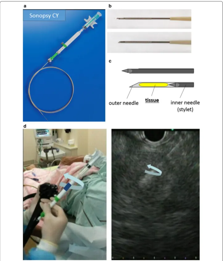
Figure 1 shows the 21-gauge Menghini-type needle (EUS Sonopsy CY), which was originally made for per-forming liver biopsies (Sonopsy C1; HAKKO Medical) [23, 24, 28]. This needle has two features of note. First, the tapered shape of the beveled-edge can facilitate obtaining adequate core tissue with just a few strokes. Second, unlike other FNA and FNB needles, keeping the inner needle attached to the plunger of the syringe within the outer needle during aspiration helps obtain high-quality tissue without crushing. After positioning at the intended puncture site, the outer barrel was quickly moved forward until the puncture needle was slightly inserted into the intended site. For aspiration, the piston of a 10-ml syringe was drawn until it was locked without removal of the inner needle (stylet). Following aspiration for at least 3 s to allow negative pressure to accumulate at the tip, the needle was pushed forward through the intended site, and then, in order to wrench out core tis-sue, we additionally turned the outer barrel until the tip of the needle was confirmed to have been rotated simultaneously within the lesion in the EUS view, just as in the liver biopsy technique [25] known as the “rolling method”. This procedure was repeated only two or three times in order to reduce the risk of contamination with blood induced by performing too many strokes, and then the needle was removed.
As the conventional EUS-FNA, the 22-gauge needles were punctured at the target lesion, and the stylets were removed. The needles were then moved back and forth within the lesion with negative pressure 15–20 times per puncture before being removed. In addition, the fanning technique was used for all 28 patients.
After performing EUS-FNA and EUS-FNB, endoscopic retrograde pancreatocholangiography (ERCP) was basi-cally performed in a single-session [29–31] to achieve pancreatographic imaging and biliary drainage if patients had obstructive jaundice.
On‑site evaluations
All samples were first assessed for cellular adequacy by cytopathologists using Papanicolau staining. After ade-quate cellularity for cytology had been confirmed, the remaining and additional samples were preserved for histological evaluations. At that time, endoscopists tried to identify whitish core tissue on a glass slide, which was so-called “MOSE” (macroscopic on-site quality evalu-ation) [32], and the procedure was performed up to six punctures.
Pathological evaluations
Two pathologists, who blinded to the type of needle, per-formed cytological and histological evaluation. A benign or malignant status was cytologically determined. As a
histological evaluation, formalin-fixed paraffin-embed-ded tissue was stained with hematoxylin–eosin (H&E) and IgG4. In addition, obliterative phlebitis (OP) was diagnosed by Elastica van Gieson staining. According to the ICDC, the LPSP findings (marked lymphoplasma-cytic infiltration, OP, storiform fibrosis [SF], abundant IgG4-positive plasma cells [> 10 cells/high-power field {HPF}]) and the IDCP findings (granulocytic infiltration of duct wall [GEL], and absent or scant IgG4-positive cells) were evaluated. Based on these findings, level 1 and 2 criteria were adopted for the diagnosis of AIP.
In addition, the total area of the acquired tissue, except for mucus or blood contamination, was accurately meas-ured under a photomicroscope (microscope, BX53, Olympus, Tokyo, Japan; camera, DP73, Olympus) using imaging software (cellSens, standard 1.8.1, Olympus).
Study outcomes
As a primary outcome, success rate of confirming his-tological diagnosis, such as AIP and pancreatitis due to chronic inflammation, was evaluated. As secondary out-comes, the amount of the acquired tissue, the histological findings, cytological diagnosis, and adverse events were also assessed. The amount of the acquired tissue was defined as the total area of the acquired tissue measured by the imaging software, as described above. Histologi-cal findings and cytologiHistologi-cal diagnosis were assessed, as described in the Pathological evaluations. Adverse events and their severity related to the procedures were defined and graded according to ASGE lexicon [33].
Statistical analyses
Continuous data are presented as medians with the range or interquartile range (IQR). Continuous variables and frequency distribution were compared with the Mann– Whitney U test and Fisher’s exact or χ2 test, respectively.
A P-value < 0.05 was considered statistically significant. All statistical analyses were performed with the Graph-Pad Prism software program, ver. 6.0 (GraphGraph-Pad Soft-ware, San Diego, CA, USA).
Results
Patients’ characteristics
Table 1 summarizes the characteristics of the 28 included patients. In this study, 14 patients each were managed using a Menghini-type needle with the rolling method and conventional-type needles. The ratio of male-to-female patients was approximately 4:1 in both groups. Based on the ICDC, the diagnosis of definitive type 1 AIP was obtainable in 23 patients (82%) but no patients had type 2 AIP before histological evaluation. No pan-creatic lesions developed malignancy during over 3-years follow-up.
Fig. 1 A 21-gauge Menghini-type needle. a Whole image of the 21-gauge endoscopic ultrasound-guided fine needle aspiration biopsy needle (EUS Sonopsy CY). b, c Image of the needle tip before (upper) and after (lower) aspiration using the piston, respectively. d After puncture with aspiration under 10 mL of negative pressure, the handle of the needle was turned until the tip of the needle was confirmed to be rotated in the lesion. To reduce the contamination of blood, this procedure was repeated only three times, and the needle was removed
Clinical findings of the enrolled patients
As shown in Table 1, segmental or focal enlargement of the pancreas was more common in patients treated with conventional-type needles than in those treated with Menghini-type needles (n = 7 [50%] and n = 3 [21%], respectively; not significant). However, a focal lesion of the pancreas head alone, which required a transduo-denal approach for EUS-FNB, was detected in 1 (7%) in both groups. Elevated serum IgG4 levels (≥ 135 mg/ dl) were seen in 75% of patients in both groups. Steroid
administration was conducted in 25 patients (89%) and was effective in all, while the remaining 3 patients showed improvement of pancreatic enlargement without steroid therapy within 6 months.
Success rate of confirming histological diagnosis and acquired tissue area by EUS‑FNB
Definitive histological diagnoses were successfully achieved in all patients treated with a Menghini-type biopsy needle, with a median of 4 (IQR: 3–5) punctures
Table 1 Patients’ characteristics and clinical findings
ICDC International Consensus Diagnostic Criteria, AIP Autoimmune pancreatitis, MPD Main pancreatic duct
a Remaining three patients improved before introduction of treatment with steroids
Menghini‑type Conventional‑type P value
(N = 14) (N = 14)
Male (n [%]) 11 (71) 12 (86) 0.622
Age (years, median [range]) 71 (50–79) 62 (54–75) 0.018
Obstructive jaundice (n [%]) 4 (29) 4 (29) 1.000
HbA1c (%, median [range]) 7.3 (5.2–11.7) 5.8 (5.1–8.7) 0.032
Diagnosis using the ICDC without histological findings of EUS-guided approach (n [%])
Definitive type 1 AIP 13 (93) 10 (71) 0.139
Not definitive type 1 AIP 1 (7) 4 (29)
Type 2 AIP 0 0
Median follow-up period (days [range]) 1116 (297–3303) 1747(683–3303) 0.028 Pancreatic imaging
Enlargement with delayed enhancement
Diffuse/Segmental or Focal 11/3 7/7 0.052
MPD narrowing
Long (≥ 1/3) or multiple/Segmental or Focal 11/2 6/4 0.115
Location of the enlarged part 0.412
Whole 8 (57) 7 (50)
Head 1 (7) 1 (7)
Body 0 1 (7)
Tail 0 2 (14)
Head and body 0 1 (7)
Body and tail 3 (21) 1 (7)
Head and tail 2 (14) 1 (7)
Serology
IgG4 (mg/dl, median [range]) 364.3 (104–1090) 267 (32–1270) 0.828
IgG4 ≥ 135 mg/dl (N [%]) 11 (79) 11 (79) 1.000
Level 1, ≥ 270 mg/dl 9 (64) 7 (50)
Level 2, ≥ 135 mg/dl but < 270 mg/dl 2 (14) 4 (29) Other organ involvement
Sclerosing cholangitis 5 (36) 6 (43) 0.699
Retroperitoneal fibrosis 4 (29) 3 (21) 0.663
IgG4-related kidney disease 3 (21) 0 0.067
Sialadenitis 2 (14) 0 0.142
Dacryoadenitis 1 (7) 0 0.309
Inflammatory bowel disease 0 0 –
(Table 2). In contrast, conventional-type needles obtained the diagnosis in 57% of cases (8/14), with a median of 3 (IQR: 1–4) punctures.
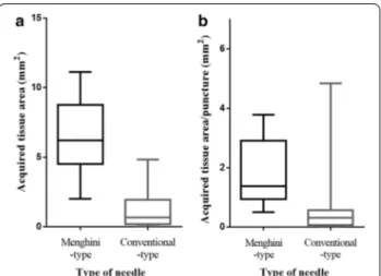
Regarding the sample area of the acquired tissue, the median sample area was significantly larger by a Meng-hini-type needle than by conventional-type needles (6.2 [IQR, 4.5–8.8] versus 0.7 [IQR, 0.2–2.0] mm2, P < 0.001)
(Fig. 2, 3, 4). In addition, the area per punctures was approximately 4 times larger by a Menghini-type needle than by conventional-type needles (1.4 [IQR: 0.9–2.9] versus 0.3 [IQR: 0.1–0.6] mm2/puncture, P < 0.001). In
contrast, the stroke lengths of the needles were not sig-nificantly different between the two approaches. Taken together, these findings show that using the 21-gauge Menghini-type needle with the rolling method was a considerably superior approach to using conven-tional 22-gauge needles for histological evaluations, resulting in obtaining abundant tissue with little blood contamination.
Histological findings by EUS‑FNB
Regarding the histopathological findings based on the ICDC, lymphoplasmacytic infiltration, abundant IgG4-postive cells, SF, and OP were found by a Menghini-type needle in 12 (86%), 9 (64%), 5 (36%), and 1 (7%) patients, respectively (Table 2, Fig. 5). Consequently, 9
patients (64%) were histopathologically diagnosed with AIP (level 1 and 2 criteria of LPSP met by 5 [36%] and 4 [29%] patients, respectively). This histological finding
Table 2 Results Associated with EUS-FNA and EUS-FNB
IQR interquartile range, HPF high-power field
Menghini‑type 21‑gauge Conventional‑type 22‑gauge P value
N = 14 N = 14
Needles Sonopsy 14 Expect 7
Sonotip 4 EchoTip 3
Route for puncture 0.231
Transgastric 12 (86) 8 (57)
Transduodenal 1 (7) 4 (29)
Transgastric and transduodenal 1 (7) 2 (14)
Number of punctures (median [IQR]) 4 (3–5) 3 (1–4) 0.016
Stroke length of needles (median [IQR]) 1.6 (1.5–2.0) 1.5 (1.5–1.9) 0.664 Successful confirmation of histological diagnosis (n [%]) 14 (100) 8 (57) 0.006 A confirmed cytological diagnosis of no malignancy (n [%]) 14 (100) 12 (86) 0.142
Adverse events (n [%]) 2 (14) 0 0.142
Histological findings
Periductal lymphoplasmacytic infiltrate without granulocytic infiltration 12 (86) 4 (29) 0.002
Obliterative phlebitis 1 (7) 0 0.310
Storiform fibrosis 5 (36) 0 0.014
Abundant (> 10 cells/HPF) IgG4-positive cells 9 (64) 0 < 0.001
Histological diagnosis for AIP < 0.001
Level 1 5 (36) 0
Level 2 4 (29) 0
Fig. 2 Comparison of the amount of acquired tissue area measured by imaging software “cellSens®”. a The median acquired sample area of the tissue, except for that with blood contamination, was markedly larger with the Menghini-type needle than with conventional-type needles (6.2 [IQR, 4.5–8.8] versus 0.7 [IQR, 0.2–2.0] mm2, P < 0.001). b
The median sample area per punctures was approximately 4 times larger (1.4 [IQR: 0.9–2.9] versus 0.3 [IQR: 0.1–0.6] mm2/puncture,
contributed to a diagnosis of definitive type 1 AIP in a patient who had not been confirmed to have definitive type 1 AIP. On the other hand, conventional-type needle did not provide any diagnosis of AIP without detectable abundant-IgG4-postive cells, though lymphoplasmacytic infiltration was seen in only 29% of cases (4/14).
Confirmed cytological diagnoses of no malignancy by EUS‑FNA
The cytological diagnosis for distinguishing between malignant and benign cases was successfully confirmed
in all patients treated with a Menghini-type needle and in 86% (12/14) of those treated with a conventional-type needle (not significant) (Table 2). The inability to make an accurate diagnosis was attributed to the acquisition of only a few cells showing degradation and the difficulty of performing a puncture due to the hardness of the pancre-atic mass.
Adverse events
Two patients (14%) treated with a Menghini-type needle with the rolling method had mild pancreatitis (Table 2).
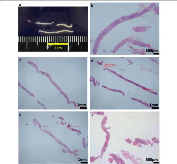
Fig. 3 Pancreatic tissue obtained by the 21-gauge Menghini-type needle with the rolling method. a A macroscopic image shows abundant whitish tissue with little blood contamination. b–f Microscopic images of the tissue with hematoxylin–Eosin (HE) staining are shown among five representative cases. Exceedingly long, high-quality pancreatic tissues can be acquired by this procedure
These patients had been punctured six times (transgas-tric and transuduodenal routes three times) in one and five times (transgastric route) in the other for the EUS procedure. Both patients had also undergone essential therapeutic-ERCP in a single session to achieve biliary drainage with pancreatographic imaging. These mild pancreatitis improved immediately with conservative therapy. No bleeding occurred in either group.
Discussion
In this study, the 21-gauge Menghini-type biopsy nee-dle with the rolling method was superior to the conven-tional 22-gauge FNA needles in terms of the histological diagnosis of AIP. We clearly demonstrated the reason was that the Menghini-type biopsy needle obtained remarkably large tissue by measurement of sample area of acquired tissue. Thus, definitive histological diagnosis was achieved in all 14 patients and the histological diag-nosis of type 1 AIP was confirmed in 64% of them accord-ing to the ICDC, by EUS-FNB usaccord-ing the Menghini-type biopsy needle with the rolling method.
The ICDC published in 2011 do not recommend EUS-FNA or EUS-FNB for the histological diagnosis of AIP, as the findings are inconclusive [2]. However, developments in EUS-FNA technology have improved the EUS-FNB-based assessment of several pancreatic masses, including pancreatic cancer, pancreatic neuroendocrine tumors, and AIP. Previous published articles on the histologi-cal diagnosis for AIP by EUS-FNB were summarized in Table 3. The first of such reports described an EUS trucut biopsy with a 19-gauge trucut needle as a safe and accu-rate procedure for preserving the tissue architecture and aiding in the diagnosis of AIP [11, 12]. Also, a 19-gauge FNA needle provided a high rate of successful histologi-cal evaluations [14]. However, these needles are quite
difficult to be handled under scope angulation and be punctured within the pancreatic mass of the head via the trasduodenal route [4, 11, 12, 26, 27], and an ex vivo study revealed that both 19-gauge needles induced greater resistance than 22- and 25-gauge needles for EUS-FNA [27], suggesting that these 19-gauge needles might be restricted to use by experts because of the han-dling difficulty.
Regarding the use of 22-gauge FNA needles for histo-logical evaluations for the diagnosis of AIP, three pro-spective studies have been already conducted. Kanno et al. reported that pancreatic tissues with more than 1 HPF were obtained in 80% (62/78) of patients, and AIP with an ICDC level of 1 or 2 was diagnosed in 58% (45/78) of the patients [17]. Cao et al. also reported that adequate tissue for histological evaluation was obtained in 93% (25/27), AIP with an ICDC level of 1 or 2 was diagnosed in 63% (17/27) of the patients and even head lesions were successfully punctured by a 22-gauge nee-dle in the majority of patients (89%; 24/27) [19]. On the other hand, Morishima et al. reported that 22-gauge nee-dles provided no patient with AIP with an ICDC level 1, and were not effective for the diagnosis of AIP due to the difficulty of obtaining pathological findings meeting three or more of the LPSP items [18]. A previous report comparing pancreatic biopsy specimens and surgically resected specimens showed that pancreatic specimens by core biopsy provided adequate histologic findings for AIP in only 22% of cases (2/9), whereas surgical speci-mens almost always provided sufficient findings for mak-ing a diagnosis [34]. In addition, the patchy distribution of specific histological findings, such as LPSP [35–37] and infiltration of IgG4-positive plasma cells [38] and the different severities [34] can affect the detection rate of histological findings. These data indicate that 22-gauge
Fig. 4 Pancreatic tissue obtained by a conventional 22-gauge needle. a A microscopic image of Hematoxylin–Eosin (HE) staining shows a rather small amount of tissue with substantial fibrin and neutrophils. This tissue proved insufficient for a histological evaluation (yellow ellipse). This case was identical to that in Fig. 3b. b A microscopic image of HE staining shows a rather small amount of tissue with substantial blood. This tissue proved insufficient for a histological evaluation (yellow rectangle). This case was identical to that in Fig. 3c
conventional FNA needles ensure relatively stable tissue collection but the amount of obtained specimen is not always sufficient for the histological diagnosis of AIP.
Recently, this 21-gauge Menghini-type biopsy needle was reported to be more useful for achieving the histo-pathological diagnosis of solid pancreatic masses than a standard 22-gauge needle [23, 24]. The most important feature of this needle is the equipment of an inner needle with plunger. Keeping the inner needle inside the outer needle during suction improves the ability to obtain
adequate core tissue with preservation of a non-crushed tissue architecture. Indeed, this report revealed that the use of this needle was strongly associated with high cel-lularity (OR = 2.99, 95% CI 0.96–9.74, P = 0.062) and low blood contamination (OR = 28.63, 95% CI 6.67–162.44,
P < 0.001) in a histological evaluation [23]. Remarkably, those good-quality tissue with less blood contamina-tion was also achieved in our cases, as shown in Fig. 3. We also consider that the unique approach of making only three passes with rotation of the needle in the lesion
Fig. 5 Histological findings for the diagnosis of AIP acquired by the 21-gauge Menghini-type needle with the rolling method. a Hematoxylin–Eosin (HE) staining shows marked lymphoplasmacytic infiltration and fibrosis in high-power fields (× 400). b IgG4 immunostaining shows abundant IgG4-positive cells (× 400). c HE staining shows storiform fibrosis with spindle-shaped cells and inflammatory cells (× 100). d, e HE and Elastica van Gieson staining show obstructive phlebitis with infiltration of inflammatory cells obstructing the vein (× 100)
Table 3 T he hist olo gic al diagnosis of AIP b y EUS -FNB and EUS -FNA NM no men tion, HPF high-po w er field a S
amples with mor
e than 10 high-po w er fields w er e c onsider ed adequa te samples b A t otal of 28 pa tien ts under w en t immunostaining c I ncluding nine pa tien ts with suspec ted OP d S
amples with mor
e than 5 high-po w er fields w er e c onsider ed adequa te samples e S
amples with mor
e than 1 high-po w er field w er e c onsider ed adequa te samples Ref er enc es Study desig n Needle Number of pa tien ts Suc cessful tissue ac quisition for hist olog ical ev alua tion Type 1 AIP Type 2 AIP Le vel 1 Le vel 2 Lympho ‑ plasmac ytic infiltr ation IgG4 ‑positiv e cells ≥ 10/ HPF St orif orm fibr osis Oblit er ativ e phlebitis Le vel 1 Le vel 2 Le vy [ 11 ] Retr ospec tiv e 19G Quick C or e 3 100% NM NM 66% NM NM NM 0 0 M izuno [ 12 ] Retr ospec tiv e 19G Quick C or e 14 100% 57% 21% 100% 64% 93% 36% 0 0 Imai [ 13 ] Retr ospec tiv e 22G NA-11 J-KB , 22G Echotip 21 100% NM NM 52% 0 0 0 0 0 Iwashita [ 14 ] Retr ospec tiv e 19G E cho Tip 44 93% NM NM 52% 11% 86% 48% 0 0 Ishik awa [ 15 ] Retr ospec tiv e 22G EZ shot 47 91% 19% 11% 34% 35% (10/28 b) 72% 0 0 6% Kanno [ 16 ] Retr ospec tiv e 22G NA-11 J-KB , 22G Echo Tip 25 80% a 56% 24% 92% 36% 80% 40% c 4% 0 Kanno [ 17 ] Pr ospec tiv e 22G Expec t 78 79% e 41% 17% 55% 24% 63% 49% 0 0 M or ishima [ 18 ] Pr ospec tiv e 22G Expec t, 22G Expec t slim, 22G Echo Tip , 22G NA-11 J-KB 45 NM 0 60% 80% 60% 0 0 6% 2% Cao [ 19 ] Pr ospec tiv e 22G E cho Tip ultra 27 93% d 19% 44% 67% 30% 67% 0 0 0 Bhattachar ya [ 20 ] Retr ospec tiv e 22G Shar kC or e 2 100% 100% 0 100% 100% 100% 0 0 0 Kur ita [ 21 ] Pr ospec tiv e 22G A cquir e 50 88% 56% 22% 84% 76% 56% 24% 0 0 20G P rocor e 51 76% 26% 20% 60% 43% 25% 14% This ar ticle Retr ospec tiv e 21G S onopsy 14 100% 36% 29% 86% 64% 36% 7% 0 0
via the “rolling method” contributed to the acquisition of wrenched core tissue with relatively little blood con-tamination, as with the liver biopsy technique using the Majima needle [25]. This method is based on the notion that a large amount of cored, good-quality tissue with lit-tle contamination may be able to be acquired by reducing the number of strokes as much as possible. In addition, even the transduodenal approach resulted in sufficient tissue collection in all 16 patients [23] as well as in 1 patient included in our study (100%), despite some rigid-ity. Taken together, these results strongly support the notion that this 21-gauge Menghini-type biopsy needle with the rolling method can be a reliable way of obtaining abundant tissue with preserved architecture for a histo-logical evaluation in order to make a diagnosis of AIP.
Another important role of EUS-FNA or EUS-FNB for patients with suspected AIP is the need to rule out the presence of malignancy. The accuracy of the cytologi-cal evaluation has recently dramaticytologi-cally improved due to the advent of rapid onsite evaluation (ROSE) as well as the development of various needles and puncture tech-niques, such as the fanning technique [39] and slow-pull technique [40]. In our study, cytological evaluations were consistently successful using both types of needles (93%: 26/28). On the contrary, confirming adequate tissue acquisition for histological evaluations during EUS-FNB is usually difficult, because the obtained sample contains mucus, blood contamination, adipose tissue as well as required pancreatic tissue. As one of solution for over-coming this issue, the identification of a macroscopic vis-ible core which consisted of whitish or yellowish pieces contributed to a high acquisition rate of the histological core with a success rate of 87% (187/216) for all passes in the MOSE study [32]. Thus, the 21-gauge Menghini-type needle combined with the MOSE method can be a pow-erful tool for confirming the histological diagnosis of AIP. Two patients treated with a Menghini-type needle developed pancreatitis after the procedure. We generally performed EUS-FNB and subsequent ERCP in a single session, based on our previous findings, which demon-strated the efficacy and safety of single-session EUS-FNB and ERCP [29–31]. Minaga et al. reported that no adverse event occurred in 47 patients with pancreatic masses treated with the Menghini-type needle [23]. On the other hand, Naito et al. reported that the incidence of post-ERCP pancreatitis was slightly lower in type 1 AIP than non-AIP cohort (1.2% vs. 5.8%, P = 0.119) [41]. The relation between this needle and pancreatitis cannot be referred to in this study, so further assessment is needed with more cases. Theoretically, the needle simply rotates in the pancreas using the additional “rolling method”. Therefore, bleeding can be considered an alarming risk associated with this method, especially if adequate core
tissue is acquired from pancreas. Fortunately, no bleed-ing occurred in any of our cases. However, further assess-ments in more cases will be needed to confirm the safety. On reviewing the previous reports described in Table 3, the utility of some FNB needles, such as the Franseen type (Acquire) and fork-tip type needles (SharkCore), has recently been reported for the diagnosis of AIP. These needles seemed to contribute to a good histo-logical assessment for diagnosing AIP. Interestingly, the Menghini-type needle with the rolling method is unlikely to be inferior to these FNB needles. In addition, one spe-cific merit of this procedure is that even a few strokes can provide a substantial amount of good-quality tissue. This merit will prove particularly beneficial for lesions that are not easy to puncture due to the scope position, helping to improve the diagnostic ability. However, which needle is the best remains unclear, and a randomized control study will be needed to clarify this point.
Several limitations associated with the present study warrant mention. First, this was a retrospective, single-center study with a small number of enrolled patients. However, the amount of specimen acquired by the Menghini-type needle with the rolling method was quite large, as shown in Figs. 2 and 3. In addition, in the two patients treated with both needles, a substantial amount of tissue was acquired by the Menghini-type needle with the rolling method, while relatively little tissue had been acquired with the previously used conventional-type needle, as shown in Fig. 4. Second, we evaluated patients treated with 22-gauge conventional-type needles as his-torical controls, and the needles were selected based on the two experts’ preference. In addition, these patients were characterized by segmental or focal enlargement of the pancreas, the adoption of a transduodenal approach for EUS, and a low number of punctures, which may have resulted in worse outcomes for these patients than for others in the present and previous studies. However, even though several prospective studies conducted in academic institutions with expert pathologists showed excellent outcomes using 22-gauge FNA needles for the diagnosis of AIP [17–19, 21], the diagnostic rate, propor-tion of Level 1 or 2 in ICDC and positive rates of spe-cific histological findings differed markedly between these studies. In reality, the biopsy-based diagnosis of AIP is challenging and remains unstandardized. Just recently, Notohara et al. advocated guidance for diagnos-ing AIP with biopsy tissue in order to standardize pathol-ogy reports of pancreatic biopsies for diagnosing type 1 AIP [42]. These differences in results are also consistent with a previous study, wherein small samples obtained by EUS-FNA induced low diagnostic agreement among pathologists from not only academic institutions but also non-academic institutions [43]. Although the acquired
sample area has not been evaluated in the prospective studies, we believe that their superb outcomes are not necessarily adapted in all institutions around the world. Third, the patients in each group underwent different biopsy techniques, whether the rolling method or the standard aspiration technique. This means that this study was not a pure comparison of both needles. We empha-size that the utility of the 21-gauge Menghini-type needle combined with the rolling method was superior to that of the conventional 22-gauge needle with the standard aspiration technique in terms of the amount of acquired tissue. However, we cannot state whether the needle or technique had a greater effect on the amount of tissue acquired in this study. To clarify this point, a randomized control study comparing the 21-gauge Menghini-type needle with and without the rolling method will need to be conducted. Fourth, all patients were type 1 AIP, so the performance of the Menghini-type needle for the diagno-sis of type 2 AIP remains unclear.
Despite these limitations, however, our study demon-strated that the outcomes in patients treated with the Menghini-type needle with the rolling method were undoubtedly preferable to the outcomes in patients treated with other approaches, as proven by the objective evaluation of the acquired tissue area. This superiority is attributed to the ability to acquire an abundant amount of tissue, which was exactly important for obtaining a histological diagnosis of AIP [22]. In addition, we empha-size that histology of larger sample would contribute to strengthened diagnostic performance and agreement among pathologists of varying expertise. However, fur-ther study is needed to be validated in a large number of AIP patients.
In conclusion, the 21-gauge Menghini-type biopsy nee-dle with the rolling method is useful for the EUS-guided histopathological diagnosis of AIP due to its abundant acquisition of good-quality tissue from the pancreas. It will significantly aid in the definitive diagnosis of AIP.
Abbreviations
AIP: Autoimmune pancreatitis; ASGE: American Society of Gastrointestinal Endoscopy; EUS-FNA: Endoscopic ultrasound-guided fine-needle aspiration; EUS-FNB: Endoscopic ultrasound-guided fine-needle biopsy; GEL: Granu-locytic infiltration of duct wall; HPF: High-power field; ICDC: International Consensus Diagnostic Criteria; IDCP: Idiopathic duct-centric pancreatitis; IQR: Interquartile range; LPSP: Lymphoplasmacytic sclerosing pancreatitis; MOSE: Macroscopic on-site quality evaluation; OP: Obliterative phlebitis; ROSE: Rapid onsite evaluation; SF: Storiform fibrosis.
Acknowledgements Not applicable. Authors’ contributions
Conceptualization: KT; Data curation: KT, TU, YN, KO1, KO2, SK, and TO; Formal analysis: KT, TU, YN, KO2, SK, HK, and HO. Writing-original draft: KT. Supervision and writing-review and editing: TU. All authors read and approved the final manuscript.
Funding Not applicable.
Availability of data and materials
The datasets used and/or analyzed during the current study are available from the corresponding author on reasonable request.
Ethics approval and consent to participate
This study was conducted in compliance with the principles of the Declara-tion of Helsinki, and the protocol was approved by the ethics committee of Fukuyama City Hospital (No. 446). Instead of written or verbal informed consent to participate, this study guaranteed the opportunity of participation refusal by disclose information about this study on our notice board in our institution (opt-out methods). The opt-out method of consent was approved by the Ethics Committee in our hospital.
Consent for publication Not applicable. Competing interests
The authors declare that they have no competing interests. Author details
1 Departments of Internal Medicine, Fukuyama City Hospital, 5-23-1, Zao-cho,
Fukuyama-City, Hiroshima 721-8511, Japan. 2 Departments of Internal
Medi-cine and Pathology, Fukuyama City Hospital, 5-23-1, Zao-cho, Fukuyama-City,
Hiroshima 721-8511, Japan. 3 Department of Gastroenterology and
Hepatol-ogy, Okayama University Hospital, 2-5-1, Shikata-cho, Kita-ku, Okayama-City,
Okayama 700-8558, Japan. 4 Department of Internal Medicine, National
Hospital Organization Fukuyama Medical Center, 4-14-17, Okinogami-cho, Fukuyama-City, Hiroshima 720-8520, Japan.
Received: 12 September 2020 Accepted: 21 December 2020
References
1. Shimosegawa T, Chari ST, Frulloni L, et al. International consensus diag-nostic criteria for autoimmune pancreatitis: guidelines of the Interna-tional Association of Pancreatology. Pancreas. 2011;40(3):352–8. 2. Kamisawa T, Zen Y, Nakazawa, et al. Advances in IgG4-related
pancreato-biliary diseases. Lancet Gastroenterol Hepatol. 2018;3(8):575–85. 3. Detlefsen S, Mortensen MB, Pless TK, et al. Laparoscopic and
percutane-ous core needle biopsy plays a central role for the diagnosis of autoim-mune pancreatitis in a single-center study from Denmark. Pancreas. 2015;44(6):845–58.
4. Sakamoto H, Kitano M, Komaki T, et al. Prospective comparative study of the EUS guided 25-gauge FNA needle with the 19-gauge Trucut needle and 22-gauge FNA needle in patients with solid pancreatic masses. J Gastroenterol Hepatol. 2009;24(3):384–90.
5. Bang JY, Hebert-Magee S, Trevino J, et al. Randomized trial comparing the 22-gauge aspiration and 22-gauge biopsy needles for EUS-guided sampling of solid pancreatic mass lesions. Gastrointest Endosc. 2012;76(2):321–7.
6. Cheng B, Zhang Y, Chen Q, et al. Analysis of needle biopsy vs. fine-needle aspiration in diagnosis of pancreatic and abdominal masses: a prospective, multicenter, randomized controlled. Trial Clin Gastroenterol Hepatol. 2018;16(8):1314–21.
7. Bang JY, Hawes R, Varadarajulu S. A meta-analysis comparing ProCore and standard fine-needle aspiration needles for endoscopic ultrasound-guided tissue acquisition. Endoscopy. 2016;48(4):339–49.
8. Mitri RD, Rimbaş M, Attili F, et al. Performance of a new needle for endo-scopic ultrasound-guided fine-needle biopsy in patients with pancreatic solid lesions: A retrospective multicenter study. Endosc Ultrasound. 2018;7(5):329–34.
9. Mukai S, Itoi T, Yamaguchi H, et al. A retrospective histological compari-son of EUS-guided fine-needle biopsy using a novel franseen needle and a conventional end-cut type needle. Endosc Ultrasound. 2019;8(1):50–7.
10. Di Leo M, Crinò SF, Bernardoni L, et al. EUS-guided core biopsies of pan-creatic solid masses using a new fork-tip needle: A multicenter prospec-tive study. Dig Liver Dis. 2019;51(9):1275–80.
11. Levy MJ, Reddy RP, Wiersema MJ, et al. EUS-guided trucut biopsy in estab-lishing autoimmune pancreatitis as the cause of obstructive jaundice. Gastrointest Endosc. 2005;61(3):467–72.
12. Mizuno N, Bhatia V, Hosoda W, et al. Histological diagnosis of autoim-mune pancreatitis using EUS-guided trucut biopsy: A comparison study with EUS-FNA. J Gastroenterol. 2009;44(7):742–50.
13. Imai K, Matsubayashi H, Fukutomi A, et al. Endoscopic ultrasonography-guided fine needle aspiration biopsy using 22-gauge needle in diagnosis of autoimmune pancreatitis. Dig Liver Dis. 2011;43(11):869–74. 14. Iwashita T, Yasuda I, Doi S, et al. Use of samples from endoscopic
ultrasound-guided 19-gauge fine-needle aspiration in diagnosis of autoimmune pancreatitis. Clin Gastroenterol Hepatol. 2012;10(3):316–22. 15. Ishikawa T, Itoh A, Kawashima H, et al. Endoscopic ultrasound-guided fine
needle aspiration in the differentiation of type 1 and type 2 autoimmune pancreatitis. World J Gastroenterol. 2012;18(29):3883–8.
16. Kanno A, Ishida K, Hamada S, et al. Diagnosis of autoimmune pancreatitis by EUS-FNA by using a 22-gauge needle based on the International Consensus Diagnostic Criteria. Gastrointest Endosc. 2012;76(3):594–602. 17. Kanno A, Masamune A, Fujishima F, et al. Diagnosis of autoimmune
pancreatitis by EUS-guided FNA using a 22-gauge needle: a prospective multicenter study. Gastrointest Endosc. 2016;84(5):797–804.
18. Morishima T, Kawashima H, Ohno E, et al. Prospective multicenter study on the usefulness of EUS-guided FNA biopsy for the diagnosis of autoim-mune pancreatitis. Gastrointest Endosc. 2016;84(2):241–8.
19. Cao L, Wang Y, Wang J, et al. The role of EUS-guided fine needle aspiration in autoimmune pancreatitis: a single center prospective study. Scand J Gastroenterol. 2018;53(12):1604–10.
20. Bhattacharya A, Cruise M, Chahal P. Endoscopic ultrasound guided 22 gauge core needle biopsy for the diagnosis of Autoimmune pancreatitis. Pancreatology. 2018;18(2):168–9.
21. Kurita A, Yasukawa S, Zen Y, et al. Comparison of a 22-gauge Franseen-tip needle with a 20-gauge forward-bevel needle for the diagnosis of type 1 autoimmune pancreatitis: a prospective, randomized, controlled, multi-center study (COMPAS study). Gastrointest Endosc. 2020;91(2):373–81. 22. Notohara K, Kamisawa T, Kanno A, et al. Efficacy and limitations of the histological diagnosis of type 1 autoimmune pancreatitis with endo-scopic ultrasound-guided fine needle biopsy with large tissue amounts. Pancreatology. 2020;20(5):834–43.
23. Minaga K, Yoshikawa T, Yamashita Y, et al. Comparison of the diagnostic performance of newly designed 21-gauge and standard 22-gauge aspiration needles in patients with solid pancreatic masses. Dig Dis Sci. 2019;64(10):2982–91.
24. Mizukawa S, Kato H, Matsumoto K, et al. Effectiveness of Menghini-type needles for endoscopic ultrasound-guided fine-needle aspiration of
pancreatic masses. Dig Dis Sci. 2020. https ://doi.org/10.1007/s1062
0-020-06628 -1.
25. Majima Y, Fujimoto T, Qwai I, et al. Histological diagnosis of hepatocellular carcinoma by a new technique of ultrasound-guided fine needle biopsy. Kanzo. 1988;29(5):628–36.
26. Itoi T, Itokawa F, Sofuni A, et al. Puncture of solid pancreatic tumors guided by endoscopic ultrasonography: a pilot study series compar-ing Trucut and 19-gauge and 22-gauge aspiration needles. Endoscopy. 2005;37:362–6.
27. Itoi T, Itokawa F, Kurihara T, et al. Experimental endoscopy: objective evaluation of EUS needles. Gastrointest Endosc. 2009;69(3 Pt 1):509–16. 28. Matsubara J, Okusaka T, Morizane C, et al. Ultrasound-guided
percu-taneous pancreatic tumor biopsy in pancreatic cancer: a comparison
with metastatic liver tumor biopsy, including sensitivity, specificity, and complications. J Gastroenterol. 2008;43(3):225–32.
29. Noma Y, Kawamoto H, Kato H, et al. The efficacy and safety of single-session endoscopic ultrasound-guided fine needle aspiration and endo-scopic retrograde cholangiopancreatography for evaluation of pancreatic masses. Hepatogastroenterology. 2014;61(134):1775–9.
30. Kawakubo K, Kawakami H, Kuwatani M, et al. Safety and utility of single-session endoscopic ultrasonography and endoscopic retrograde cholan-giopancreatography for the evaluation of pancreatobiliary diseases. Gut Liver. 2014;8(3):329–32.
31. Ascunce G, Ribeiro A, Rocha-Lima C, et al. Single-session endoscopic ultrasonography and endoscopic retrograde cholangiopancreatog-raphy for evaluation of pancreaticobiliary disorders. Surg Endosc. 2010;24(6):1447–50.
32. Iwashita T, Yasuda I, Mukai T, et al. Macroscopic on-site quality evalua-tion of biopsy specimens to improve the diagnostic accuracy during EUS-guided FNA using a 19-gauge needle for solid lesions: a single-center prospective pilot study (MOSE study). Gastrointest Endosc. 2015;81(1):177–85.
33. Cotton PB, Eisen GM, Aabakken L, et al. A lexicon for endoscopic adverse events: report of an ASGE workshop. Gastrointest Endosc. 2010;71(3):446–54.
34. Zamboni G, Lüttges J, Capelli P, et al. Histopathological features of diagnostic and clinical relevance in autoimmune pancreatitis: a study on 53 resection specimens and 9 biopsy specimens. Virchows Arch. 2004;445(6):552–63.
35. Kawaguchi K, Koike M, Tsuruta K, et al. Lymphoplasmacyticsclerosing pancreatitis with cholangitis: a variant of primary sclerosing cholangitis extensively involving pancreas. Hum Pathol. 1991;22(4):387–95. 36. Notohara K, Burgart LJ, Yadav D, et al. Idiopathic chronic pancreatitis with
periductal lymphoplasmacytic infiltration: clinicopathologic features of 35 cases. Am J Surg Pathol. 2003;27(8):1119–27.
37. Suda K, Takase M, Fukumura Y, et al. Histopathologic characteristics of autoimmune pancreatitis based on comparison with chronic pancreatitis. Pancreas. 2005;30(4):355–8.
38. Chu KE, Papouchado BG, Lane Z, et al. The role of Movat pentachrome stain and immunoglobulin G4 immunostaining in the diagnosis of auto-immune pancreatitis. Mod Pathol. 2009;22(3):351–8.
39. Bang JY, Magee SH, Ramesh J, et al. Randomized trial comparing fanning with standard technique for endoscopic ultrasound-guided fine-needle aspiration of solid pancreatic mass lesions. Endoscopy. 2013;45(6):445–50. 40. Nakai Y, Isayama H, Chang KJ, et al. Slow pull versus suction in endoscopic ultrasound-guided fine-needle aspiration of pancreatic solid masses. Dig Dis Sci. 2014;59(7):1578–85.
41. Naitoh I, Nakazawa T, Okumura F, et al. Endoscopic retrograde cholangio-pancreatography-related adverse events in patients with type 1 autoim-mune pancreatitis. Pancreatology. 2016;16(1):78–82.
42. Notohara K, Kamisawa T, Fukushima N, et al. Guidance for diag-nosing autoimmune pancreatitis with biopsy tissues. Pathol Int. 2020;70(10):699–711.
43. van Riet PA, Cahen DL, Biermann K, et al. Agreement on endoscopic ultrasonography-guided tissue specimens: Comparing a 20-G fine-needle biopsy to a 25-G fine-fine-needle aspiration fine-needle among academic and non-academic pathologists. Dig Endosc. 2019;31(6):690–7. Publisher’s Note
Springer Nature remains neutral with regard to jurisdictional claims in pub-lished maps and institutional affiliations.