inducing nickel-allergy in mice.
著者
Kanan Bando, Toshinobu Kuroishi, Shunji
Sugawara, Yasuo Endo
journal or
publication title
Clinical and experimental allergy : journal of
the British Society for Allergy and Clinical
Immunology
volume
49
number
10
page range
1362-1373
year
2019-05-20
URL
http://hdl.handle.net/10097/00130827
doi: 10.1111/cea.13467Interleukin-1 and histamine are essential for inducing nickel-allergy in mice
Short running title: IL-1 and histamine in nickel allergy
manuscript : 4,875 words tables : 2
figures : 9
Kanan Bando1, Toshinobu Kuroishi2, Shunji Sugawara2, Yasuo Endo3
1Division of Orthodontics and Dentofacial Orthopedics, Graduate School of Dentistry,
Tohoku University, 4-1 Seiryo-machi, Aoba-ku, Sendai 980-8575, Japan
2Division of Oral Immunology, Department of Oral Biology, Graduate School of Dentistry,
Tohoku University, 4-1 Seiryo-machi, Aoba-ku, Sendai 980-8575, Japan
3Division of Oral and Maxillofacial Surgery, Graduate School of Dentistry, Tohoku
University, 4-1 Seiryo-machi, Aoba-ku, Sendai 980-8575, Japan
Correspondence
Kanan Bando, Division of Orthodontics and Dentofacial Orthopedics, Graduate School of Dentistry, Tohoku University, 4-1 Seiryo-machi, Aoba-ku, Sendai 980-8575, Japan. E-mail: [email protected]
Abstract
Background: We previously reported that (i) lipopolysaccharide (LPS) is a potent adjuvant for
inducing Nickel (Ni)-allergy in mice at both the sensitization and elicitation steps, (ii) LPS induces Interleukin-1 (IL-1) and histidine decarboxylase (HDC, the histamine-forming enzyme), and IL-1 induces HDC, (iii) Ni-allergy is induced in mast-cell-deficient, but not IL-1-deficient (IL-1-KO) or HDC-KO mice.
Objective: To examine the roles of IL-1 and HDC (or histamine) and their interrelationship
during the establishment of Ni-allergy.
Methods: Ni (NiCl2) 1 mM containing IL-1 and/or histamine was injected intraperitoneally
(sensitization step). Ten days later, test substance(s) were intradermally injected into ear-pinnas (elicitation step), and ear swelling was measured.
Results: In wild-type mice, Ni+LPS or Ni+IL-1 injection at sensitization step followed by Ni alone at elicitation step induced Ni-allergy. In IL-1-KO, injection of Ni+IL-1 (but not
Ni+histamine) was required at both sensitization and elicitation steps to induce Ni-allergy. In HDC-KO, Ni+IL-1+histamine at sensitization step followed by Ni+histamine at elicitation step induced Ni-allergy. In histamine-H1-receptor-deficient mice, IL-1 induced HDC, but was ineffective as an adjuvant for inducing Ni-allergy. In wild-type mice, injection into ear-pinnas of Ni 10 mM alone or Ni 1 mM+LPS induced IL-1, HDC, and a prolonged swelling of ear-pinnas. In non-sensitized mice, injection of IL-1 by itself into ear-pinnas in IL-1-KO mice induced prolonged ear swelling. Ni augmented IL-1 production (both IL-1 and IL-1) and HDC induction in wild-type mice sensitized to Ni.
Conclusions: In mice: (i) For inducing Ni-allergy, IL-1 is essential at both the sensitization and
receptor is also essential for inducing Ni-allergy at both sensitization and elicitation steps, and (iii) the “sensitization to Ni” state may be a state where tissues are primed for augmented production of IL-1 and/or IL-1 in response to Ni. (within 300 words, now 300)
1. INTRODUCTION
Nickel (Ni) is the most frequent metal allergen producing contact hypersensitivity (CHS). However, few adequate murine models exist of Ni-induced CHS or Ni-allergy. Indeed, in animal experiments Ni induces Ni-allergy only at very high concentrations. We previously found that bacterial or chemical inflammatory substances can act as adjuvants for the induction of Ni-allergy .1-5 Among such adjuvants, lipopolysaccharide (LPS), a cell-wall component of
Gram-negative bacteria, exhibits the strongest adjuvant activity, and LPS markedly reduces the concentration of NiCl2 needed to induce Ni-allergy at both the sensitization and elicitation steps
(Table 1),6 and LPS thereby greatly enhances the sensitivity to Ni. However, it is not clear what
mechanisms are involved in this sensitization or in the adjuvant effect of LPS, or indeed such effects by other inflammatory substances.
Histamine is a well-known mediator of allergic inflammation. Histamine formation from the amino acid histidine is catalyzed by the enzyme histidine decarboxylase (HDC). Schayer, Kahlson and their coworkers found a marked increase in HDC activity in various tissues of mice or rats under conditions such as inflammatory reactions,7,8 anaphylaxis,9 injection of various
inflammatory stimulants,10-12 rapid growth,13 repair of wounds,13 and tissue transplantation.14
They emphasized that “newly formed histamine” via enhanced HDC activity might play important roles under conditions such as those listed above.12,13,15At the time of that pioneering
research, however, little was known about the newly formed histamine itself, because of the lack of adequate methods to measure it. Moreover, pre-formed, long-lived mRNA had been thought to be responsible for enhancing HDC activity because actinomycin D (inhibitor of RNA synthesis) was either ineffective or actually potentiated HDC activity.8
histamine is detectable in various tissues and blood during the induction of HDC, indicating its release from cells during the induction of HDC.16,17 (ii) HDC is induced via the formation of
short-lived HDC mRNA.18 (iii) HDC is induced by various inflammatory cytokines including
IL-1, TNF, IL-12, and IL-1819-21 and hematopoietic cytokines (G-CSF, GM-CSF, and IL-3).22-25
(iv) Various bacterial components induce or promote the induction of HDC.26-28 (v) HDC
induction by LPS or IL-1 is independent of mast cells, and the major HDC-inducing cells are non-mast cells (including macrophages and endothelial cells).29 (vi) In mice sensitized to an
antigen, antigen challenge induces HDC in mast cells and non-mast cells and, in this case, the major HDC-inducing cells are mast cells.30 Thus, it should be noted that in addition to its release
from mast cells and/or basophils, histamine is provided via induction of HDC in other cells. We have previously reported the following findings. (i) Among exogenous substances, LPS is particularly potent at inducing HDC in mice,16,26 and C3H/HeJ mice, which lack active receptors
for LPS [i.e., Toll-like receptor 4 (TLR4)], are considerably less responsive to LPS with regard to HDC induction26 and C3H/HeJ mice are insensitive to the adjuvant effect of LPS in Ni-allergy.1
(ii) Among endogenous substances, IL-1 is particularly potent at inducing HDC in mice19,24 and
Ni-allergy is barely induced at all in mice deficient in IL-1 (IL-1-KO),1 HDC (HDC-KO),1,5,31 or
histamine H1 receptors (H1R-KO).31 (iii) Ni-allergy occurs in mast-cell-deficient W/Wv mice,
but not in macrophage-depleted mice.1
LPS is a potent (possibly the strongest) inducer of IL-1,32,33 and IL-1 is primarily associated
with innate immunity.34 Based on these findings and those described above, we earlier
hypothesized that IL-1 and histamine may be essential endogenous molecules mediating the adjuvant effect of LPS or other inflammatory substances for the induction of Ni-allergy in mice at either sensitization or elicitation step.1,5,31 In the present study, on mice, we examined the roles of
or Ni-induced CHS.
2. METHODS 2.1. Animals
Wild-type (WT) BALB/c and C57BL/6N mice were purchased from SLC (Shizuoka, Japan). IL-1-KO mice (deficient in both IL-1 and IL-1) were established from original IL-1-KO and IL-1-KO BALB/c mice.35 HDC-KO and C57BL/6 H1R-KO mice (both C57BL/6) were
established as previously described.36,37 All mice were raised in standard aluminum cages (with a
lid made of stainless steel wires) and allowed standard food pellets (LabMR Stock; Nihon Nosan Inc, Yokohama, Japan) and tap water ad libitum (the latter from a plastic bottle through a stainless steel tube) in an air-conditioned room at 23 ± 1°C and 55 ± 5% relative humidity with a standard cycle of 12 h light and 12 h dark (lights on at 07:00 a.m.). Female or male mice were used in the various experiments. All animal procedures were approved by the Institutional Animal Care and Use Committee of Tohoku University (approval number: 2018DnA-014). All experiments complied with Regulations for Animal Experiments and Related Activities at Tohoku University.
2.2. Reagents
E. coli O55:B5 LPS (prepared by phenol extraction) was purchased from Sigma (St. Louis, MO, USA). Recombinant mouse IL-1β was purchased from BioLegend (CA, USA). NiCl2-6H2O
(99.999 %) was purchased from Sigma-Aldrich (St. Louis, MO, U.S.A.). All other reagents were purchased from Wako Pure Chemical Ind. Ltd. (Osaka, Japan), unless otherwise indicated.
2.3. Sensitization to Ni and elicitation of Ni-allergy
intraperitoneal (i.p.) injection of a solution containing 1 mM NiCl2 (Ni) alone or an indicated
concentration of a test substance(s). Ten days after the sensitization injection, a solution
containing 1 mM Ni alone or an indicated test substance(s) was intradermally (i.d.) injected into ear-pinnas (termed “challenge”), and ear swelling was measured (using a Peacock dial thickness gauge; Ozaki MFG Co. Ltd, Tokyo, Japan), and the induced difference (after vs. before the challenge) was recorded. Throughout this study, a test solution was i.p. injected into a mouse at 0.1 ml/10 g body weight, while 20 l of a test solution was used for i.d. injection into an ear-pinna.
In some experiments, histamine was i.p. or i.d. injected. Injected histamine is known to be rapidly cleared from the circulation. So, in order to keep the effects of the injected histamine, we tested the following concentrations. The concentration of histamine used for i.p. injection (0.1 ml/10 g body weight) was 180 mM, resulting in 1/10 of the LD50. For i.d. injection into
ear-pinnas (20 l/ear), 45 mM histamine was used, the amount of histamine included in such an injection corresponding to about 100-times the histamine normally present within a single ear-pinna (see Section 3.4). These concentrations of histamine did not induce any toxic effects, such as bristling, shock, or swelling.
Incidentally, we showed previously that an epicutaneous injection (Into flanks or ear-pinnas) of a Ni solution containing LPS as an adjuvant is effective at inducing Ni-allergy, and that
injections of Ni and LPS via different routes were similarly effective.1,5 We think that either local
or systemic inflammatory stimuli that produce IL-1 may contribute to the establishment of
sensitization to Ni. In our previous and present studies, we used i.p. injection at sensitization step.
Tissues (ear and/or spleen) were removed after decapitation, and HDC activity was assayed using our previously described method.26,29 HDC activity is expressed as nmol of histamine formed
during a 1 h period of incubation by the enzyme contained in 1 g (wet weight) of tissue (nmol/h/g).
2.5. Measurement of IL-1 in ear-pinnas and blood serum
Test materials were injected i.d. into ear-pinnas (20 l/ear) and whole ear-pinnas were removed after the indicate time. In other experiments, test materials were i.p. injected into mice (0.1 ml/10 g body weight of mouse), and their blood was collected directly into test tubes following
decapitation. Serum was recovered by centrifugation at 2000 g at 4 C, then stored at -80 C until used. Frozen tissue or serum was homogenized in RPMI 1640 solution containing Triton X-100 (5 l/ml), HEPES (10 mol/ml), bovine serum albumin (100 g/ml), gentamicin sulfate (50 g/ml), and proteinase inhibitor cocktail (10 l/ml). The protease inhibitor cocktail contains 4-(2-aminoethyl)benzenesulfonyl fluoride, aprotinin, leupeptin, bestatin, pepstatin A, and E-64. The supernatant obtained by centrifugation (10,000 g for 10 min at 4 C) of the homogenate was subjected to the measurement of cytokines. IL-1 was measured using ELISA kits from Thermo Scientific (Waltham, MA, USA). The procedures used were essentially the same as those described previously,38,39 although the ELISA kits and proteinase inhibitor cocktail came from
different makers than those used previously.
2.6. Culture of peritoneal cells
Thioglycol solution (3 %) was i.p. injected into mice (3 ml/mouse). Three days later, peritoneal cells were collected after i.p. injection of cooled saline (5 ml). The cells were cultured (2 x 10⁵
cells/well in a 96-well plate) under the presence of LPS (100 ng/ml) or NiCl2 (0.5 mM) for 24
hours, and IL-1β in the supernatant was measured.
2.7. Data and Statistical analysis
In this study, each experiment was performed twice, using three male or female mice, and the results confirmed that there are no essential differences between males and females. So, data obtained from experiments using females are presented in the figures, to be consistent with our previous studies. Experimental values are given as the mean ± SD. The statistical significance of differences was analyzed using a Bonferroni multiple comparison test after ANOVA with the aid of InStat software (InStat, Scottsdale, AZ, U.S.A.). P < 0.05 was considered to indicate
3. RESULTS
3.1. IL-1β acts as an adjuvant for inducing Ni-allergy
As shown in Table 1, LPS markedly reduces the minimum allergy-inducing concentration of Ni at both sensitization and elicitation steps. We first examined whether IL-1 can exhibit such an adjuvant effect at sensitization step. In this experiment, a 1 mM Ni solution containing various amounts of IL-1 was i.p. injected into mice, and 10 days later, the mice were i.d. challenged into their ear-pinnas with 1 mM Ni. As shown in Fig. 1A, ear swelling did not occur in mice given either 1 mM NiCl2 alone or 50 µg/ml IL-1 alone at sensitization step, while ear swelling did
occur in mice given both 1 mM Ni and 50 µg/ml IL-1 at sensitization step, indicating that IL-1, when co-injected with 1 mM Ni, is effective as an adjuvant for inducing Ni-allergy. As shown in Fig. 1B, IL-1 was effective even at 5 g/ml (50 g/kg).
3.2. IL-1β induces HDC
LPS is a potent inducer of HDC.16 Indeed, intravenously injected LPS, even at 1 g/kg body
weight, induces HDC within a few hours in various tissues (e.g. spleen, liver, and lung).16 I.p.
injection of LPS (20 g/kg) also induces HDC in various tissues.24 Therefore, we examined
whether i.p. injected IL-1 can induce HDC in ear pinnas. As shown in Fig. 2, IL-1 induced HDC in spleen, as well as ear pinnas, even at 0.04 g/ml (0.4 g/kg), and 1 mM Ni had no significant effect on the IL-1-induced HDC activity. Thus, we supposed that IL-1 might exert its adjuvant effect via an induction of HDC. In the following sections, we examined this
hypothesis by using IL-1-KO and H1R-KO mice.
Ni-allergy
IL-1-KO mice are non- or low-responder mice as regards the Ni-allergy induced by Ni + LPS,1
and LPS and IL-1 are potent inducers of HDC.16,19 Thus, we examined whether co-injection of
IL-1β and 1 mM Ni might induce HDC and Ni-allergy in IL-1-KO mice. As shown in Fig. 3A, i.p. injection of 4 g/ml IL-1β induced HDC in spleen and ear-pinnas similarly between WT and IL-1-KO mice. As shown in Fig. 3B, co-injection of 1 mM NiCl2 and 4 g/ml IL-1β at
sensitization step followed by co-injection of 1 mM Ni and 2.5 g/ml IL-1β at elicitation step induced Ni-allergy in IL-1-KO mice. However, co-injection of Ni and histamine 45 mM, or injection of Ni alone, at elicitation step was ineffective at inducing Ni-allergy in IL-1-KO mice. These results suggest that for Ni-allergy to be induced, IL-1 is required as an adjuvant at both the sensitization step and the elicitation step (i.e., not just at one of them), supporting our previous study.5 As IL-1 has the ability to induce HDC, we examined the relationship between the adjuvant
effect of IL-1 and the induction of HDC (i.e., newly formed histamine), as described in the following sections.
3.4. IL-1β by itself, but not histamine, has the ability to induce allergy-like prolonged ear swelling
The ear-pinna is the tissue containing the highest amount of histamine (ca. 500 nmol/g) in mice.30 The elevation of blood histamine by its release from mast cells after an antigen challenge
in mice previously sensitized to the antigen is rapid, but its duration is short (ending within 3 hours of the antigen challenge).30 In contrast, the elevated blood histamine that occurs via
induction of HDC after i.p. injection of LPS is slow but long-lasting (more than 3 hours).29 Thus,
20 l of a high-concentration histamine solution (45 mM) was injected into each ear (i.e., 900 nmol histamine/ear pinna), i.e., about 100-fold greater than the histamine normally contained within ear pinnas. It should be noted that there was no detectable difference in ear swelling between Ni 1 mM alone and Ni 1 mM + histamine 45 mM, indicating that histamine by itself does not induce allergy-like (long-lasting) ear swelling. However, unexpectedly and importantly, we found that 1 mM Ni + 2.5 g/ml IL-1β induced a prolonged (albeit small magnitude) ear swelling in non-sensitized IL-1-KO mice (Fig. 3B), and IL-1β alone also induced such a swelling (data not shown), indicating that IL-1β by itself has the ability to induce allergy-like prolonged ear swelling.
3.5. The adjuvant activity of IL-1β is mediated by H1R
As described above, IL-1β is effective as an adjuvant in the induction of Ni-allergy via induction of HDC, i.e., via formation of new histamine. Histamine H1 receptors are known to be involved in inflammatory reactions. Thus, we examined whether IL-1β + Ni at sensitization step can induce Ni-allergy in H1R-KO mice. As shown in Fig. 4A, IL-1β induced HDC similarly between WT and H1R-KO mice. However, IL-1β was not effective as an adjuvant for inducing Ni-allergy in H1R-KO mice (Fig. 4B). These results indicate that the adjuvant effect of IL-1β in the
induction of Ni-allergy is mediated by stimulation of H1R by the histamine newly formed via induction of HDC.
3.6. IL-1ß may exhibit its adjuvant activity via an additional effect(s) other than HDC induction
Ni-allergy. Incidentally, the LD50 of i.p. injected histamine is reported to be around 2,020 mg (18
mmol/kg) in mice.40 So, in the present experiment, histamine was i.p. injected at a dose of 1.8
mmol/kg (1/10 of LD50) (i.e., 180 mM histamine solution was i.p. injected at 0.1 ml/10 g body
weight) into WT mice, and these mice showed no detectable toxic signs (such as bristling or shock). Such i.p. injection of histamine, either alone or in combination with 1 mM Ni, did not increase serum IL-1β (Fig. 5A) and was not effective as an adjuvant at sensitization step for inducing Ni-allergy (Fig. 5B). These results indicate that histamine alone is not sufficient as an adjuvant for inducing Ni-allergy, i.e., IL-1ß may exhibit its adjuvant activity via an additional effect(s) as well as by HDC induction.
3.7. In HDC-KO mice, Ni + IL-1β + histamine at sensitization step and Ni + histamine (without IL-1) at elicitation step induces Ni-allergy
Ni-allergy is not induced in HDC-KO mice1,5,31. In the present study, IL-1β exhibited an LPS-like
adjuvant effect (see above), leading us to suppose that the adjuvant effect of IL-1β may be (at least partly) mediated by histamine newly formed via induction of HDC. Hence, we tried to induce Ni-allergy in HDC-KO mice by using combinations of Ni, IL-1β, and/or histamine at both sensitization and elicitation steps. In this experiment, 180 mM histamine was used at sensitization step as in Fig. 5 (where it was ineffective as an adjuvant), and 45 mM histamine at elicitation step as in Fig. 3 (where it did not augment the inflammatory effect of Ni). As shown in Fig. 6, in HDC-KO mice treated at sensitization step with Ni + IL-1β + histamine (but not Ni alone or Ni + IL-1β), challenge with Ni + histamine at elicitation step without IL-1β did induce Ni-allergy.
3.8. Ni by itself has the potential to induce IL-1β and HDC, effects that are augmented by LPS and lead to ear-swelling
(i) Systemic effects of Ni and/or LPS As described above, induction of IL-1 and release of
histamine are required as adjuvants for producing Ni-allergy at both sensitization and elicitation steps, and the adjuvant effect of LPS may be due to its ability to induce IL-1 and HDC. However, as shown in Table 1, Ni alone, albeit at a high concentration (i.e., 10 mM), can sensitize mice to Ni, suggesting that Ni by itself may exhibit an adjuvant effect even if only at high concentrations, possibly via induction of IL-1 and HDC. Thus, we examined the effect of Ni 10 mM. As shown in Fig. 7A, i.p. injected 10 mM Ni significantly increased IL-1 in serum and induced HDC in spleen. However, the effects of 1 mM Ni were very slight, if any. These results indicate that i.p. injected Ni by itself can, at a high concentration, have an adjuvant effect via systemic inductions of IL-1 and HDC in various tissues. Interestingly, combined i.p. injection of 1 mM Ni with LPS augmented the inductions of IL-1 and HDC.
(ii) Local effects of Ni and/or LPS Fig. 7B shows the local effects induced by Ni (1 or 10
mM) and/or LPS (1 mg/ml) when i.d. injected into ear pinnas in WT mice. The local effects of Ni and/or LPS on IL-1 and HDC were, as a pattern, broadly similar to the systemic effects shown in Fig. 7A. Interestingly, Ni 10 mM alone or a combination of Ni 1 mM with 1 mg/ml LPS induced ear swelling. LPS 1 mg/ml alone induced increases in IL-1 and HDC activity that were comparable to those induced by 10 mM Ni (Fig. 7B). However, the ear swelling induced by 1 mg/ml LPS was far smaller than that induced by Ni 10 mM or Ni 1 mM + LPS 1 mg/ml (see Discussion for explanation). It should be noted that a value suggesting a significant amount of IL-1 was detected even in the ears of IL-1-KO mice. This suggests that the values for IL-1 obtained using the present method may include a significant amount of non-IL-1 substances, which might interfere with our ability to detect a small increase in IL-1 (see Section 4.2).
production of IL-1 in vitro by peritoneal cells, although it markedly augmented the LPS-induced production of IL-1.
Collectively, the findings shown in Fig. 7 may be summarized as follows. (i) Ni by itself has the potential to induce IL-1 and HDC (although the present method is limited in its ability to detect their levels). (ii) The combined presence of Ni and LPS leads to augmented inductions of IL-1 and HDC. (iii) Such augmented inductions of IL-1 and HDC may lead to ear swelling.
3.9. Inductions of IL-1 (both and ) and HDC are augmented in mice sensitized to Ni Lastly, we examined the effects of Ni on the inductions of IL-1, IL-1, and HDC in mice
(C57BL/6) sensitized to Ni (Fig. 8). Although IL-1ß tended to increase, no increase in IL-1 or in HDC activity was induced by 1 mM Ni in mice pretreated with 1 mM Ni + LPS (i.e.,
Ni-sensitized mice). However, 5 mM Ni significantly increased all of IL-1, IL-1, and HDC in mice sensitized to Ni, but not in non-sensitized mice. These results indicate that the Ni-induced productions of IL-1 and/or IL-1 and induction of HDC may be augmented in mice sensitized to Ni.
4. DISCUSSION
4.1. Summary of the findings
We previously reported that (i) LPS is a potent adjuvant for inducing Ni-allergy in mice at both sensitization and elicitation steps, (ii) Ni-allergy is not induced in IL-1-KO mice or HDC-KO mice, although Ni-allergy is induced in mast-cell-deficient mice, and (iii) LPS induces IL-1 and HDC, and IL-1 induces HDC. In the present study, we found the following. (a) By supplying IL-1β and/or histamine to IL-1-KO mice and HDC-KO mice, respectively, Ni-allergy can be induced in these mice (Table 2). (b) IL-1, when co-injected with 1 mM Ni, is effective as an adjuvant for inducing Ni-allergy, and the adjuvant effect of IL-1β is mediated via an induction of HDC (i.e., newly formed histamine) and subsequent stimulation of histamine H1R at both sensitization and elicitation steps. (c) IL-1 exhibits its adjuvant effect via additional actions as well as HDC induction (i.e., histamine alone is not sufficient for inducing Ni-allergy) (Table 2). (d) Ni by itself, at a high concentration (10 mM), exhibits activities inducing IL-1 and HDC, and combined injection of 1 mM Ni and LPS augments such inductions of IL-1 and HDC (both systemically and locally) (Fig. 7). (e) The productions of IL-1 and IL-1 by Ni are augmented in mice sensitized to Ni (Fig. 8). (f) When i.d. injected into the ear pinnas of mice not sensitized to Ni , IL-1β (2.5 g/ml) (Fig. 3) or LPS (1 mg/ml) (Fig. 7B) by themselves induce ear swelling, albeit of small magnitude, while a high concentration of Ni (10 mM) alone or Ni (1 mM) + LPS (1 mg/ml) causes marked ear swelling (Fig. 7B). We now discuss these findings.
4.2. Adjuvant effect of IL-1
Like LPS, resin monomers and nitrogen-containing bisphosphonates exhibit adjuvant effects in the induction of Ni-allergy,3,5 and they also stimulate productions of IL-1 and IL-1,5,33,41
although they have no adjuvant effects either in IL-1-KO mice or in macrophage-depleted mice.5
In addition, cell-transfer experiments in IL-1-KO mice have indicated that both sensitization and elicitation steps depend on IL-1.5 In mouse models of the CHS induced by organic haptens such
as dinitrofluorobenzene (DNFB) and trinitrochlorobenzene (TNCB), IL-1 has been shown to be involved in sensitization step. Enk and Katz (1992)42 reported that painting skins with DNFB
stimulates a rapid production of IL-lβ mRNA in Langerhans cells, this being the fastest of the tested cytokine mRNAs (within 15 min after the application of DNFB). Shornick et al. (1996)43
reported that “IL-lβ”-KO (not IL-1-KO) mice showed defective CHS responses to topically applied TNCB, and this defect in IL-1β-KO mice could be overcome either by application of very high doses of TNCB or by local intradermal injection of recombinant IL-1β immediately before the TNCB application, this being similar to the findings concerning Ni-allergy made in the present study. Recently, we reported a mouse model of CHS to hydroquinone (a widespread chemical in our environment) in which hydroquinone stimulated the productions of IL-1 and IL-1, and the CHS to hydroquinone was not induced in IL-1-KO mice.44 These findings,
collectively, strongly support the idea that IL-1 is essential for the establishment of the CHS to either metals or organic haptens.
In the present study, the ear swelling induced by LPS 1 mg/ml alone was very small, although there were increases in IL-1 and HDC activity (Fig. 7B). It should be noted that the IL-1 antibody used in the method for measuring IL-1 cannot distinguish mature IL-1 (active) from pro-IL-1 (inactive). For that reason, we speculate that the LPS-induced IL-1 may be largely inactive pro-IL-1. We intend to examine this in a future study.
Mast cells are well-known providers of histamine, and the present study suggests that histamine is essential as an adjuvant for Ni-allergy. This could be of particular interest for an understanding of how mast cells are involved in contact hypersensitivity. However, the roles of mast cells in hypersensitivity are controversial (including both positive and negative regulations), possibly because of the use of SCF or Kit mutant mice for such studies on mast cells. Dudeck et al. (2011)47 pointed out that Kit mutant W/Wv mice develop various immunologically unusual
features -- such as macrocytic anemia, neutropenia, decreased T cells, and a high rate of
spontaneous dermatitis -- because Kit is also expressed in cells other than the mast cell lineage (in particular, hematopoietic stem cells and precursor cells of most lineages). In our Ni-allergy model induced by LPS + Ni, allergic ear-swelling also occurs in W/Wv and is markedly prolonged in
these mice,1 although allergic swelling in our model can be strongly reduced by prior treatment
with a histamine H1-receptor antagonist (pyrilamine).31 Interestingly, such a prolonged
ear-swelling also occurs in DNFB-induced contact hypersensitivity in W/Wv mice.47 Based on the
results obtained from experiments using mast cell-deficient mice produced by mast cell-specific gene inactivation, Dudeck et al. (2011)47 concluded that (i) the CHS induced by organic haptens
(including DNFB) depends on mast cells, (ii) an early innate inflammatory response to haptens (i.e., inflammatory effects of haptens themselves) is essential for the induction of CHS, and (iii) the early innate inflammatory response to haptens is dependent on mast cell-derived histamine, and consequently the DNFB-induced CHS is reduced by pyrilamine. Surprisingly, however, it should be noted that in mice in which 90 % or more of mast cells have been constitutively depleted, allergic ear-swelling still occurs, although its magnitude is about 50 % of that seen in control mice.47 As described in the above section, organic haptens can stimulate a rapid
that (i) organic haptens can induce HDC even in mast cell-deficient mice, and (ii) in addition to mast cell-derived histamine, the free histamine newly produced via induction of HDC is also involved in the mediation of organic hapten-induced CHS. Therefore, IL-1-induced HDC induction may be a prerequisite for the establishment of CHS in response to either organic haptens or Ni.
In the present study, we showed that histamine did not induce IL-1, and that histamine itself was not effective as an adjuvant in the induction of Ni-allergy (Fig. 5). In IL-1-KO mice, Ni + IL-1 (but not Ni + histamine) was effective at both sensitization and elicitation steps for inducing Ni-allergy (Fig. 3). In HDC-KO mice, Ni + IL-1 + histamine at sensitization step and Ni + histamine at elicitation step induced Ni-allergy (Fig. 6). These results indicate that both IL-1 and histamine are involved at both sensitization and elicitation steps in the induction of Ni-allergy. This implies that the adjuvant effect of IL-1 is due not only to an induction of HDC, but also to additional effects. At present, although we have no data to indicate what such additional effects might be, we speculate that IL-1 may have effects such as increased expressions of TLR4 and/or Ni-binding proteins (such as CXCL445 and heat shock protein 90β46), and activation of immune
competent cells, which may lead to augmented production of IL-1 and/or IL-1.
4.4. Comparison between mice and humans
In 2007, we reported that TLR4 (the receptor for LPS) is important in the induction of Ni-allergy in mice1. Later, Schmidt et al. (2010)48 reported that (i) Ni binds to human TLR4 but not to
murine TLR4, because their TLR4 structures are different (due to an amino-acid displacement), and (ii) Ni-allergy can be induced by Ni alone (i.e., without the addition of any adjuvant) in mice in which human TLR4 is expressed. These findings might be thought to suggest that the
mechanism underlying Ni-allergy is different between mice and humans. However, those findings may also indicate that TLR4 is essential not only in mice but also in humans. Moreover, it should be noted that in the study by Schmidt et al., a high concentration of Ni (10 mM) was used at both the sensitization and elicitation steps to stimulate TLR4 (compare to the concentrations of Ni shown in Table 1), suggesting that the ability of Ni to stimulate TLR4 is much weaker than that of LPS (i.e., Ni stimulates TLR4 only at high concentrations). Thus, stimulation of TLR4 by LPS may be important as an adjuvant effect in humans, too, and thus the mechanism underlying the induction of Ni-allergy may be essentially the same between mice and humans. If so, mouse models may provide data that are directly applicable to the development of strategies against human Ni-allergy.
4.5. Conclusion
On the basis of the above results and discussion, we conclude that (i) IL-1 is essential for inducing Ni-allergy at both the sensitization step and the elicitation step, and HDC induction is involved in the effect of IL-1, (ii) stimulation of H1 receptors is also essential for inducing Ni-allergy at both sensitization and elicitation steps, and (iii) the state described as “sensitization to Ni” may be a state in which tissues are primed for augmented production of IL-1 and/or IL-1 in response to Ni (Fig. 9).
ACKNOWLEDGEMENT
This work was supported by Tohoku University (Sendai, Japan) and Grants from the Japan Society for the Promotion of Science [16K11672 (to Endo)] and [18K17240 (to Bando)]. We are grateful to Dr. Robert Timms for editing the manuscript.
CONFLICT OF INTEREST
REFERENCES
1. Sato N, Kinbara M, Kuroishi T, Kimura K, Iwakura Y, Ohtsu H, Sugawara S, Endo Y. Lipopolysaccharide promotes and augments metal allergies in mice, dependent on innate immunity and histidine
decarboxylase. Clin Exp Allergy. 2007;37:743-751.
2. Sato N, Oizumi T, Kinbara M, Sato T, Funayama H, Sato S, Matsuda K, Takada Sugawara S, Endo Y. Promotion of arthritis and allergy in mice by aminoglycoglycerophospholipid, a membrane antigen specific to Mycoplasma fermentans. FEMS Immunol Med Microbiol. 2010;59:33-41.
3. Takahashi H, Kinbara M, Sato N, Sasaki K, Sugawara S, Endo Y. Nickel allergy-promoting effects of microbial or inflammatory substances at the sensitization step in mice. International Immunopharmacol. 2011;11:1534-1540.
4. Huang L, Kinbara M, Funayama H, Takada H, Sugawara S, Endo Y. The elicitation step of nickel allergy is promoted in mice by microbe-related substances, including some from oral bacteria. International Immunopharmacol. 2011;11:1916-1924.
5. Bando K, Takahshi H, Kinbara M, Tanaka Y, Kuroishi T, Sasaki K, Takano-Yamamoto T, Sugawara S, Endo Y. Resin monomers act as adjuvants in Ni-induced allergic dermatitis in vivo. J Dental Res. 2014;93:1101-1107.
6. Kinbara M, Sato N, Kuroishi T, Takano-Yamamoto T, Sugawara S, Endo Y. Allergy-inducing nickel concentration is lowered by lipopolysaccharide at both the sensitization and elicitation steps in a murine model. Br J Dermatol. 2011;164:356-362.
7. Schayer RW, Ganley OH. Adaptive increase in mammalian histidine decarboxylase activity in response to nonspecific stress. Am J Physiol. 1959;196:295-298.
8. Schayer RW, Reilly MA. Suppression of inflammation and histidine decarboxylase by protein synthesis inhibitor. Am J Physiol. 1968;215:472-476. 9. Kahlson G, Rosengren E, Thunberg R. Accelerated histamine formation in hypersensitivity reactions. Lancet. 1966;i:782-784.
10. Schayer RW, Ganley OH, Rahway NJ. Relationship of increased histidine decarboxylase activity to Bordetella pertussis vaccine sensitization of mice. J
Allergy. 1961;32:204-213.
11. Schayer RW. Relationship of induced histidine decarboxylase activity and histamine synthesis to shock. Am J Physiol. 1960;198:1187-1192.
12. Schayer RW. Evidence that induced histamine is an intrinsic regulator of the microcirculatory system. Am J Physiol. 1962;202:66-72.
13. Kahlson G, Rosengren E. New approaches to the physiology of histamine. Physiol Rev. 1968;48:155-196.
14. Moore TC, Schayer RW. Histidine decarboxylase activity of autografted and allografted rat skin. Transplantation. 1969;7:99-104.
15. Schayer RW. Histamine and circulatory homeostasis. Fedn Proc. 1965;24:1295-1297.
16. Endo Y. Simultaneous induction of histidine and ornithine decarboxylases and changes in their product amines following the injection of Escherichia coli lipopolysaccharide into mice. Biochem Pharmacol. 1982;31:1643-1647.
17. Shiraishi M, Hirasawa N, Koayashi Y, Oikawa S, Murakami A, Ohuchi K. Participation of
mitogen-activated protein kinase in thapsigargin- and TPA-induced histamine production in murine macrophage 264.7 cells. Br J Pharmacol. 2000;129:515-524.
18. Kikuchi H, Watanabe M, Endo Y. Induction by interleukin-1 (IL-1) of the mRNA of histidine
decarboxylase, the histamine-forming enzyme, in the lung of mice in vivo and the effect of actinomycin D. Biochem Pharmacol. 1997;53:1383-1388.
19. Endo Y. Induction of histidine and ornithine decarboxylase activities in mouse tissues by recombinant interleukin-1 and tumor necrosis factor. Biochem Pharmacol. 1989;38:1287-1292.
20. Yamaguchi K, Motegi K, Endo Y. Induction of histidine decarboxylase, the histamine-forming enzyme, in mice by interleukin-12. Toxicology. 2000;156:57-65.
21. Yamaguchi K, Motegi K, Kurimoto M, EndoY. Induction of the activity of the histamine-forming enzyme, histidine decarboxylase, in mice by IL-18 and by IL-18 plus IL-12. Inflammation Res. 2000;49:513-519. 22. Dy M, Lebel B, Schneider E. Histamine-producing cell stimulating factor (HCSF) and interleukin 3
(IL-3): evidence for two distinct molecular entities. J lmmunol. 1986;136:208-212.
23. Schneider E, Pollard H, Lepault F, Guy-Grand D, Minkowski M, Dy M. Histamine-producing
cell-stimulating activity. Interleukin 3 and granulocyte-macrophage colony-stimulating factor induce de novo synthesis of histidine decarboxylase in hemopoietic progenitor cells. J Immunol.
1987;139:3710-3717.
24. Endo Y, Takeda Y, Nitta Y, Rikiishi H, Kumagai K. GM-CSF and G-CSF stimulates the synthesis of histamine and putrescine in the hematopoietic organs in vivo. Immunology Letters. 1992;33:9-14. 25. Dy M, Schneider E. Histamine-cytokine connection in immunity and hematopoiesis. Cytokine Growth
Factor Rev. 2004;15:393-410.
26. Endo Y. Induction of histidine decarboxylase in mouse tissues by mitogens in vivo. Biochem Pharmacol. 1983;32:3835-3838.
27. Ando T, Endo Y, Abe M, Kumagai K. Stimulation of the synthesis of histamine and putrescine in mice by a peptidoglycan of Gram-positive bacteria. Microbiol Immunol. 1994;38:209-215.
28. Funayama H, Huang L, Asada Y, Endo Y, Takada H. Enhanced induction of a histamine-forming enzyme, histidine decarboxylase, in mice primed with NOD1 or NOD2 ligand in response to various Tool-like receptor agonists. Innate Immunity. 2010;16:265-272.
29. Wu X, Yoshida A, Sasano T, Iwakura Y, Endo Y. Histamine production via mast cell-independent induction of histidine decarboxylase in response to lipopolysaccharide and interleukin-1. International Immunopharmacol. 2004;4:513-520.
30. Deng X, Wu X, Yu Z, Arai I, Sasano T, Sugawara S, Endo Y. Inductions of histidine decarboxylase in mouse tissues following systemic antigen challenge: contributions made by mast cells, non-mast cells and IL-1. Int Arch Allergy Immunol. 2007;144:69-78.
31. Kinbara M, Bando K, Shiraishi D, Kuroishi T, Nagai Y, Ohtsu H, Takano-Yamamoto T, Sugawara S, Endo Y. Mast cell histamine-mediated transient inflammation following exposure to nickel promotes nickel allergy in mice. Exp Dermatol. 2016;25:466-471.
32. Dinarello CA, Interleukin-1. Rev Infect Dis. 1984;6:51-95.
a potent inhibitor of bone resorption, on lipopolysaccharide-induced production of interleukin-1 and tumour necrosis factor in mice. Br J Pharmacol. 1998;125:735-740.
34. Dinarello CA. Overview of the IL-1 family in innate inflammation and acquired immunity. Immunol Rev. 2018;281:8-27.
35. Horai R, Asano M, Sudo K, Kanuka H, Suzuki M, Nishihara M, Takahashi M, Iwakura Y. Production of mice deficient in genes for interleukin (IL)-1, IL-1, IL-1/, and IL-1 receptor antagonist shows that IL-1 is crucial in turpentine-induced fever development and glucocorticoid secretion. J Exp Med. 1998;187:1463-1475.
36. Ohtsu H, Tanaka S, Terui T, Hori Y, Makabe-Kobayashi Y, Pejler G,
Tchougounova E, Hellman L, Gertsenstein M, Hirasawa N, Sakurai E, Buzás E, Kovács P, Csaba G, Kittel A, Okada M, Hara M, Mar L, Numayama-Tsuruta K, Ishigaki-Suzuki S, Ohuchi K, Ichikawa A, Falus A, Watanabe T, Nagy A. Mice lacking histidine decarboxylase exhibit abnormal mast cells. FEBS lett. 2001:502:53-56.
37. Inoue I, Yanai K, Kitamura D, Taniuchi I, Kobayashi T, Niimura K, Watanabe T, Watanabe T. Impaired locomotor activity and exploratory behavior in mice lacking histamine H1 receptors. Proc Natl Acad Sci USA. 1996:93:13316-13320.
38. Deng X, Yu Z, Funayama H, Shoji N, Sasano T, Iwakura Y, Sugawara S, Endo Y. Mutual augmentation of the induction of the histamine-forming enzyme, histidine decarboxylase, between alendronate and immuno-stimulants (IL-1, TNF, and LPS), and its prevention by clodronate. Toxicol Appl Pharmacol. 2006;213:64-73.
39. Shikama Y, Nagai Y, Okada S, Oizumi T, Shimauchi H, Sugawara S, Endo Y. Pro-IL-1ß accumulation in macrophages by alendronate and its prevention by clodronate. Toxicol Lett. 2010;199:123-128.
40. O’Neil MJ, Smith A, Heckelman P.E. The Merck Index. 13 th edition;2001. 842 p.
41. Yamaguchi K, Motegi K, Iwakura Y, Endo Y. Involvement of interleukin-1 in the inflammatory actions of aminobisphosphonates in mice. Br J Pharmacol. 2000;130:1646-1654.
Acad Sci USA. 1992;89:1398-1402.
43. Shornick LP, De Togmi P, Mariathasan S, Goellner J, Strauss-Schoenberger J, Karr PW, Ferguson TA, Chaplin DD. Mice deficient in IL-1 manifest impaired contact hypersensitivity to trinitrobenzene. J Exp Med. 1996;183:1427-1436.
44. Bando K, Tanaka Y, Kuroishi T, Sasaki K, Takano-Yamamoto T, Sugawara S, Endo Y. Mouse model of hydroquinone hypersensitivity via innate and acquired immunity, and its promotion by combined reagents. J Invest Dermatol. 2017;137:1082-1093.
45. Kuroishi T, Bando K, Tanaka Y, Shishido K, Kinbara M, Ogawa T, Muramoto K, Endo Y, Sugawara S. CXCL4 is a novel nickel-binding protein and augments nickel allergy. Clin Exp Allergy.
2017;47:1069-1078.
46. Asakawa S, Onodera R, Kasai K, Kishimoto Y, Sato T, Segawa R, Mizuno N, Ogasawara K, Moriya T, Hiratsuka M, Hirasawa N. Nickel ions bind to HSP90β and enhance HIF-1α-mediated IL-8 expression. Toxicology. 2018;395:45-53.
47. Dudeck A, Dudeck J, Scholten J, Petzold A, Surianarayanan S, Köhler A, Peschke K, Vöhringer D, Waskow C, Krieg T, Müller W, Waisman A, Hartmann K, Gunzer M, Roers A. Mast cells are key promoters of contact allergy that mediate the adjuvant effects of haptens. Immunity. 2011;34:973-984. 48. Schmidt M, Raghavan B, Müller V, Vogl T, Fejer G, Tchaptchet S, Keck S, Kalis C, Nielsen PJ, Galanos C,
Roth J, Skerra A, Martin SF, Freudenberg MA, Goebeler M. Crucial role for human Toll-like receptor 4 in the development of contact allergy to nickel. Nat Immunol. 2010;11:814-819.
Figure 1. IL-1β acts as an adjuvant for inducing Ni-allergy in WT mice. Sensitization and
challenge were carried out as indicated in WT C57BL/6 mice. Each value is the mean SD (n = 6 ears). *P < 0.05 vs. Ni 1mM alone.
Figure 2. IL-1β induces HDC in WT mice. Saline or the indicated concentration of Ni and/or
IL-1 was i.p. injected into WT C57BL/6 and, 4 h later, spleen and ears were subjected to measurement of HDC activity. Each value is the mean SD (n = 3 spleens or 6 ears). *P < 0.05.
Figure 3. In IL-1-KO mice, injected IL-1β induces HDC and is effective for inducing Ni-allergy. (A) Saline or the indicated concentration of IL-1 was i.p. injected into IL-1-KO Balb/c or WT Balb/c mice. Then, 4 h later, the spleen and ears were subjected to measurement of HDC activity. Each value is the mean SD (n = 3 spleens or 6 ears). *P < 0.05. (B) Sensitization and challenge were carried out in IL-1-KO mice as indicated. Each value is the mean SD (n = 6 ears). *P < 0.05 vs. (a).
Figure 4. In H1R-KO mice, IL-1β induces HDC, but it is not effective for inducing
Ni-allergy. (A) Saline or the indicated concentration of IL-1 was i.p. injected into WT C57BL/6 or H1R-KO C57BL/6 mice, and, 4 h later, the spleen and ears were subjected to measurement of HDC activity. Each value is the mean SD (n = 3 spleens or 6 ears). *P < 0.05. (B) Sensitization and challenge were carried out as indicated. Each value is the mean SD (n = 6 ears). *P < 0.05 vs. same time in WT.
Figure 5. In WT mice, injected histamine is not effective as an adjuvant for inducing Ni-allergy. (A) The indicated concentration of Ni and/or histamine was i.p. injected into WT
C57BL/6 mice and, 4 h later, the blood was subjected to measurement of IL-1. (B) The sensitization and elicitation were carried out as indicated. Each value is the mean SD (n = 3 serum samples or 6 ears).
Figure 6. In HDC-KO mice, co-injected histamine and IL-1β act as adjuvants for inducing Ni-allergy. Sensitization and challenge were carried out as indicated. Each value is the mean SD (n = 6 ears). *P < 0.05 vs. Ni 1mM alone.
Figure 7. In vivo (i.p. or i.d.) and in vitro effects of Ni on IL-1 and HDC and effects of LPS
in WT C57BL/6 mice. (A) Indicated concentrations of Ni and/or LPS were i.p. injected into WT
mice, and 4 h later IL-1 and HDC activities were measured. (B) Indicated concentrations of Ni and/or LPS were i.d. injected into ear pinnas of WT mice. Then, 24 h later, IL-1, HDC activity, and ear swelling were measured. Each value is the mean SD (n = 3 serum samples, 3 spleens, or 6 ears). (C) Peritoneal cells were treated as described in Methods. The results were expressed as the mean ± SD of a single experiment performed in triplicate. *P < 0.05. Note: For reference, basal levels of IL-1 in the serum and ear of IL-1-KO mice are shown in A and B.
Figure 8. Effects of i.d. injected Ni on IL-1 ( and ) production and HDC induction in
ear-pinnas of WT mice sensitized or non-sensitized to Ni. Saline or Ni 1 mM containing LPS 1
g/ml was i.p. injected into C57BL/6 mice, and 10 days later Ni (1 or 5 mM) was i.d. injected into their ear-pinnas. IL-1 ( and ) and HDC activities were measured 48 h after the challenge.
Each value is the mean SD (n = 6 ears). *P < 0.05. Note: For reference, basal levels of IL-1 and IL-1 in the ear of IL-1-KO mice are shown in A and B.
Figure 9. Roles of IL-1 and HDC in inducing Ni-allergy. Inflammatory stimuli associated with
infection or exposure to chemicals induce IL-1 and/or IL-1 (IL-1/) and HDC (the induced IL-1/ by itself also induces HDC). The histamine (H) newly formed by the HDC induction stimulates H1R. Under the presence of IL-1/ and the newly formed histamine, sensitization to Ni is established. It is supposed that “sensitization to Ni ” is the primed state for inducing a large amounts of IL-1 (and thus HDC), and their augmented production after the second exposure to Ni results in allergic inflammation. As described in the text, it should be noted that (i) at a high concentration, Ni by itself induces IL-1 and HDC [in humans, Ni reportedly acts as an agonist of TLR4 (see Ref. 48)], (ii) the presence of an inflammatory substance(s) augments the induction of IL-1/ and HDC and reduces the allergy-inducing concentration of Ni (Table 1), and (iii) the adjuvant effect of IL-1/ is not due only to the induction of HDC, but also to other unidentified effect(s).
Table 1. Summary of the effects of LPS on the minimum allergy-inducing concentration of NiCl2 (Ni) for
Concentrations of solutions containing LPS and/or Ni At sensitization step* At elicitation step** LPS (g/ml) Ni LPS (g/ml) Ni 0 10 mM 0 10 M 1 1 mM 0.01 2 nM 10 10 M 0.1 20 pM 50 2 M 1 2 pM
* Mice were challenged by 5 mM Ni to induce Ni-allergy
**Mice were sensitized to Ni by a solution containing Ni 0.5 mM and LPS 0.5 g/ml.
In the experiments shown in this table, a solution containing Ni alone or Ni plus LPS (Ni-LPS solution) was injected at sensitization and elicitation steps. At sensitization step, 250 l of a Ni or Ni-LPS solution was i.p. injected. At elicitation step (10 days after the injection for sensitization), 20 l of a Ni or Ni-LPS solution was i.d. injected into each ear-pinna. The table shows that, for example, without using LPS, 10 mM and 10 M of Ni is needed at sensitization and elicitation steps, respectively, while at sensitization step, if 1 g/ml LPS is included, 1 mM Ni is sufficient for sensitizing mice to Ni.
Note:
(i) LPS acts as an adjuvant at markedly low concentrations at elicitation step.
(ii) Concentrations of Ni and LPS used at sensitization step do not affect the concentrations of LPS and Ni required at elicitation step.
(iii) Reported concentrations of Ni in humans who have contact with metals are 0.1-1 mM in sweat, 0.01 ~ 1 M in saliva, and ~1 M in blood (see Ref. 6).
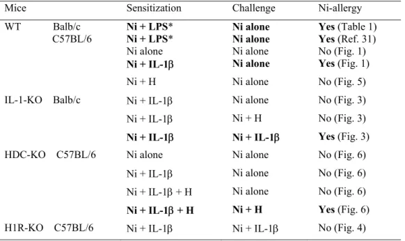
1. Table 2. Summary of the effects of IL-1 and histamine (H) on the induction of Ni-allergy in mice. Test solutions were i.p. injected (sensitization), and 10 days later test solutions were i.d. injected into ear-pinnas (challenge). In these experiments, 1 mM Ni was used for both the sensitization and challenge.
Mice Sensitization Challenge Ni-allergy WT Balb/c C57BL/6 Ni + LPS* Ni + LPS* Ni alone Ni alone Ni alone Ni alone Yes (Table 1) Yes (Ref. 31) No (Fig. 1) Ni + IL-1 Ni alone Yes (Fig. 1) Ni + H Ni alone No (Fig. 5) IL-1-KO Balb/c Ni + IL-1 Ni alone No (Fig. 3) Ni + IL-1 Ni + H No (Fig. 3) Ni + IL-1 Ni + IL-1 Yes (Fig. 3) HDC-KO C57BL/6 Ni alone Ni alone No (Fig. 6) Ni + IL-1 Ni alone No (Fig. 6) Ni + IL-1 + H Ni alone No (Fig. 6) Ni + IL-1 + H Ni + H Yes (Fig. 6) H1R-KO C57BL/6 Ni + IL-1 Ni + IL-1 No (Fig. 4)
*LPS concentrations are 1 or 0.5 g/ml.
Note: (i) HDC is induced in either IL-1-KO or H1R-KO mice by IL-1 (Figs. 3 and 4), and (ii) Ni + IL-1 induces allergy-like prolonged ear swelling in non-sensitized IL-1-KO mice (Fig. 3).