Title
Ventilatory Failure and Successful Management for a Dog with
Severe Cervical Meningioma(Surgery)( 本文(Fulltext) )
Author(s)
ASANO, Kazushi; KADOSAWA, Tsuyoshi; MORI, Takashi;
MIYAMOTO, Toru; UENO, Hiroshi; WATANABE, Kazuhiro;
FUJINAGA, Toru
Citation
[The journal of veterinary medical science] vol.[67] no.[6]
p.[599]-[602]
Issue Date
2005-06-25
Rights
The Japanese Society of Veterinary Science (社団法人日本獣医
学会)
Version
出版社版 (publisher version) postprint
URL
http://hdl.handle.net/20.500.12099/29728
NOTE Surgery
Ventilatory Failure and Successful Management for a Dog with Severe Cervical
Meningioma
Kazushi ASANO1), Tsuyoshi KADOSAWA2), Takashi MORI3), Toru MIYAMOTO4), Hiroshi UENO5),
Kazuhiro WATANABE6) and Toru FUJINAGA7)
1)Laboratory of Veterinary Surgery, Department of Veterinary Medicine, College of Bioresource Sciences, Nihon University, 1866
Kameino, Fujisawa 252–8510, 2)Department of Companion Animal Medicine, School of Veterinary Medicine, Rakuno Gakuen University,
582 Bunkyodai-Midorimachi, Ebetsu 069–8501, 3)Hokuai Animal Hospital, 2–10 Kita 12 Higashi 13, Higashiku, Sapporo 065–0012, 4)Laboratory of Veterinary Surgery, Department of Veterinary Sciences, Faculty of Agriculture, Miyazaki University, 1–1 Nishi,
Gakuen-kibanadai, Miyazaki 889–2192, 5)Department of Clinical Veterinary Science, Obihiro University of Agriculture and Veterinary Medicine,
2–11 Nishi, Inada, Obihiro 080–8555, 6)Department of Veterinary Surgery, Faculty of Applied Biological Sciences, Gifu University, 1–1
Yanagido, Gifu 501–1193 and 7)Laboratory of Veterinary Surgery, Department of Veterinary Clinical Sciences, Graduate School of
Veterinary Medicine, Hokkaido University, Kita 18 Nishi 9, Kitaku, Sapporo 060–0818, Japan (Received 9 September 2004/Accepted 8 February 2005)
ABSTRACT. A 12-year-old intact male mongrel dog with a weight of 22 kg was referred with a complaint of progressive tetraparesis.
Cer-vical myelography revealed an intradural-extramedullary mass at the second cerCer-vical vertebra. After computed tomography (CT) under general anesthesia, the patient showed dyspnea and cyanosis caused by insufficient movement of the chest wall. Positive pressure ven-tilation was therefore initiated. Hemilaminectomy and partial mass removal were performed 12 hr after the CT. The mass was histo-pathologically diagnosed as meningioma. Gradual weaning from the mechanical ventilation lasted for 80 hr after the operation. The patient eventually recovered from the ventilatory failure and the tetraparesis at approximately 6 and 14 days after the operation, respec-tively.
KEYWORDS: canine, cervical meningioma ventilatory failure.
J. Vet. Med. Sci. 67(6): 599–602, 2005
Fatal respiratory paralysis and quadriplegia have been described in animals with complete transverse myelopathy of the cervical spinal cord [6]. There have been few reports on canine patients with ventilatory failure caused by cervi-cal spinal cord disorders [4, 5]. This article describes the complete recovery of a dog with severe and progressive cer-vical meningioma with tetraparesis in which respiratory paralysis appeared after computed tomography (CT) under general anesthesia; the treatment methods included surgical intervention and perioperative positive pressure ventilation (PPV).
A 12-year-old intact male mongrel dog with a weight of 22 kg was referred for tetraparesis that had lasted 4 days. Two months prior to referral, the patient exhibited motor ataxia and partial muscle spasm; his condition had gradually deteriorated despite treatments with steroidal and nonsteroi-dal anti-inflammatory drugs, eventually resulting in tetra-paresis. On initial physical examination, the patient was conscious and retained voluntary control of micturition and defecation. The patient had normal visual responses with normal light reflex and no anisocoria. Tachypnea was present, but cyanosis was not visible. On neurological examination, postural reactions were absent in both thoracic limbs and the right hindlimb and quite weak in the left hind-limb. Spinal reflexes of all limbs were normal to slightly exaggerated. Responses to deep painful stimuli were present in all limbs. Based on the neurological examina-tions, the location most suspected for the lesion was the cer-vical region. The results of hematology and routine serum
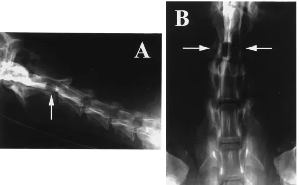
biochemical tests were within normal ranges. Survey radi-ography of the cervical region showed no abnormalities. Cervical myelography was carried out after premedication and intubation under general anesthesia with sevoflurane. Iohexol (240 mg/ml of iodine concentration, 0.4 ml/kg) as a contrast medium was injected into the subarachnoid space via cisternal puncture after removal of an equal volume of cerebrospinal fluid. An intradural-extramedullary mass having a golf-tee appearance was detected on the right side and in the caudal area at the second cervical vertebra (C2)
(Fig. 1). After the cervical myelography, the neurological status of the patient was stable. Cerebrospinal fluid analysis was normal.
CT was performed 1 week after the cervical myelogra-phy, when the clinical signs were almost unchanged. After premedication with flunitrazepam (0.03 mg/kg, intravenous infusion), anesthesia was induced by mask inhalation of sevoflurane, and the dog was intubated, and general anes-thesia was maintained with sevoflurane (1 to 2%) and oxy-gen (2 l/min) for 30 min. CT under contrast enhancement with meglumine iotalamate (20 ml of 480 mg/ml of iodine concentration, intravenous injection) revealed a mass occu-pying most of the right side of the spinal canal and severely compressing the spinal cord toward the left at the caudal portion of C2 (Fig. 2). The patient exhibited cyanosis and
abnormal respiration after anesthesia, as the thoracic cavity hardly expanded despite nostril movements. The patient was therefore re-intubated, and ventilatory management with volume-limited PPV was initiated. Isoflurane (0.5 to
K. ASANO ET AL.
600
1.0%) was used for sedation and restraint. The ventilator settings were controlled to satisfy the following criteria: PaO2>60 mmHg, PaCO2<50 mmHg, and SaO2 (or
SpO2)>90% (Table 1). Simultaneously, fluid therapy and
intravenous steroid therapy (hydrocortisone sodium succi-nate 5 mg/kg, every 8 hr, and dexamethasone 0.5 mg/kg, every 12 hr) were initiated.
Hemilaminectomy and durotomy on the right side of the axis was carried out 12 hr after the beginning of the ventila-tory management. The discolored mass causing the com-pression/deviation of the spinal cord did not involve the nerve root, and it was removed. The patient’s condition did
Fig. 1. Lateral (A) and ventrodorsal (B) myelograms of the cervical region of the patient. The well-defined intradural-extramedullary mass at the right side and the caudal portion in the C2 vertebra had a golf-tee appearance (arrows).
Fig. 2. Transverse (A) and coronal (B) post-contrast CT images of the caudal area within the C2 vertebra of the patient. The enhanced mass was shown to occupy mostly the right side of the spinal canal at the C2 vertebra (arrows).
Table 1. Ventilatory settings on the mechanical ventila-tion
Parameter Range
Respiratory rate 12 breaths/minute
Tidal volume 18–23 mL/kg
Peak airway pressure 15–20 mmHg
Inspiration : expiration ratio 1:2 Insiratory O2 concentration 30–50 % Positive end-expiratory pressure Not used
601
VENTILATORY FAILURE AND MANAGEMENT FOR A DOG
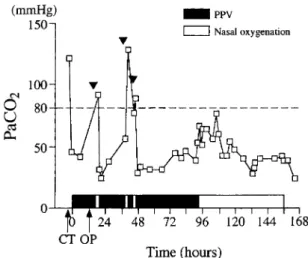
not permit extubation; hence, PPV was continued postoper-atively. Weaning from PPV and alternative oxygenation via a nasal catheter were challenged, but PaCO2 increased to
above 80 mmHg (Fig. 3), despite PaO2 and SaO2 remaining
within their reference ranges. Complete weaning from PPV was possible 80 hr from the operation. Chest wall move-ments had improved, and PaO2, PaCO2, and SaO2 were 88
mmHg, 45 mmHg, and 96%, respectively, with oxygen sup-plied via a nasal catheter. The nasal catheter was removed 144 hr after the operation and the patient’s respiratory func-tion was satisfactory without oxygenafunc-tion. The voluntary control of micturition was preserved during the ventilatory management. The patient was discharged 8 days after the beginning of mechanical ventilation.
The patient gradually recovered from tetraparesis, and could stand and walk 1 week after discharge from the hospi-tal. The patient still had an insufficient gait 10 days after the discharge; however, the postural reactions were improved in all limbs, and chest wall movements were normal. In addi-tion, the patient preserved normal respiratory function with-out oxygen supplement (PaO2, PaCO2, SaO2 and pH were 91
mmHg, 33 mmHg, 96% and 7.41, respectively). The mass surgically removed was histopathologically diagnosed as meningioma. Adjunctive radiation therapy was performed, since the histopathological findings revealed incomplete resection of the tumor margin. Six weeks after the opera-tion, orthovoltage radiation therapy (4 Gy, two times/week) was carried out for 4 weeks (total doses 32 Gy). The patient showed normal respiration and no neurological problems 27 months after the operation.
A complete transverse myelopathy in the upper cervical spinal cord has been documented to result in fatal respira-tory paralysis due to interruption of descending respirarespira-tory
motor pathways or damage to motor neurons of the phrenic nerve [6]. Previous reports have described two dogs with respiratory failure attributable to acute cervical disk pro-lapse [5], and a dog with compression fracture-luxation of the C2-C3 vertebra developed respiratory paralysis
follow-ing dorsal laminectomy of the cervical vertebrae [4]. In the present study, catastrophic deterioration from tachypnea to ventilatory failure in a canine patient with severe progres-sive cervical meningioma was thought to have been trig-gered by hypoperfusion to the impaired cervical spinal cord due to general anesthesia, handling of the patient during CT, side effects (causing irritation or inflammation to the tumor) of intravenous contrast material, and/or tumor embolism or thrombosis after CT. Thus, extreme care is required for han-dling and contrast material injection of dogs with severe cer-vical spinal cord impairment under general anesthesia.
Meningiomas are central nervous system tumors that develop most frequently as benign intradural-extramedul-lary masses in the upper cervical region [3, 7, 10]. The prognosis in dogs with spinal meningioma is reportedly good to excellent [3, 7]. However, it has been suggested that the prognosis might depend on tumor size, degree of neuro-logical impairment at the time of surgery, tumor location, and degree of invasiveness [10]. Despite a severity that caused respiratory paralysis, the cervical spinal cord impair-ment in our case might have been reversible because surgi-cal decompression was performed only about 12 hr after the patient’s ventilatory failure appeared. The patient then recovered from ventilatory failure and tetraparesis at approximately 6 and 14 days after the operation, respec-tively. Therefore, it might be of advantage to perform surgi-cal decompression even in severe cases of respiratory paralysis attributable to cervical spinal cord disorders.
A few previous reports have described mechanical venti-lation in veterinary medicine [1, 2, 5, 8, 9]. In this case, the ventilatory settings were controlled according to the modi-fied guidelines reported previously [1]. When cessation of PPV was attempted prior to 80 hr, PaCO2 exceeded 80
mmHg, despite PaO2 and SaO2 remaining within their
nor-mal ranges. Continued PPV was therefore needed in the patient to prevent intra-alveolar accumulation of CO2.
Withdrawal of PPV was possible after 80 hr.
In conclusion, we have described a dog with cervical meningioma showing ventilatory failure after CT under general anesthesia. Surgical decompression with removal of the tumor and perioperative ventilatory management with PPV enabled the patient’s recovery from ventilatory failure and tetraparesis at approximately 6 and 14 days after the operation, respectively. Prompt surgery and respiratory support are therefore suggested to play an important thera-peutic role in severe cases of ventilatory failure and tetra-paresis secondary to cervical spinal cord disorder.
REFERENCES
1. Bistner, S. I. and Ford, R. B. 1995. pp. 78–83. In: Handbook of Veterinary Procedures and Emergency Treatment, 6th ed., W. Fig. 3. Change in PaCO2 of the patient during ventilatory
support. Time 0 marks the beginning of positive pressure ventilation (PPV). Weaning from PPV was attempted three times (arrowheads), but failed each time due to increase in PaCO2 above 80 mmHg. Withdrawal of PPV was possible 80 hr after the operation. CT: Time at which CT scan was performed. OP: Time at which surgery was performed.
K. ASANO ET AL.
602
B. Saunders, Philadelphia.
2. Bjorling, D. E. 1986. Vet. Surg. 15: 399–406.
3. Fingeroth, J. M., Prata, R. G. and Patnaik, A. K. 1987. J. Am. Vet. Med. Assoc. 191: 720–726.
4. Gambardella, P. C. 1974. J. Am. Vet. Med. Assoc. 165: 542. 5. King, L. G. and Hendricks, J. C. 1994. J. Am. Vet. Med. Assoc.
204: 1045–1052.
6. LeCouteur, R. A. and Child, G. 1995. pp. 629–696. In: Text-book of Veterinary Internal Medicine, 4th ed. (Ettinger, S. J. and Feldman, E. C. eds.), W. B. Saunders, Philadelphia.
7. Levy, M. S., Kapatkin, A. S., Patnaik, A. K., Mauldin, G. N. and Mauldin, G. E. 1997. J. Am. Anim. Hosp. Assoc. 33: 307– 312.
8. Moon, P. F. and Concannon, K. T. 1992. pp. 98–104. In: Cur-rent Veterinary Therapy XI (Kirk, R. W. and Bonagura, J. D. eds.), W. B. Saunders, Philadelphia.
9. Pascoe, P. J. 1988. Semin. Vet. Med. Surg. (Small Anim.) 3: 202–209.
10. Tomlinson, J. 1996. Semin. Vet. Med. Surg. (Small Anim.) 11: 225–234.