切歯交差咬合を発症する短肢症マウス頭蓋骨の骨計
測に関する研究
著者
藤森 修, 赤木 充宏, 平林 義章
雑誌名
名古屋学院大学論集 人文・自然科学篇
巻
47
号
2
ページ
31-45
発行年
2011-01-31
URL
http://doi.org/10.15012/00000389
Summary
Brachymorphic mice are characterized by disproportionally short limb and trunk, and short and thick tail. This disorder is induced by the bm gene. The bm gene is recessive autosomal gene and induces the point mutation in the sulfate groups convert enzymes in the sulfation pass way. Therefore, bm gene induces the undersulfation in many tissues, resulting chondrodystrophia in cartilage tissues. The mice in approximately 3―5% of this strain are with anterior transverse malocclusion. Consequentially, we examined the morphological differences between normal and brachymorphic mice with or without the anterior transverse malocclusion.
To demonstrate the deformation of craniofacial complex and mandible, dry skeletons were prepared and a series of the length and width were measured. All data were compiled and the statistically significant differences between the data were determined by t-test. And moreover, we plotted each biometric point on the graphs.
As a result, when comparing between normal and brachymorphic mice,
1) Total length of craniofacial complex in brachymorphic mice was shorter than that of normal mice (approximately 20%). Especially, the decrease in the length of nasal and occipital region of craniofacial complex in the brachymorphic mice was prominent in comparison with the normal mice.
2) The length of mandible in brachymorphic mice was decreased approximately 10―15% compared to that of normal mice. Especially, the difference in the length of LB-G was noted between normal and brachymorphic mice.
3) When comparing the dental arch of normal and brachymorphic mice, the differences in the length of UB-M3 in the maxilla were more conspicuous than the differences in the length of LB-M3 in the mandible.
4) No significant differences were observed in the width of mandible (CO-CO and G-G) and molars (M1-M1 and M3-M3 in maxilla and mandible) in normal and brachymorphic mice.
切歯交差咬合を発症する短肢症マウス頭蓋骨の
骨計測に関する研究
藤 森 修
1,赤 木 充 宏
1,平 林 義 章
21 名 古 屋 学 院 大 学 リ ハ ビ リ テ ー シ ョ ン 学 部 理 学 療 法 学 科 Nagoya Gakuin University, Faculty of Rehabilitation Science, Department of Physical Therapy
2 名古屋文理大学健康生活学部健康栄養学科 Nagoya Bunri University, Faculty of He-alth and Human Life, Department of Health and Nutrition
はじめに 短肢症マウスは軟骨形成不全による異常に短 縮した四肢,尾および体幹をもつ小人症を自然 発症するマウスである1)。この短肢症は単一の 常染色体劣性遺伝子であるbm遺伝子によって 引き起こされる。bm遺伝子はATP sulfurylase とadenosine-phosphosulfate kinaseの2つの作 用をもつ酵素のポイントミューテーションを起 こし,硫酸基供与体の低形成を介して種々の組 織,特に軟骨組織の低硫酸化を生じる。軟骨組 織の低硫酸化は骨形成,特に置換骨の形成を障 害するため,長骨や頭蓋骨底の形成障害を起こ す2―7)。 本研究に用いたBALB/c-bm/bmマウスは, 短肢症を自然発症するB57BL系bm/bmマウス と正常BALB/cマウスを自然交配することに よりbm遺伝子をBALB/cマウスに導入したも のである8)。この短肢症を自然発症するBALB/ c-bm/bmマウスのうち約10%のものが不正咬 合(切歯部左右交差咬合)を自然発症すること が報告されている9―10)。不正咬合は,その発症 過程において,先天的要因と後天的要因が複雑 に作用する他因子疾患と考えられているが,そ の発症機序については確定されておらず不明な 点が多い。従来,短肢症マウスに関する報告 は,四肢や体幹の骨形成に関するものが大部分 で,頭蓋骨に関するものは少ない3―5)。また, 不正咬合の発症に関する報告においては,実験 動物に器具等を用いて人為的に発症させた方法 や11),口蓋裂に併発して自然発症した例が報告 されているが12),不正咬合単独の自然発症例を 報告した例は見られない。 本研究では,BALB/c系の正常マウスと短肢 症マウスの頭部の乾燥骨格標本を作製し,骨計 測を行って,正常マウスと短肢症マウス間の頭 蓋骨の各部位の距離の比較,また不正咬合の有 無と頭蓋骨の計測値の比較を行い不正咬合発症 の要因について検討した。 材料および方法 1.実験動物 正常マウス(BALB/c--/-)の雌,13週齢 (正常マウス13週齢群)と26週齢(正常マウ ス23週齢群)を各5匹ずつ,正常咬合の短肢 症マウス(BALB/c-bm/bm)の雌,13週齢(正 常咬合短肢症マウス13週齢群)と26週齢(正 常咬合短肢症マウス26週齢群)を各15匹ずつ, 下顎切歯が左方に変位した不正咬合をもつ短肢 When comparing between normal occlusion and anterior transverse malocclusion in the brachymorphic mice,
1) No significant differences were observed in most of the length and width between normal occlusion and anterior transverse malocclusion in the brachymorphic mice.
2) In the brachymorphic mice with anterior transverse malocclusion, the deformation in the incisive (premaxillary) bone was observed.
3) In the brachymorphic mice with anterior transverse malocclusion, the deformation in the mandibular bone was hardly observed.
4) The shift to the left of lower incisor in mandible was caused by the shift to the right of the incisive (premaxillary) bone.
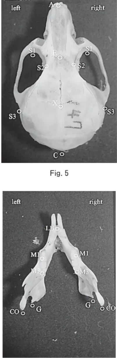
症マウスの雌,13週齢(左方変位不正咬合短 肢症マウス13週齢群)と26週齢(左方変位不 正咬合短肢症マウス26週齢群)を各7匹,下 顎切歯が右方に変位した不正咬合をもつ短肢症 マウスの雌,13週齢(右方変位不正咬合短肢 症マウス13週齢群)と26週齢(右方変位不正 咬合短肢症マウス26週齢群)各5匹を用いた (Fig. 1)。 2.乾燥骨標本作製法 マウスをネンブタール麻酔下に屠殺後,断頭 し頭部の皮膚を隔離した。頭部を10分から20 分水で煮沸し,軽く水洗後,付着している筋肉 片を水中にて実態顕微鏡下で除去した。除肉し た骨格(頭蓋骨および下顎骨)を0.2~0.5%の パパイン溶液に入れ,40~50℃で30分超音波 洗浄を行った後,流水で10~30分洗浄した。 次に骨格を10%ホルマリン溶液に30~60分浸 漬してからに流水で30~60分洗浄後,37℃の 乾燥器で一晩乾燥した13)。 3.写真撮影法 写真撮影は,Kvinnsland14)が提唱した pala-teal planeを基準平面とした。マウス頭蓋骨の 頭蓋底の口蓋骨を水平位に保ち,横に0.5mm 目盛の定規を置いた後,頭蓋骨上面(顔面頭蓋) および下面(上顎)の写真撮影を行った。また, 下顎は下顎骨体下縁を水平位に保ち,上方より 写真撮影を行った。各写真は5倍に引き伸ばし たものを骨計測に用いた。 4.分析方法 1)実測長計測法 計測点および計測部位は関本ら15)の報告に準 じて行った(Figs. 2―13)。 計測点 顔面頭蓋 A:鼻骨最前点 E:前頭鼻骨縫合と矢状縫合の交点 X:頭頂前頭骨縫合と矢状縫合の交点 O:後頭骨最後点 S1:頬骨弓内縁再前部 S2:前頭骨再陥凹部 S3:側頭骨鱗部起始点 上顎 UB:上顎口蓋側歯槽突起正中部先端 F:左右口蓋孔後端中点 M1:上顎第一臼歯近心頬側第一裂溝部 M3:上顎第三臼歯頬側最大豊隆部 OC:外後頭隆起後端 FM:大後頭孔外側縁 下顎 LB:下顎舌側歯槽突起正中部先端 G:下顎角最突出部 CO:顎関節頭先端
Fig. 1 Brachymorphic mouse (BALB/c-bm/bm) with anterior transverse malocclusion at age of 13 weeks. Lower incisors of mandible shift to the right.
M1:下顎第一臼歯近心頬側第一裂溝部 M3:下顎第三臼歯頬側最大豊隆部 計測部位 頭蓋骨部の測定部位 長径 A-E:頭蓋前方部 E-X:頭蓋中央部 X-O:頭蓋後方部 A-O:頭蓋全体 幅径 S1-S1:頭蓋前方部 S2-S2:頭蓋中央部 S3-S3:頭蓋後方部 Fig. 3 Fig. 4
Figs. 2―4 Normal mouse (BALB/c- -/-) at age of 13 weeks, normal occlusion. Fig. 2
下顎骨部の測定部位 長径 LB-CO:顎関節頭までの距離で下顎骨長 LB-G:下顎角までの距離で下顎体長 幅径 CO-CO:左右顎関節頭間 G-G:左右下顎角間 歯列弓の計測 上顎歯列弓 長径 UB-M3:歯列弓長径 幅径 M1-M1:歯列幅径(前方) M3-M3:歯列幅径(後方) Fig. 7
Figs. 5―7 Brachymorphic mouse (BALB/c-bm/ bm) at age of 13 weeks, normal occlusion.
Fig. 6 Fig. 5
下顎歯列弓 長径 LB-M3:歯列弓長径 M1-M1:歯列幅径(前方) M3-M3:歯列幅径(後方) 各実験群の長径および幅径の検定は,それぞれ の平均値にT検定を行って有意差を評価した。 2)座標分析法 顔面頭蓋部および上顎部の計測 左右のS3を結ぶ線分をX軸とし,この線分 の垂直二等分線をY軸として,顔面頭蓋部お よび上顎部の各計測点のX・Y座標を計測し, Excelでグラフ化して,比較検討した(Figs. 2, 3)。 Fig. 8 Fig. 9 Fig. 10
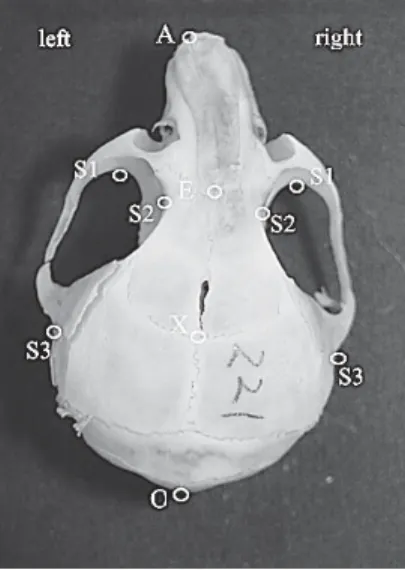
Figs. 8―10 Brachymorphic mouse (BALB/c-bm/ bm) at age of 13 weeks, anterior transverse malocclusion (left shift).
下顎骨の計測 左右のCOを結ぶ線分をX軸とし,この線分の 垂直二等分線をYとして,下顎骨部の各計測点 のX・Y座標を計測し(Fig. 4),Excelでグラ フ化して,比較検討した。 結果 1)実測長計測法 正常マウスの比較 13週齢と26週齢の正常マウスを比較すると (Table 1 A, B, Table 2 Group A-B),顔面頭蓋 では頭蓋全体の長径(A-O)に有意差があった。 頭蓋中央部(E-X)および後方部(X-O)の長
Fig. 11 Fig. 12
Fig. 13
Figs. 11―13 Brachymorphic mouse (BALB/c-bm/ bm) at age of 13 weeks, anterior transverse malocclusion (right shift)
径には有意差は見られず,主にこの差異は頭蓋 前方部(A-E)によるものであった。また,上 顎歯列弓の長径(UB-M3)にも有意差が見ら れた。幅径においては,頭蓋中央部(S2-S2), 上顎および下顎歯列幅径(前方)(M1-M1)に おいて有意差が見られたのみであった(Table
Table 1: Measurements (mm) of craniofacial complex and mandible of mice. Strain: BALB/c - / - BALB/c bm/bm
Occlusion: Normal occlusion Normal occlusion Malocclusion (left shift) Malocclusion (right shift) Age: 13W(A) 26W(B) 13W(C) 26W(D) 13W(E) 26W(F) 13W(G) 26W(H)
Mean S.D. Mean S.D. Mean S.D. Mean S.D. Mean S.D. Mean S.D. Mean S.D. Mean S.D. Craniofacial complex A-E: 7.66 0.23 8.14 0.30 6.60 0.29 7.03 0.42 6.29 0.46 6.45 0.27 5.88 0.58 6.94 0.41 E-X: 7.08 0.30 7.28 0.13 6.63 0.33 6.46 0.51 6.35 0.50 6.35 0.27 6.46 0.16 6.47 0.08 X-O: 8.68 0.21 8.78 0.15 6.40 0.34 6.39 0.41 6.15 0.32 6.33 0.26 6.15 0.08 6.28 0.24 A-O: 23.38 0.31 24.20 0.32 19.63 0.55 19.89 0.51 18.79 1.14 19.12 0.16 18.48 0.65 19.68 0.53 S1-S1 7.28 0.11 7.45 0.17 7.16 0.32 7.19 0.25 6.87 0.32 7.35 0.29 6.90 0.11 7.16 0.07 S2-S2 3.97 0.05 4.10 0.07 4.00 0.10 4.06 0.11 3.87 0.10 3.96 0.13 3.86 0.22 3.98 0.10 S3-S3 11.18 0.16 11.18 0.09 11.42 0.20 11.32 0.26 11.11 0.48 11.16 0.18 11.09 0.07 11.26 0.33 Mandible LB-CO: right 12.61 0.50 13.08 0.21 12.00 0.40 12.08 0.46 11.54 0.64 12.00 0.25 11.73 0.33 12.68 0.86 LB-CO: left 12.72 0.33 13.21 0.21 11.97 0.43 12.01 0.42 11.56 0.48 11.99 0.25 11.60 0.33 12.25 0.60 LB-G: right 12.26 0.25 12.46 0.29 10.89 0.30 11.03 0.28 10.58 0.49 10.67 0.48 10.79 0.26 11.23 0.33 LB-G: left 12.20 0.20 12.54 0.35 10.94 0.35 10.91 0.25 10.50 0.40 10.90 0.26 10.57 0.28 10.86 0.28 CO-CO 9.72 0.24 9.90 0.22 10.45 0.30 10.37 0.58 9.86 0.65 9.92 0.41 9.89 0.31 10.15 0.51 G-G 7.49 0.26 7.56 0.17 7.96 0.35 7.94 0.45 7.20 0.65 7.03 0.55 7.50 0.30 7.72 0.46 Dental arch (Maxilla) UB-M3: right 10.22 0.12 10.61 0.21 8.69 0.29 8.91 0.23 8.45 0.71 8.72 0.13 8.45 0.30 9.00 0.36 UB-M3: left 10.18 0.16 10.59 0.09 8.68 0.25 8.87 0.25 8.47 0.48 8.89 0.21 8.14 0.31 8.79 0.32 M1-M1 4.97 0.10 5.22 0.10 4.90 0.21 4.97 0.15 4.82 0.20 5.10 0.13 4.85 0.10 5.05 0.10 M3-M3 4.67 0.16 4.74 0.07 4.74 0.18 4.64 0.14 4.36 0.66 4.70 0.18 4.58 0.09 4.55 0.15 Dental arch (Mandible) LB-M3: right 6.70 0.35 6.74 0.17 6.26 0.26 6.26 0.30 6.24 0.23 6.40 0.15 6.32 0.10 6.48 0.33 LB-M3: left 6.67 0.25 6.76 0.19 6.67 1.51 6.22 0.26 6.17 0.22 6.34 0.23 6.32 0.17 6.43 0.37 M1-M1 4.85 0.15 5.11 0.09 4.75 0.20 4.78 0.22 4.60 0.33 4.74 0.33 4.48 0.25 4.81 0.21 M3-M3 4.76 0.20 4.79 0.15 4.67 0.27 4.55 0.32 4.35 0.33 4.39 0.30 4.35 0.20 4.49 0.28 S.D.: standard deviation
1 A, B, Table 2 Group A-B)。 短肢症マウスの比較
13週齢と26週齢の正常咬合短肢症マウス を 比 較 す る と(Table 1 C, D, Table 2 Group C-D),頭蓋前部の長径(A-E)に有意差が あったが,頭蓋全体の長径(A-O)には影響 を及ぼしていなかった。これは,24週齢の マウスの頭蓋前部の長径にばらつきがあった ためと考えられる。下顎骨の長径にも有意差 は見られなかった。加えて,上顎歯列弓長 径(UB-M3)に有意差があったが,下顎歯列
Table 2: Statistically significant difference between the groups as determined by T-test.
Groups A-B C-D E-F G-H A-C B-D A-E B-F A-G B-H C-E D-F C-G D-H E-G F-H Craniofacial complex A-E: + ++ n.s. + +++ +++ +++ +++ +++ ++ n.s. ++ +++ n.s. n.s. n.s. E-X: n.s. n.s. n.s. n.s. + ++ + +++ + +++ n.s. n.s. n.s. n.s. n.s. n.s. X-O: n.s. n.s. n.s. n.s. +++ +++ +++ +++ +++ +++ n.s. n.s. n.s. n.s. n.s. n.s. A-O: ++ n.s. n.s. + +++ +++ +++ +++ +++ +++ + ++ ++ n.s. n.s. + S1-S1 n.s. n.s. + + n.s. n.s. + n.s. ++ + n.s. n.s. n.s. n.s. n.s. n.s. S2-S2 + n.s. n.s. n.s. n.s. n.s. n.s. n.s. n.s. n.s. + n.s. n.s. n.s. n.s. n.s. S3-S3 n.s. n.s. n.s. n.s. + n.s. n.s. n.s. n.s. n.s. + n.s. ++ n.s. n.s. n.s. Mandible LB-CO: right n.s. n.s. n.s. n.s. + +++ + +++ + n.s. n.s. n.s. n.s. n.s. n.s. n.s. LB-CO: left + n.s. n.s. n.s. ++ +++ ++ +++ ++ + n.s. n.s. n.s. n.s. n.s. n.s. LB-G: right n.s. n.s. n.s. n.s. +++ +++ +++ +++ +++ ++ n.s. + n.s. n.s. n.s. n.s. LB-G: left n.s. n.s. n.s. n.s. +++ +++ +++ +++ +++ +++ + n.s. n.s. n.s. n.s. n.s. CO-CO n.s. n.s. n.s. n.s. +++ n.s. n.s. n.s. n.s. n.s. + n.s. ++ n.s. n.s. n.s. G-G n.s. n.s. n.s. n.s. + n.s. n.s. n.s. n.s. n.s. ++ ++ + n.s. n.s. n.s. Dental arch (Maxilla) UB-M3: right + + n.s. n.s. +++ +++ +++ +++ +++ +++ n.s. n.s. n.s. n.s. n.s. n.s. UB-M3: left ++ + n.s. + +++ +++ +++ +++ +++ +++ n.s. n.s. n.s. n.s. n.s. n.s. M1-M1 ++ n.s. + + n.s. ++ n.s. n.s. n.s. n.s. n.s. n.s. n.s. n.s. n.s. n.s. M3-M3 n.s. n.s. n.s. n.s. n.s. n.s. n.s. n.s. n.s. n.s. n.s. n.s. n.s. n.s. n.s. n.s. Dental arch (Mandible) LB-M3: right n.s. n.s. n.s. n.s. ++ ++ + ++ n.s. n.s. n.s. n.s. n.s. n.s. n.s. n.s. LB-M3: left n.s. n.s. n.s. n.s. n.s. +++ + + n.s. n.s. n.s. n.s. n.s. n.s. n.s. n.s. M1-M1 + n.s. n.s. n.s. n.s. + n.s. n.s. + n.s. n.s. n.s. n.s. n.s. n.s. n.s. M3-M3 n.s. n.s. n.s. n.s. n.s. n.s. n.s. n.s. + n.s. + n.s. n.s. n.s. n.s. n.s. +: P<0.05, ++: P<0.01, +++: P<0.001, n.s.: no significance
弓長径(LB-M3)や全ての幅径には有意差が 見られなかった(Table 1 C, D, Table 2 Group C-D)。 13週齢と26週齢の左方変位不正咬合短肢 症マウスを比較すると(Table 1 E, F, Table 2 Group E-F),全ての長径において有意な差異 は見られなかった。幅径においては,頭蓋中央 部の幅径(S2-S2)および上顎歯列幅径(前部) (M1-M1)に有意差が見られた(Table 1 E, F,
Table 2 Group E-F)。
13週齢と26週齢の右方変位不正咬合短肢 症マウスを比較すると(Table 1 G, H, Table 2 Group G-H),頭蓋前部の長径(A-E)にやや 有意差が見られたが,その他の長径では顕著 や差異は見られなかった。幅径では頭蓋前方 部(S1-S1)や前方の歯列幅径(M1-M1)にお いて有意差が見られた(Table 1 G, H, Table 2 Group G-H)。 正常マウスと短肢症マウスの比較 13週齢および26週齢の両群において,正 常マウスと短肢症マウスを比較すると(Table 1, Table 2 Group A-C, B-D, A-E, B-F, A-G, B-H),短肢症マウスの頭蓋骨の顔面頭蓋の前 方部,中央部,後方部,頭蓋全体の各部位の長 径が不正咬合の有無にかかわらず有意に短縮し ていた。特に頭蓋前部(A-E)および頭蓋後部 (X-O)において短縮が顕著であった。また下 顎骨長(LB-CO)および下顎骨体長(LB-G) も短肢症マウスにおいて著しく短縮していた。 長径に比較して幅径は正常マウスと短肢症マ ウスの間で差異は少なかった(Table 1, Table 2 Group A-C, B-D, A-E, B-F, A-G, B-H)。 歯列弓を比較すると,歯列弓長径(UB-M3, LB-M3)は短肢症マウスにおいて有意に短縮 していた。特に,下顎(LB-M3)よりも上顎 (UB-M3)の歯列長径が顕著に短縮していた。 歯列幅径に関しては,短肢症マウスの上顎およ び下顎の前部歯列幅径(M1-M1)においてや や短縮が見られるものが散見されたが(Table 1 A, B, D, G, Table 2 Group B-D, A-G),正常 マウスおよび短肢症マウスの両者でほとんど 有意な差異は見られなかった(Table 1, Table 2 Group A-C, B-D, A-E, B-F, A-G, B-H)。 正常咬合短肢症マウスと不正咬合短肢症マウス の比較
13週齢および26週齢の両週齢において,正 常咬合短肢症マウスと不正咬合短肢症マウス を比較すると(Table 1 C, D, E, F, G, H, Table 2 Group C-E, D-F, C-G, D-H, E-G, F-H),13週 齢の正常咬合短肢症マウスと不正咬合短肢症 マウスの間でのみ,不正咬合短肢症マウスの 頭蓋骨の長径にやや短縮が見られた(Table 1 C, E, G, Table 2 C-E, C-G)。また,13週齢な らびに26週齢の両週齢において,不正咬合短 肢症マウスの頭蓋骨の幅径(S2-S2, S3-S3)や 下顎骨の左右顎関節頭間(CO-CO)や左右下 顎角間(G-G)が,正常咬合短肢症マウスより も短縮していた(Table 1 C, E, G, Table 2 C-E, C-G)。
左方変位不正咬合短肢症マウスと右方変位不
正咬合短肢症マウスを比較すると(Table 1 E, F,
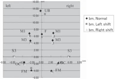
G, H, Table 2 Group E-G, F-H),全ての部位の 長径および幅径において顕著な差異は見られな かった。 2)座標分析法 13週齢の正常咬合短肢症マウス,左方変位 ならびに右方変位不正咬合短肢症マウスの3群 間における計測点を比較すると,顔面頭蓋(Fig. 14)では,鼻骨最前点(A)が,正常咬合短肢
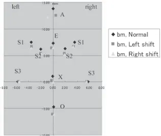
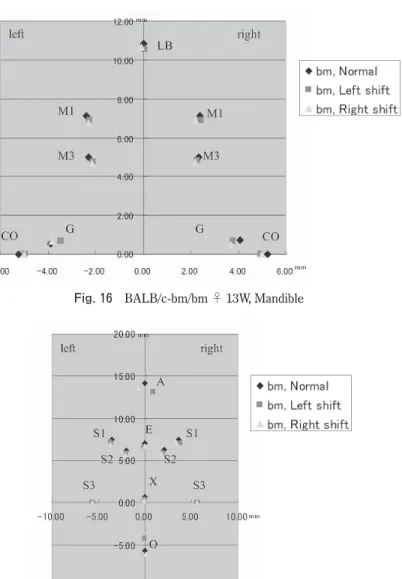
症マウスではほぼ正中部に位置するのに対し て,左方変位不正咬合短肢症マウスでは右方に 変位し,右方変位不正咬合短肢症マウスでは左 方に変位していた。他の計測点は,検索した3 群の間でほぼ同じ位置であった。 上顎部(Fig. 15)では,上顎口蓋側歯槽突 起正中部端(UB)が,正常咬合短肢症マウス において,ほぼ正中部に位置するのに対して, 左方変位不正咬合短肢症マウスでは右方に変位 し,右方変位不正咬合短肢症マウスでは左方に 変位していた。顔面頭蓋の場合と同じく,他の 計測点については,検索した3群の間でほぼ同 じ場所に位置した。 下顎骨(Fig. 16)については,検索した3群 の間で全ての計測点がほぼ同位置にあった。 26週齢の正常マウス,左方変位ならびに右 方変位不正咬合マウスの3群間における計測点 を比較すると,13週齢における結果と全く同
Fig. 14 BALB/c-bm/bm ♂ 13W, Craniofacial complex
じであった(Figs. 17, 18, 19)。 考察 実測長計測法によれば,13週齢および26週 齢の両週齢において,正常マウス群に比較して 短肢症マウス群の頭蓋骨の長径は,前方部,中 央部,後方部,頭蓋全体の全ての部位において 短縮していた。塚本ら16―17)は,短肢症マウス では頭蓋底を形成する成長中心の一つである蝶 形骨間軟骨結合および蝶形後頭軟骨結合の成長 軟骨が低硫酸化を示し,軟骨形成が障害をう けていることを報告している。この部位は頭蓋 骨の前後長の成長に関与する部位なので,頭蓋 骨の特に後方部の成長障害に関与しているもの と考えられた。また,いくつかの報告によれ ば,鼻中隔軟骨が上顎形成のペースメーカーと して重要な役割を演じていることが知られてい る18―20)。軟骨の低硫酸化による鼻中隔軟骨の形 成障害は頭蓋前方部の発育不良を引き起こすと
Fig. 17 BALB/c-bm/bm ♀ 26W, Craniofacial complex Fig. 16 BALB/c-bm/bm ♀ 13W, Mandible
推測される。 今回行った実測長計測法では,正常咬合短肢 症マウスと不正咬合短肢症マウス間,または左 方変位および右方変位不正咬合短肢症マウス間 において,頭蓋骨ならびに下顎骨各部位の長径 および幅径の有意差を検出することができな かった。今後,不正咬合短肢症マウスの頭蓋骨 ならびに下顎骨の実測長計測を行うときは,新 たな計測部位を考慮する必要性があると考え る。 座標分析法によれば,下顎切歯の左方変位は 上顎部の右方変位によること,逆に下顎切歯の 右方変位は上顎部の左方変位によることを示し た。本結果によれば,BALB/c系短肢症マウス に見られる切歯部の左右交差咬合は,下顎の変 位によるものではなく,上顎の変位により形成 されるものと解された。 不正咬合の発症は先天的な要因のみならず,
Fig. 19 BALB/c-bm/bm ♀ 26W, Mandible Fig. 18 BALB/c-bm/bm ♀ 26W, Maxilla
後天的な種々の要因が複雑に絡み合って発症 すると考えられる9―10)。低硫酸化マウスでは軟 骨などの結合組織成分のみならず,上皮性組織 の硫酸化糖タンパク質など広く硫酸化複合糖 質を低硫酸化させていることが確かめられてい る8, 21)。軟骨のみならず,骨組織や歯周靱帯に 含まれるグリコサミノグリカンも低硫酸化して いると推測される。歯周靱帯の低硫酸化はそこ に存在する膠原線維の結合性を低下させ,歯根 を歯槽骨に支持機能を低下させるものと考えら れる22)。この歯周靱帯の支持機能の低下は,本 来,歯周靱帯により吸収されるべき咬合圧を切 歯の側方変位への力へと変化させる可能性が考 えられる。 本研究では,不正咬合を自然発症する短肢症 マウスのマクロ的な骨計測により,不正咬合の 原因について検討し,切歯部左右交差咬合が下 顎の左右方向の変位によるものではなく,頭蓋 前部の左右への変位によるものであることを解 明した。今後さらにこの不正咬合の発症過程を 解明するためには,本肢症マウスを用いて不正 咬合発症途中のマウスの頭蓋前部,特に鼻中隔 および切歯歯周靱帯の生後発生に関して,組織 学的ならびに組織化学的検索をすることが必要 であると考える。 本研究の一部は2008年度名古屋学院大学研 究奨励金を得て行った。 参考文献
1) Lane P, Dickie M: Three recessive mutations producing disproportionate dwarfing in mice. J Hered 59, 300―308, 1968.
2) Kurima K, Warman ML, Krishnan S, Komowicz M, Krueger-JR RC, Deyrup AD, Schwartz NB: A member of a family of sulfate-activating
enzymes causes murine brachumorphism. Proc Natl Acad Sci USA 95, 8681―8685, 1998 3) Orkin RW, Williams BR, Cranley RE, Poppke
DC, Brown KS: Defects in the cartilaginous growth plate of brachymorphic mice. J Cell Biol 73, 287―299, 1977
4) Greene RM, Brown KS, Pratt RM: Autoradiographic analysis of altered grlycosaminoglycan synthesis in the epiphyseal cartilage of neonatal brachy-morphic mice. Anat Rec 191, 19―30, 1978 5) Yamada K, Shimizu S, Brown KS, Kimata K:
The histochemistry of complex carubohydrates in certain organs of homozygous brachymorphic (bm/bm) mice. Histohem J 16, 587―599, 1984 6) Wikstrom B, Gay R, Gay S, Hjerpe A,
Menga-relli S, Reinholt FP, Engfeldt B: Mor-phorogical studies of the epiphyseal growth zone in the brachymorphic (bm/bm) mouse. Virchows Arch (Cell Pathol) 47, 167―176, 1984 7) Wezeman FH, Bollnow MR: I m m u n o h i s -tochemical localization of fibroblast growth factor―2 in normal and brachymorphic mouse tibial growth plate and articular cartilage. Histochem J 29, 505―514, 1997
8) Hirabayashi Y, Fujimori O, Shimizu S: Bruch’s membrane of the brachymorphic mouse. Med Electron Microsc 36, 139―146, 2003
9) Kajii TS, Sugawara Y, Hirabayashi Y, Fujimori O, Sato Y, Iida J: Brachymorphic mice induced by the bm gene exhibit crossbites. Dentistry in Japan 40, 76―79, 2004
10) Kajii TS, Hirabayashi Y, Fujimori O, Tsukamoto Y, Oonshi Y, Sugawara Y, Iida J: Histlogical and biochemical evaluation of temporomandibular j o i n t s o f B A L B C / c - b m / b m m o u s e t h a t spontaneously induces anterior transverse crossbite. Dentistry in Japan 42, 187―190, 2006.
11) Fuentes MA, Opperman LA, Buschang P, Bellinger LL, Carlson DS, Hinton RJ: Lateral functional shif t of the mandible: Part 1.
Effects on condylar cartilage thickness and proliferation. Am J Orthod Dentofacial Orthop 123, 153―159, 2003
12) Nagata M, Amin N, Kannari Y, Hayatsu M, Ohashi Y, Oguro A: Isolated maxillary bending in CL/FR strain mice: observation of craniofacial deformity and inheritance pattern. Cleft Palate Craniofac J 34, 101―105, 1997 13) 八谷 昇,大泰司紀之:骨格標本作製法
Meth-ods of preparing osteal specimens, 北海道大学 図書刊行会,1994
14) Kvinnsland S: The changes in craniofacial growth following papain administration in young mice. Acta Odontol Scand 32, 291―356, 1974
15) 関本恒夫,大竹章夫,鈴木啓之,柏木朗男,宮 尾真知子,河野寿一,原秀一,河内慶子,服部 吉秀,渋井尚武:ラットの顎・顔面頭蓋の成長 発育に関する研究。歯学 65, 647―667, 1997 16) Tsukamoto Y, Kajii TS, Oonishi Y,
Sugawara-Kato Y, Hirabayashi Y, Iida J: Histological and histochemical study of the spheno-occipital synchondrosis of the cranial base on BALB/ c-bm/bm mouse. Orthodontic Waves 65, 166― 172, 2006.
17) Tsukamoto Y, Kajii TS, Oonshi Y, Sugawara-Kato Y, Hirabayashi Y, Iida J: Growth and development of the cranial base in mice that
spontaneously develop anterior transverse crossbite. Americal Journal of Orthodontics and Dentofacial Orthopedics 134, 676―683, 2008
18) Scott JH: The growth of the human face. Proccedings of the Royal Society of Medicine 47, 91―100, 1954
19) Sarnat BG, Wexler MR: Postnatal growth of the nose and face after resection of septal cartilage in the rabbit. Oral Surgery, Oral Medicine, and Oral pathology 26, 712―727, 1968
20) Latham RA: Maxillary development and growth: the septopremaxillary ligament. J Anato 107, 471―478, 1970
21) Maruyama H, Hirabayashi Y, El-Malky M, Okamura S, Aoki M, Itagaki T, Nakamura-Uchiyama N, Nawa Y, Shimada S, Ohta N: Strongyloides venezuelensis: longitudinal distribution of adult wor ms in the host intestine is influenced by mucosal sulfated carbohydrates. Experimental Parasitolgy 100, 179―185, 2002
22) Fujii T, Hirabayashi Y: Histochemical studies of glycosaminoglycans in developing periodontal ligaments of ICR mice. Anat Rec 254, 465―473, 1999