Variation in Lipid Compositions during the
Larval Development of the Prawn (Penaeus
japonicus)
著者
TESHIMA Shin-ichi, KANAZAWA Akio
journal or
publication title
鹿児島大学水産学部紀要=Memoirs of Faculty of
Fisheries Kagoshima University
volume
31
page range
205-212
別言語のタイトル
クルマエビ幼生期における脂質成分の変化
Variation in Lipid Compositions during the Larval
Development of the Prawn (Penaeus japonicus)
Shin-ichi Teshima and Akio Kanazawa*1
Abstract
This paper presents the variation in lipid compositions during the larval development of the prawn, Penaeus japonicus. The lipid concentrations were high in the ovaries, decreased to low levels at nauplius and zoeai stages, and then remained roughly constant during the subsequent stages to
post-larvai. The marked decrease in lipid concentrations during the period of ovaries to early zoeal stages was mainly due to those in both phospholipids (PL) and triglycerides (TG) which were the
major lipid classes throughout the larval developmental stages. The free sterol concentrations did
not vary notably during the larval development from nauplius to post-larvai. As for total lipids, the
proportions of highly unsaturated fatty acids such as 20: 5 « 3,22:6 «> 3, and 20:4 ^ 6 increased during
the period of ovaries to zoeai stage, whereas those of saturated and monoene fatty acids, specifically 16: 0 and 16:1, decreased during the same period. Moreover, the decrease in the proportions of 16: 1 and 18:1 during the above period was attributable mainly to those in both TG and PL, but the
increase in the proportion of 20: 5^3 and 22: 6<*>3 was due mainly to those in PL.
Many papers have been presented up to date with the nutritional requirements of juvenile prawns, Penaeus japonicus1®'. Quite recently, the knowledge on the nutritional requirements of larval P. japonicus has also been obtained by the introduction of artificial diets3_6). Kanazawa4) has shown the necessity of some phospholipids (PL) and ^3-series of fatty acids in the diets for normal growth and survival of P. japonicus larvae. The essentiality of dietary sources of PL such as phosphatidylcholine has also been revealed in the juvenile American lobster, Homarus
americanus1®. Furthermore, Teshima et al9). have demonstrated that the survival of P. japonicus
larvae was notably improved by the addition of cholesterol or some other sterols to a sterol-deficient diet, indicating the necessity of sterols for the larval development of P. japonicus and also the probability of conversion of some C28 and C29 sterols to cholesterol in their bodies.
Thus, the nutritional studies have shown the important role of lipids in the larval development of P. japonicus. On the other hand, however, only a little biochemical studies on the larval development of P. japonicus and other crustacean species have been conducted. As part of investigating lipid metabolism relating to the larval development, the present study is designed to examine the variation in lipid compositions of P. japonicus larvae reared with a live food.
Laboratory of Fisheries Chemistry, Faculty of Fisheries, University of Kagoshima. 4-50-20
206 Mem. Fac. Fish., Kagoshima Univ. Vol. 31 (1982)
Materials and Methods
Mother prawns were caught by the fishermen and transported to this laboratory. Eggs of the prawn, P. japonicus, were hatched on June 5 and September 15, 1981. The prawn larvae were reared in a 500-/ tank at 27~28°C with Chaetoceros gracilis during zoeal stages and both Artemia salina nauplii and C. gracilis for mysis stages as described previously3,5). The sea water (500 /) was fertilized every day with 500 ml of the salt solution containing KNOs (100g), KH2PO4 (10 g), Clewat-Ca (15 g), and Clewat-32 (15 g). Developmental stages of the larval prawn were estimated on 10 random samples as described by HudinagaI0).
Lipids were extracted with chloroform-methanol-water by the method of Bligh and DyerM) from the ovaries and larvae and then separated into lipid classes such as hydrocarbons (HC), steryl esters (SE), triglycerides (TG), free fatty acids (FFA), diglycerides (DG), free sterols (FS; mostly cholesterol), monoglycerides (MG), and phospholipids (PL). The separation of total lipids into the lipid classes was achieved by thin-layer chromatography on Kieselgel G with the solvents,2): 1st, isopropyl ether-acetic acid (96: 4); 2nd, petroleum ether-ether-acetic acid (90: 10: 1) as mentioned previously13'. The concentrations of total lipids and lipid classes were determined by the method of Amenta,4). Fatty acid compositions were analysed by gas-liquid chromatography using 2 columns, 10% DEGS (column temperature, 180°C) and 5% Shinchrom E~71 (column temperature, 225°C) as described previously15'.
Results and Discussion
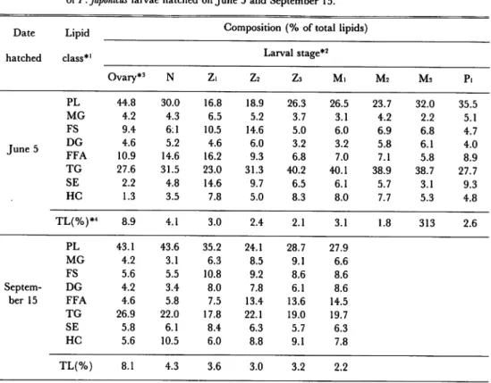
In the present study, 2 feeding trials using the prawn larvae hatched in June and September were carried out to clarify the variation in lipid compositions during the larval development. Fig.l shows the variation in lipid concentrations (% of wet weight) of the ovaries and whole body of larvae during the larval development. Similar results were obtained on the variation in lipid concentrations of the larvae hatched in June and September, indicating no significant difference between the 2 experiments. The lipid concentrations were high in the ovaries as found in the previous study16), decreased to low levels at nauplius and zoeai stages, and then remained roughly constant during the subsequent stages to post-larvai. Table 1 shows the variation in lipid class distributions during the larval development. PL and TG were the major lipid classes in the ovaries and larvae at every stage (Table 1), and both 2 lipid classes were thought to be mainly responsible for a marked decrease in lipids during the period of ovaries to early zoeal stages (Figs. 2 and 3). These results suggest that the PL and TG accumulated in the ovaries are probably utilized during the period of embryogenesis to nauplius as energy sources and as the constituents of the cells. The prawn larvae at nauplius stage feed no food until they grow up to zoeai, so both PL and TG are suspected to play important roles in the metamorphosis of nauplius to zoea.
40 r 30 20 10 t: June samples o: September samples M, Mo •: June samples 0: September samples M, ,., ,2 .3 ^ n2 ..3 r}
Developmental Stages of Larvae
Fig. 1. Variation in lipid concentrations during the larval development of P. japonicus. O, ovaries; N, nauplius; Zi, zoeai; Z2, zoea2; Z3, zoea3; Mi, mysisi; M2, mysis2; M3, mysis3; Pi, post-larvai. Lipid concentration: percentage of wet weight
M, Mo Mo
<-! '-z ^3 nl "2 "3 Developmental Stages of Larvae
Fig. 2. Variation in phospholipid concentrations during the larval development of P.japonicus.
208 Mem. Fac. Fish., Kagoshima Univ. Vol. 31 (1982)
Table 1. Variation in lipid concentrations and lipid class compositions during the development
of P. japonicus larvae hatched on June 5 and September 15.
Date Lipid class*1
Composition(% of total lipids)
hatched Larval stage*2
Ovary*3 N Z. Z2 Z3 M. M2 M3 P. PL 44.8 30.0 16.8 18.9 26.3 26.5 23.7 32.0 35.5 MG 4.2 4.3 6.5 5.2 3.7 3.1 4.2 2.2 5.1 FS 9.4 6:1 10.5 14.6 5.0 6.0 6.9 6.8 4.7 June 5 DG 4.6 5.2 4.6 6.0 3.2 3.2 5.8 6.1 4.0 FFA 10.9 14.6 16.2 9.3 6.8 7.0 7.1 5.8 8.9 TG 27.6 31.5 23.0 31.3 40.2 40.1 38.9 38.7 27.7 SE 2.2 4.8 14.6 9.7 6.5 6.1 5.7 3.1 9.3 HC 1.3 3.5 7.8 5.0 8.3 8.0 7.7 5.3 4.8 TL(%)*4 8.9 4.1 3.0 2.4 2.1 3.1 1.8 313 2.6 PL 43.1 43.6 35.2 24.1 28.7 27.9 MG 4.2 3.1 6.3 8.5 9.1 6.6 FS 5.6 5.5 10.8 9.2 8.6 8.6 Septem DG 4.2 3.4 8.0 7.8 6.1 8.6 ber 15 FFA 4.6 5.8 7.5 13.4 13.6 14.5 TG 26.9 22.0 17.8 22.1 19.0 19.7 SE 5.8 6.1 8.4 6.3 5.7 6.3 HC 5.6 10.5 6.0 8.8 9.1 7.8 TL(%) 8.1 4.3 3.6 3.0 3.2 2.2
*' PL, phospholipids; MG, monoglycerides; FS, free sterols; DG, diglycerides; FFA, free fatty acids; TG, triglycerides; SE, steryl esters; HC, hydrocarbons; TL, total lipids.
*2 Larval stages examined are as follows: N, nauplius; Zi, zoeai; Z2, zoea2; Z3, zoeas; Mi, mysisi; M2,
mysis2; M3, mysis3; Pi, post-larvai.
*3 The ovaries were taken out from the prawns with the following gonadosomatic indices (GSI): June
samples, GSI = 10.1; September samples, GSI = 9.7. External obserbation showed that the female prawns had well-mature ovaries.
30 20 10 #: June samples o: September samples Ll Lz Ll "l "2 Developmental Stages of Larvae
1
Fig. 3. Variation in triglyceride concentrations during the larval development of
P. japonicus.
Crustaceans are incapable of synthesizing cholesterol de novo and require dietary sources of
cholesterol or other sterol for their growth1718*. The free sterol concentrations decreased between the period of ovaries to nauplii, but they remained relatively constant levels throughout the larval stages from nauplius to post-larvai (Fig. 4). Whitney19* have shown in the blue crab, Callinectes
sapidus, that the amount of total sterols did not change significantly throughout development of
eggs, but the percentage of steryl esters decreased steadily and significantly. He has assumed that rapid tissue growth may necessitate a source of free sterols for new membrane formation which was provided at the expense of the steryl ester pool,9). In the present study, the
concentration (mg/g of wet weight) of steryl estersdecreased from 4.70 to 2.60 between the ovary
and nauplius stages in the case of September samples, but it remained almost constant in June
samples (ovaries, 1.96; nauplii, 1.97). The difference betweenJune and September samples may be attributable to the indivual variation on the steryl ester concentrations as pointod out by
Whitney19*.
§: June samples o: September samples
U .1 ^ i.2 ^ n-, n2
Developmental Stages of Larvae
Fig. 4. Variation in cholesterol concentrations during the larval development of
P. japonicus.
210 Mem. Fac. Fish., Kagoshima Univ. Vol. 31 (1982)
MG, DG, FFA, and HC were the minor lipid classes (less than about 15% of total lipids) in the ovaries and larvae at every stage (Table 1). The concentrations of these lipid classes did not
vary markedly throughout the larval developmental stages, except for a decrease in the FFA concentration during the period of ovaries to zoeal stages.
As mentioned above, a notable variation in lipid and lipid class concentrations was observed
during the period of ovaries to early zoeal stages. On September samples, therefore, the variation in fatty acid compositions of lipids, PL, and TG were investigated. The results are given in Tables 2 and 3. The total lipids of the ovaries, nauplii, and zoeae contained palmitic (16:0), palmitoleic (16: 1), oleic (18: 1^9)and eicosapentaenoic (20: 5 *> 3)acids as the major
fatty acids (Table 2). These 4 fatty acids were also predominant in the PL from the ovaries and larvae. The TG of the ovaries and larvae also contained 16:0, 16:1, and 18:1^9 predominantly but involved small amounts, of 20: 5^3. During the period of ovaries to zoeai stage, the proportions of 20: 5^3, arachidonic (20: 4^6), and docosahexanoic (22: 6^3) acids in total lipids increased, whereas those of saturated and monoene fatty acids such as 14: 0, 16: 0, 16: 1, and 18: 1^9 decreased during the same period. These results suggest that the saturated
arid monoene fatty acids, especially 16:0 and 16:1, are utilized as energy sources for
embryogenesis and growth of nauplii.
The decrease in the proportions of 16: 1 and 18: 1<° 9 during the period of ovaries to early zoeal stages was due to those in not only TG but also PL, whereas the increase in the proportions of
20: 5 *» 3 and 22: 6^3 mainly come from those in PL (Table 3). Since PL are important as tissue
lipids, the increase in the 20:5^3 and 22: 6^3 levels in PL may assume that the PL rich in these highly unsaturated fatty acids are preferentially utilized as cellular and subcellular
Table 2. Variation in fatty acid compositions of total lipids during the larval development of P. japonicus hatched on September 15.
Main
Composition (%) of fatty acids*'
fatty acid Larval stage Ovary N Z. Z2 Z3 14:0 4.7 4.3 2.8 6.1 5.6 16:0 21.7 22.7 15.8 19.4 21.7 16: 1 12.0 10.3 5.0 7.3 14.4 18:0 6.7 6.7 5.8 8.2 9.1 18: 1">9 21.0 20.0 17.5 13.9 9.5 18:2">6 0.8 0.1 0.7 0.1 0.1 18:3*>3 1.1 0.9 1.1 0.9 0.9 20: 1*>9 4.4 3.0 3.5 1.5 1.0 20:4«>6 3.4 4.7 7.1 3.6 5.7 20:5*>3 5.1 7.1 11.3 15.9 12.3 22:6*>3 2.7 6.0 8.3 6.6 8.6
ment of P. japonicus hatched on September 15.
Composition (%) of fatty acid lviam
Lipid class Larval stage
fatty acid Ovary N Zi Z2 Zs 14:0 2.1 1.8 1.4 5.8 4.9 16:0 14.6 15.2 16.8 18.2 23.6 16: 1 12.6 11.9 5.6 8.0 10.6 18: 1<*>9 19.8 18.5 14.8 10.5 10.9 18:2"6 18:3*>3 0.6 1.2 4.6 3.8 3.0 0.1 0.6 0.9 0.6 0.8 20: 1^9 5.2 3.5 1.7 1.6 1.7 20:4«>6 6.3 8.6 7.1 3.6 3.3 20:5^3 8.6 11.2 13.9 20.4 15.3 22:6^3 3.7 4.9 10.4 9.5 7.6 14:0 7.7 6.2 5.0 7.2 14.7 16:0 27.6 23.7 23.5 22.5 21.3 16: 1 12.6 12.4 10.8 7.2 14.2 18:0 4.9 5.5 4.0 5.0 3.6 18: 1*>9 10.4 18.6 21.6 34.3 9.2 Tp 18:2*>6 18:3^3 0.9 0.5 3.5 6.4 1.8 0.1 0.1 0.9 0.4 0.1 20: 1"9 4.9 5.2 4.4 3.2 1.3 20:4«>6 2.2 1.6 1.7 0.5 0.6 20:5«>3 4.1 3.3 3.5 1.1 0.6 22:6^3 2.4 4.2 2.3 0.6 1.4
components during embryogensis and metamorphosis.
As for insects belonging to the same phylum as crustaceans, many papers have revealed the vital importance of lipids for oogenesis and embryogenesis; generally the lipid content of ovaries increases markedly during oogenesis with a maximum level before ovulation and fertilization, and the embryonic lipids decreases remarkably during embryogenesis20*. In the previous study16*, we have demonstrated that the ovarian lipid concentration of P. japonicus was increased at the yellow period (gonadosomatic indices, about 7~8%) and decreased to low levels at the spent ovaries, suggesting the movement of hepatopancreatic lipid to the developing ovaries. The results of the present study shows the notable decrease in lipid concentrations between the period of ovaries to nauplius and zoea stages. These data on P. japonicus suggest the important roles of lipids during the maturation of ovaries and embryogenesis as found in insects. During the embryonic development of insects, Gilbert and O'Connor20* have observed the increasing proportion of PL and concomitant decrease in the proportion of glycerides, suggesting the synthesis of PL and degradation of glycerides. In the present study, however, the proportions of PL and TG did not change markedly between the lipids of ovaries and nauplii (Table 1). This assumes that the
212 Mem. Fac. Fish., Kagoshima Univ. Vol. 31 (1982)
lipid metabolism during embryogenesis of P. japonicus may differ from that in insects. On the other hand, Waldock and Holland21* have shown that the TG levels of the free-swimming cypris (final stage of larval development) of the barnacle, Balanus balanoides, decreased during the metamorphosis to juveniles, but the ratio of fatty acids in the TG remained
constant throughout metamorphosis, suggesting no selective utilization of particular fatty
acids. Differing from the case of B. balanoides, P.japonicus did not show the marked decrease
in TG levels during the metamorphosis of nauplius to zoea, mysis, and post-larva (Fig.3).
Unfortunately, however no information comparable to these data are available with crustaceans.2) 3) 4; 5; 6; 7 8 9 10 11 12 13 14; 16; 17 is; 19; 20: References
Kanazawa, A.: Mar. Sci., 12, 864-871 (1980).
Deshimaru, O.: Mem. Kagoshima Pre/. Fish. Exptl. St., 12, 1-118 (1981).
Jones, D.A., A. Kanazwa, and S. Abdel Rahman: Aquaculture, 17, 33-43 (1979).
Kanazawa, A.: Proc. 2nd Int.Conf. Aquaculture Nutrition, held at Delaware, October, 1981, in press. Kanazawa, A., S. Teshima,H. Sasada, and S. Abdel Rahman: Bull. Japan. Soc. Sci. Fish., 48, 195-199
(1982).
Teshima, S. and A. Kanazawa: Min. Rev. Data File Fish. Res., Kagoshima Univ., 2,67-86 (1982).
Conklin, D.E., L.R. D'Abramo, C.E. Bordner, and N.A. Baum: Aquaculture, 21, 243-249 (1980). D'Abramo, L.R., C.E. Bordner, D.E. Conklin, and N.A. Baum: J. Nutr., Ill, 425-431 (1981). Teshima, S., A. Kanazawa, and H. Sasada: Aquaculture, in press.
Hudinaga, M.: Japan. J. Zool., 10, 305-593 (1942).
Bligh, E.G. and W.J. Dyer: Can. J. Biochem. physiol., 37, 911-917 (1959).
Skipsi, V.S., A.F. Smolower, R.C. Sulivan, and M. BARCLAY: Biochim. Biophys. Acta, 106, 386-396
(1965).
Teshima, and A. Kanazawa: Bull. Japan. Soc Sci. Fish., 44, 925 (1978). Amenta, J.S.: J. Lipid Res., 5, 270-272 (1964).
Teshima, S., A. Kanazawa, and H. Okamoto: Mem. Fac. Fish. Kagoshima Univ., 25, 41-46 (1976). Teshima, S. and A. Kanazawa: Bull. Japan. Soc Sci. Fish., in contribution.
TESHIMA, S.: Mem. Fac Fish. Kagoshima Univ., 21, 69-147 (1972).
Teshima, S.: Proc. 2nd Int. Conf. Aquaculture Nutrition, held at Delaware, October, 1981, in press. Whitney, J.O.: Acta Embry. Exptl., 1, 111-121 (1969).
Gilbert, L.I. and J.D. O'Connor: in "Chemical Zoology" (ed. by Florkin, M. and B.T. Scheer), Vol. V, Part A, Arthropoda, Academic Press, New York, 1970, pp. 229-253.