INTRODUCTION
Aquaporins (AQPs) are the water channel pro-teins that have been identified in virtually all living organisms, and they are generally responsible for rapid water movement across the plasma membrane in almost all cells (1, 2). Among the 13 mammalian aquaporins identified so far, i.e., AQP0-AQP12 (2), AQP5 is reported to be expressed in the cell mem-brane of multiple secretory glands, including the lac-rimal, salivary, and airway submucosal glands, as well as in type 1 alveolar cells (3, 4), sweat glands (5), corneal epithelium (6), and duodenal Brunner’s gland (7). The essential role of AQP5 has been
shown in the study of AQP5 gene knockout mice, which demonstrated significantly increased lethality of the embryo and problems related with water ho-meostasis (8, 9). In addition, defective cellular traf-ficking was noted in the lacrimal gland and salivary gland biopsies from patients with Sjögren’s syn-drome but not in patients with non-Sjögren’s dry eye and dry mouth (10, 11), although there are some opposite reports (12).
In a murine lung epithelial cell line (MLE-12), a cAMP analog, 8-(4-chlorophenylthio)-cAMP (cpt-cAMP), was found to induce the AQP5 mRNA and protein expression (13). It is also reported that such stimulations induce translocation of AQP5 from the intracellular storage sites to the apical membrane (14 -17) ; i.e., AQP5, majority of which is distributed in the cytoplasm, can be translocated to the plasma membrane in response to cpt-cAMP in MLE-12 cells (13). AQP5 was also targeted to the cell membrane after incubation with cpt-cAMP in Madin-Darby
ORIGINAL
Trafficking of GFP-AQP5 chimeric proteins conferred
with unphosphorylated amino acids at their PKA -
tar-get motif (
152SRRTS) in MDCK-II cells
Mileva Ratko Karabasil
1, Takahiro Hasegawa
1, Ahmad Azlina
1, Nunuk Purwanti
1,
Javkhlan Purevjav
2, Chenjuan Yao
1, Tetsuya Akamatsu
1, and Kazuo Hosoi
1 1Department of Molecular Oral Physiology, and2
Department of Periodontology and Endodontology, Institute of Health Biosciences, the University of Tokushima Graduate School, Tokushima, Japan
Abstract : Three constructs having mutated PKA-target motif at152SRRTS of AQP5, an
exocrine type water channel, were prepared and fused to C-terminus of green fluores-cence protein cDNA to examine the effects of blocking of phosphorylation at152SRRTS (a
consensus PKA-target motif of AQP5) on translocation or trafficking of the chimeric pro-teins expressed in the Madin-Darby canine kidney-II (MDCK-II) cells. H-89 treatment increased translocation of wild-type GFP-AQP5 to the apical membrane. All 3 mutant molecules translocated 1.5 to 2 times more than the control wild-type GFP-AQP5. Col-chicine but not cytochalasin B inhibited the translocation of wild-type GFP-AQP5. Present results suggest dephosphorylation of this consensus sequence increase GFP-AQP5 translo-cation, and that microtubules but not microfilaments are involved in this event. J. Med. Invest. 56 : 55-63, February, 2009
Keywords : aquaporin-5, PKA-consensus sequence, trafficking
Received for publication January 5, 2009 ; accepted January 14, 2009.
Address correspondence and reprint requests to Prof. Kazuo Hosoi, Department of Molecular Oral Physiology, Institute of Health Biosciences, the University of Tokushima Graduate School, Kuramoto - cho, Tokushima 770 - 8504, Japan and Fax : +81 - 88 - 633 - 7324.
canine kidney cells (MDCK) cells (14). In contrast, Sidhaye, et al. reported a biphasic effect of cpt-cAMP on AQP5 translocation in MLE-12 cells ; they showed that short-term exposure to this second messenger reduces membrane expression of AQP5, which is subsequently recovered by long-term exposure (18). However, these contradictive results may be mainly due to apparent difference in the basal level of AQP5 expression in the cell membrane ; i.e., the mem-brane expression of AQP5 was far greater in the later study (18). On the contrary, Woo, et al. found that AQP5 membrane targeting may not be regu-lated solely by PKA phosphorylation and that it can be regulated by more than one mechanism besides that by cAMP dependent phosphorylation (19).
The present study was aimed to determine the role of PKA phosphorylation site at the152SRRTS in
the regulation of AQP5 trafficking. For this purpose we prepared 3 GFP-AQP5 constructs containing mutated PKA target site where one or more of Ser and Thr are replaced with Ala and Val, respectively so that phosphorylation of this residue is restricted or completely prohibited when they were expressed in the host cells. Here, we showed that AQP5 can be expressed on the plasma membrane of MDCK-II cells even phosphorylation of PKA-target sequence is restricted or prohibited, suggesting the existence of PKA-independent trafficking.
MATERIALS AND METHODS
Reagents
Dulbecco’s modified Eagle’s medium (DMEM) and cytochalasin B were obtained from Sigma -Aldrich (St.Louis, MO). Lipofectamine 2000, Opti-MEM I, and Alexa Fluor 594-conjugated streptavidin were purchased from Invitrogen (Carlsbad, CA). Sulfosuccinimidyl 6-(biotinamido) hexanoate (sulfo-NHS-LC-biotin) was obtained from Pierce Biotech-nology (Rockford, IL). Restriction enzymes, Xho I and EcoR I, came from Takara Bio. Inc (Shiga, Ja-pan). Ligation-Convenience Kit was from Nippon Genetech Co., Ltd (Tokyo, Japan). pEGFP-C2 vector (Cat. No. 6083 - 1) was obtained from Clontech (Palo Alto, CA). pGEM-T Easy vector was bought from Promega (Madison, WI). Transwell polycarbonate filter culture chambers (Cat. No. 3413) were ob-tained from Corning Costar (Lowell, MA). Micro slide glass with 70 μm-thickness frame (Cat. No. S 2193A7), and micro cover glasses were products of Matsunami Glass (Osaka, Japan). H-89 came from
Seikagaku Corporation (Tokyo, Japan). Colchicine was obtained from Wako Pure Chemical Industries (Osaka, Japan). The QuickChange Site-Directed Mutagenesis Kit was obtained from Stratagene (La Jolla, CA).
PCR cloning and preparation of the pcDNA 3.1/Hy-gro (+) construct containing wild-type (wt) AQP5 cDNAs
Total RNA was isolated from the submandibular gland (SMG) of rats by using TRI Reagent. A full-length AQP5 cDNA was synthesized by RT-PCR with the SuperScript One-Step RT-PCR System in a thermal cycler (Takara Thermal Cycler MP, Model TP 3000). To a final volume of 25 μl, the following components were mixed on ice : 12.5 μl of 2-times concentrated reaction mixture, 5 pmol of each primer, 0.5 μl of a mixture of reverse transcriptase and Taq DNA polymerase and 1 μg of template RNA. The RT reaction (cDNA synthesis) was carried out at 45!! for 30 min. The reaction mixture was then incubated at 94!!for 2 min to inactivate the enzyme and dena-ture the RNA/cDNA hybrid. The DNA amplification by PCR was next performed for 30 cycles, each cy-cle consisting of denaturation at 94!!for 15 s, primer annealing at 55!!for 30 s, and extension at 72!!for 1.5 min, followed by 1 cycle of extension at 72!!for 5 min. The primer set used was 5’-AAGCTTCCCC-AAGGCACCATGAAAAA-3’ (sense) and 5’-CTCG-AGTCACGAATCTCTGAGGTCTG-3’ (anti-sense), which had Hind III and Xho I restriction sites (un-derlined) at 5’ and 3’ end, respectively (20). The AQP5 cDNA (1073 bp) obtained by RT-PCR were cloned into the pGEM-T Easy vector, from which the insert was next cut by Hind III and Xho I restric-tion enzymes and subcloned into a multiple cloning site of the pcDNA 3.1/Hygro (+) vector. The resul-tant plasmid was termed as wild - type AQP5 - Hygro (AQP5(wt) - Hygro). This construct was used for preparation of AQP5 genes with mutated PKA-con-sensus sequence.
Preparation of AQP5-Hygro having mutated PKA-target motif at deduced amino acids 152 - 156 (152
SRRTS) by site-directed mutagenesis
The AQP5-Hygro cDNAs having mutated PKA-target motif at nucleotides 454-468 (5’-CCTCCAC- CGACTCTCGCCGAACCAGCCCTGTGGGCTC-3’), which corresponds to the amino acid sequence
152SRRTS, were prepared by using a QuickChange
site-directed mutagenesis kit. First, AQP5(wt)-Hygro plasmid described above was used as a template,
and artificial mutations were introduced. AQP5’s with mutated nucleotide sequences were next sub-cloned into pEGFP-C2. The following is the experi-mental design employed : 3 primers, PK-1, PK-3, and PK-5 were synthesized ; the sequence of PK-1 was 5’-CCTCCACCGACGCTCGCCGAACCGCCC-CTGTGGGCTC-3’, which had 3 mutated nucleotides (indicated by the underline), resulting in152ARRTA.
The sequence of PK-5 was 5’-CCACCGACTCTCG-CCGAGTCAGCCCTGTGG-3’, which had 2 mu-tated nucleotides, resulting in152SRRVS. Lastly, the
sequence of PK-3 was 5’-CCACCGACGCTCGCC-GAGTCGCCCCTGTGG-3’, which had 5 mutated nucleotides, resulting in152ARRVA. PK-2, PK- 4, and
PK- 6 were complementary DNAs of PK-1, PK- 3, and PK-5, respectively. The AQP5-Hygro plasmids having these mutated sequences were designated as AQP5(S152A/S156A) Hygro, AQP5(T155V) -Hygro, and AQP5(S152A/T155V/S156A) - -Hygro, respectively. In this nomenclature, S152A, T155V, and S156A denote that serine (S) at position 152, threonine (T) at position 155, and serine (S) at po-sition 156 were replaced with alanine (A), valine (V), and alanine (A), respectively. Plasmid, AQP5(S152A /S156A) - Hygro was prepared directly by using AQP5(wt)-Hygro as a template and the set of prim-ers, PK-1 and PK-2. Similarly, AQP5(T155V)-Hygro was synthesized by using the same plasmid and the primer set, PK-5 and PK-6. Plasmid, AQP5(S152A/ T155V/S156A) - Hygro was prepared from AQP5 (S152A/S156A)-Hygro used as a template and the set of primers, PK-3 and PK-4. The brief experimen-tal procedure was as follows. To a final volume of 50 μl, the following components were mixed : 5 μl of 10 x reaction buffer, each set of primers (125 ng/3 μl), 1 μl of dNTP mixture, 50 ng of the template plasmid, 1 μl of PfuTurbo DNA polymerase, and dis-tilled water. The PCR was performed for 18 cycles, each cycle consisting of denaturation at 95!!for 30 s, primers annealing at 55!!for 1 min, and ex-tension at 68!!for 13 min. The PCR products were examined by electrophoresis on 1% agarose gel to ensure sufficient amplification. The PCR product was then treated with Dpn I endonuclease at 37!! for 1 h to digest methylated, parental DNA tem-plates, and purified by electrophoresis on 1% agarose gel. The plasmids with mutated AQP5 sequences were next used for transformation and introduced into E.coli JM109 ; and the sequences of cloned plasmids were then verified by DNA sequencing. The AQP5 - Hygros having a mutated PKA - target motif were subcloned into pEGFP-C2 as described
below.
Construction of chimeras, GFP-AQP5(wt) and GFP-AQP5s having mutated PKA-target motif
For transfection experiments, we prepared the constructs of chimeric genes of AQP5 and EGFP fol-lowing the report which used GFP-AQP2 for study of AQP2 trafficking (21). Complementary DNAs of AQP5’s were connected to 3’-end of EGFP DNA in the pEGFP-C2 plasmid because AQP5 connected to N-terminus of EGFP is known to be expressed con-stitutively at the plasma membrane (14) and is not suitable for the trafficking study. Wild-type (wt) and mutant AQP5 inserts in the Hygro vector were re-covered by digestion with Xho I and EcoR I and sub-cloned into the multiple cloning site of pEGFP- C2 plasmid. The resultant plasmids were termed GFP -AQP5(wt), GFP-AQP5(S152A/S156A), GFP-AQP5 (T155V), and GFP - AQP5(S152A/T155V/S156A), respectively. The sequence of AQP5 inserts in all constructs was verified by use of an ABI PRISM 3100-Avant genetic analyzer (Applied Biosystems, Foster City, CA).
Cell culture, transient transfection, cell surface biotinylation, and observation under a confocal la-ser scanning microscope
MDCK - II cells, kindly provided by Dr. Mikio Furuse (Kobe University), were cultured in DMEM supplemented with 10% fetal bovine serum (FBS), 100 U/ml penicillin, and 100 μg/ml streptomycin sulfate at 37!!in 5% CO2; they were supplemented
with fresh medium every third day. For experi-ments, MDCK-II cells were plated at the cell den-sity of 6
!
104cells/well on 6.5-mm polycarbonatemembranes 48 h prior to transfection. By use of Lipofectamine 2000, the cells were transfected with GFP-AQP5(wt) or GFP-AQP5 having mutated PKA-target motif ; i.e., the cells were covered with a mixture of 0.5 μg of DNA and 2 μl of Lipofectamine 2000 in a final volume of 100 μl of Opti-MEM I me-dium per well as described by the manufacturer’s protocol (Invitrogen). The transfection medium was removed after 5.5 h of incubation and replaced with fresh DMEM containing 10% FBS without antibiot-ics. After cultivation for 24 h following transfection, in which time point MDCK II cells had been cultured for 3 days in total (22), polycarbonate membranes containing cell monolayers were washed with phos-phate - buffered saline (PBS) 3 times, fixed with 3% paraformaldehyde for 20 min, washed, and incubated with 50 mM NH4Cl for 15 min. For biotinylation,
cells were washed, and blocked with 1% BSA (frac-tion V) in PBS for 1 h at room tenperature and re-acted with cell-impermeable biotinylation reagent (2 mM sulfo-NHS-LC-biotin) in PBS containing 1 mM MgCl2, 0.1 mM CaCl2, pH 7.4 at room
tem-perature for 40 min. After having been washed, cells were incubated with 50 mM NH4Cl in PBS to
quench excess biotinylation reagent. Cells were next reacted with Alexa Fluor 594-conjugate strep-tavidin (1 : 200), washed, and mounted with Vec-tashield mounting medium (Vector Laboratories, CA, USA) on slide glasses with frames of 70 - μm thickness. They were covered with a coverslip, and sealed with nail polish. Samples were stored in the dark at 4!!until they were examined under a con-focal laser scanning microscope (Leica TCN NT, Heidelberg, Germany) with excitations at 488 nm (for FITC) and 568 nm (for Alexa Fluor 594).
The samples were examined by scanning at hori-zontal directions for 32 times at different vertical planes (0.5 μm intervals). The cells expressing GFP-AQP5 were selected and their transverse images were saved. These data were examined to verify whether AQP5 was completely targeted to apical cell membrane. This can be determined by merging 2 pictures, GFP fluorescence (green) and biotin (red) as the cells expressing AQP5 at cell membrane show the yellow signal. For the sake of convenience, the cell membrane facing to the medium will be re-ferred to as the “apical membrane,” and the one facing to the polycarbonate membrane, the “basal membrane.” In all transfection experiments, 4 - 6 wells were prepared and means!S.E. were calcu-lated.
Treatment with H-89, colchicine, and cytochala-sin B
For examination of the effects of inhibitors of PKA, microtubules, and microfilaments, cells cul-tured on polycarbonate membranes for 48 h were transfected with the chimeric gene, GFP-AQP5(wt) as described above and cultured under a growth-ar-rest condition (14), i.e., DMEM containing 0.5% FBS, for 18 h. The medium was then replaced with fresh medium containing 0.5% FBS and either 30 μM H-89 (14), 10 μM colchicine or 10 μM cytocha-lasin B (17) ; and the cells were cultured for another 6 h (14). During 6 h-culture, medium was replaced with fresh ones containing same inhibitors once at 3 h. The cells were then fixed, biotinylated, and ex-amined under a confocal laser scanning microscope as described above.
Statistics
Data are expressed as means!SE. For statistical analysis of results, Mann-Whitney U-test was ap-plied.
RESULTS
AQP5 expression in MDCK-II cells
We examined physiological behavior of AQP5 in MDCK-II cells, since these cells are known to pro-vide a well-defined polarized cell model during culti-vation on a polycarbonate membrane, and are widely utilized in studies of epithelial cell polarity and in-tracellular protein trafficking (23). Thus trafficking and/or translocation of particular proteins, includ-ing AQP5 protein (22), toward apical or basolateral membranes can be studied in this model system. MDCK-II cells were transfected with plasmids gen-erating chimeric proteins for GFP-AQP5(wt) or GFP-AQP5 proteins with mutations in PKA consen-sus sequence, and cytoplasmic or apical-membrane localization of these gene products was analyzed (Figs. 1 and 3). In Fig. 1, typical MDCK-II cells ex-pressing GFP-AQP5(wt) presenting prominent GFP signals and indicating the localization of the AQP5 chimeric protein in them are shown (Fig. 1A). We counted the number of cells that expressed AQP5(wt) and those showing localization of GFP-AQP5(wt) at apical membrane (Fig. 1B) ; mem-brane localization of the chimeric protein was con-firmed by yellow signal which verified its co - local-ization with surface-labeled biotin.
At 6 h after transfection, GFP-AQP5(wt) was al-ready expressed but it stayed in the cytosol until 12 h. At 24 h after transfection, 33% of the cells among those expressing GFP-AQP5(wt) showed localiza-tion at the apical membrane. This value increased to 73% at 48 h after transfection.
Effects of H-89, colchicine, and cytochalasin B on translocation of GFP-AQP5(wt)
We used several reagents known to stimulate or inhibit cell dynamics in order to understand and con-firm the machinery involved in the translocation of GFP-AQP5(wt) in the MDCK-II cells.
Thus, at first, we determined whether PKA ac-tivity was required for GFP-AQP5(wt) transloca-tion. An experiment to examine the possible in-volvement of microfilaments or microtubules was also concomitantly performed. MDCK-II cells, plated
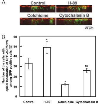
and transiently transfected as described above, were treated with either H-89 (30 μM ; 13, 14), colchicine or cytochalasin B (10 μM, each) at 18 h after trans-fection ; they were cultured further for 6 h under a growth-arrest condition as described above. In good accordance with our previous finding (17), col-chicine, an inhibitor of cytoskeleton assembly, in-hibited strongly the translocation of the chimeric proteins ; i.e., the number of cells expressing the GFP-AQP5(wt) at their apical membrane was only 38% of that of the non-treated control cultures (Fig. 2), suggesting that translocation of AQP5-bearing vesicles, required the microtubule system. A micro-filament inhibitor, cytochalasin B had no or only little effect on the trafficking of the GFP-AQP5(wt) (81% of control).
On the other hand, percentage of the cells that expressed GFP-AQP5(wt) at their apical membrane among GFP-AQP5(wt) positive cells increased in the presence of the PKA inhibitor H-89, as compared with that for the non-treated cells. The percentage
increase in apical translocation by H-89 was 1.5 times of that for non-treated control (Fig. 2).
The above results suggest that the translocation of AQP5-bearing vesicles towards the apical mem-brane was increased by decreasing the phosphoryla-tion of Ser/Thr of AQP5. From these initial observa-tions, we hypothesized that the phosphorylation it-self is probably not crucial for membrane expression of AQP5. The phosphorylation target site blocked by H-89 may be amino acid 152-156 (SRRTS), since this is a PKA-target motif found in AQP5 (3). We, therefore constructed 3 GFP-AQP5 mutants having mutated consensus PKA-target motifs to examine the role of this PKA-target motif in membrane ex-pression of AQP5.
Effects of mutation in PKA-target motif (152SRRTS)
on translocation of the AQP5 chimeric proteins
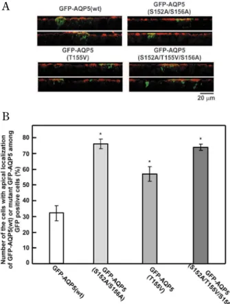
Twenty-four hours after transfection of MDCK-II cells with GFP-AQP5 (S152A/S156A), GFP-AQP5 (T155V) or GFP - AQP5 (S152A/T155V/S156A), A
A
B B
Fig. 1 AQP5 expression in polarized MDCK - II cells. Cultures expressing chimeric proteins were observed under the confocal microscope. A, Vertical pictures (x - z vertical sections ; see MA-TERIALS AND METHODS section for detail methods) of cells in typical cultures at 6, 12, 24, and 48 h post- transfection, express-ing the wt chimeric protein, are shown. The merge technique was used to visualize the co localization of biotin/Alexa Fluor 594 -conjugated streptavidin (red) and GFP signal of GFP - AQP5 (green) in the apical cell membrane which appears as yellow ar-eas in cells. Bar, 20 μm. B, Confocal images shown in “A” were assessed, and the histogram showing the percentages of the cells expressing GFP - AQP5(wt) protein at their apical membrane among GFP - positive cells at different post transfection times is presented. The number of sample wells analyzed was 4 to 6.
A A
B B
Fig. 2 Effects of H- 89, colchicine, and cytochalasin B on the apical translocation of GFP - AQP5(wt). MDCK - II cells were trans-fected as described in the text. Eighteen hours after transfection, the cells were incubated under growth- arrest conditions (incu-bation in the medium containing 0.5% FBS) for the next 6 h in the presence or absence of either H- 89 (30 μM), colchicine (10 μM) or cytochalasin B (10 μM) and examined under the confocal laser scanning microscope. A, Vertical pictures of GFP - AQP5 positive cells. Bar, 20 μm. B, Histogram showing the percentage of the cells expressing GFP - AQP5(wt) at apical membrane is presented as in Fig. 1. *p!0.05, significantly different from the GFP - AQP5(wt) group. NS, not significantly different from the GFP - AQP5(wt). The number of sample wells analyzed was 4 to 6.
the number of cells expressing each mutant chi-meric proteins at cell membrane were 76, 57, and 74%, respectively of the GFP-AQP5 positive cells. These numbers were significantly greater compared to the 33% for the cells expressing GFP-AQP5(wt) at the cell membrane (Fig. 3).
DISCUSSION
The salivary glands are innervated by both sym-pathetic and parasymsym-pathetic branches of the auto-nomic nervous system. The parasympathetic stimu-lation and activation of M3 muscarinic receptors on the acinar cells produces the largest increase in the salivary flow rate (24). The presence of an exocrine-type water channel, such as AQP5, in acinar cells is
essential at least for transcellular water transport as shown by the results of its knockout experiment (8, 9). Trafficking of AQP5 is thought to be involved in the regulation of salivary water secretion via this channel protein ; e.g., in the parotid gland, AQP5 was reported to be trafficked from the intracellu-lar vesicles to the apical membrane in response to stimulation by muscarinic receptors in vitro (15). In human salivary gland cells (HSG cells) trans-fected with AQP5 cDNA, the AQP5 protein is traf-ficked to the plasma membrane following an in-crease in [Ca2+]
i(17). All of these reports imply that
trafficking of AQP5 from intracellular vesicles to the apical membrane is provoked by secretogogue-induced [Ca2+]
iincrease leading to an increase in
water permeability at the apical membrane. From the aspect of molecular phylogenic comparison, AQP5 is known to be closest to AQP2, suggesting that their physiological and biochemical properties would be close as well (3, 25). AQP5 is trafficked from intracellular vesicles toward the apical mem-brane in response to a muscarinic agonist (15), which response is similar to the vasopressin - in-duced trafficking of AQP2 to the plasma membrane (26). A key event that triggers signals for prompting AQP2 trafficking is phosphorylation of the amino acid residue located at the carboxyl terminus, Ser-256, by protein kinase A (27). For instance, van Balkom, et al. (28) constructed several AQP2 genes having a mutation at either putative casein kinase II-target motifs (Ser-148, Ser-229, Thr-244), a pro-tein kinase C (PKC) - target motif (Ser - 231), or a PKA-target motif (Ser-256) ; these constructs were then expressed in MDCK cells. All of these mutant proteins, except Ser-256 mutant, trafficked from the intracellular vesicles to the apical membrane via a forskolin-sensitive mechanism, similarly as wild-type AQP2 did, suggesting that phosphorylation of Ser-256 is essential in this cellular event (28).
GFP has been used for studying the behavior and localization of particular proteins in the living sys-tem. For example, GFP-AQP2 chimera (GFP fused to the amino-terminus of AQP2) expressed in cul-tured porcine kidney epithelial cells (LLC-PK1cells)
was found to traffick in a regulated pathway from the intracellular vesicles toward the basolateral plasma membrane in response to vasopressin or forskolin stimulation (21). Similar to 2 different constructs of GFP with AQP2, GFP-AQP5 was localized primarily in the intracellular vesicles, while AQP5-GFP was predominantly localized on plasma membranes un-der non-stimulated condition (14).
A A
B B
Fig. 3 Effects of mutation of PKA- target motif,152SRRTS, in GFP - AQP5 on the apical trafficking of the chimeric proteins. GFP - AQP5 plasmids with a mutated PKA- target motif, i.e., GFP AQP5 (S152A/S156A), GFP AQP5 (T155V), and GFP -AQP5 (S152A/T155V/S156A), as well as GFP - -AQP5(wt), were used to transfect MDCK - II cells, as described in Fig. 1. A, Verti-cal pictures (x - z vertiVerti-cal section) of cells at 24 h post- transfection, expressing the wt chimeric protein or chimeric protein with mu-tated PKA- target motif, are shown. The merge technique was used as it is described above. Bar, 20 μm. B, confocal images shown in “A” were assessed, and a histogram showing the per-centages of the cells expressing wt AQP5 or AQP5 with a mutated PKA motif at their apical membrane at 24 h post- transfection is presented. *p!0.05, significantly different from the GFP-AQP5 (wt) group. The number of sample wells analyzed was 4 to 6.
Although present and previous inhibitor experi-ments suggested that blocking the PKA-dependent phosphorylation increased the trafficking of AQP molecule toward the apical membrane, this does not necessarily mean that the inhibitor blocked phos-phorylation of the PKA-target motif of AQP5 mole-cules ; it might have blocked proteins to which AQP5 may be associated. The present in vitro mutagene-sis study for the first time explored that blocking the PKA-target motif of AQP5 molecule increases the trafficking of this molecule toward the apical mem-brane.
Our study was conducted to examine the effects of the PKA phosphorylation site of the AQP5 mole-cule on its intracellular translocation or trafficking. The consensus sequence of the PKA-target motif is located in cytoplasmic loop D of AQP5 at amino acid residues 152-156 (Ser-Arg-Arg-Thr-Ser ; 3). Since there are not many reports which describe the function of this motif in the trafficking of AQP5, we investigated the cell physiological properties of rat AQP5 expressed in MDCK-II cells used as a model system. We prepared GFP-AQP5 constructs in which PKA consensus sequence motif was mu-tated to Ala and/or Val to generate the un-phos-phorylated state and these constructs were used to transfect MDCK-II cells.
The amino-terminal fusion chimera, GFP-AQP5 (wt), was shown to be translocated to the apical membrane from the intracellular compartments in polarized MDCK-II cells during cultivation for more than 24 h after transfection. In cells transfected with GFP-AQP5(wt) or mutant GFP-AQP5s, all chimeric proteins were expressed on the plasma membrane ;
i.e., even PKA - dependent phosphorylation was
blocked, GFP-AQP5 was trafficked toward apical membrane. By the experiment using GFP-AQP5 with replacement of various amino acids at152SRRTS
(S152A/T155V/S156A), we found that blocking the phosphorylation of AQP5 at this PKA-target motif increased its translocation to the apical membrane. Also, the cells expressing the wild-type molecule at their apical membrane increased in number by treat-ment with H-89 comparing to non-stimulated con-ditions. This membrane trafficking of GFP-AQP5 (wt) required the involvement of microtubules.
In the present study, we have demonstrated here that GFP-AQP5 chimeric molecules having muta-tion at their PKA consensus sequence can be ex-pressed at the apical membrane. In contrast with phosphorylation-dependent translocation of AQP2 (28), our data suggest that AQP5 can be trafficked
toward the cell membrane irrespective of phospho-rylation of PKA-target motif. Thus the present study imply that a mechanism(s) independent of phospho-rylation of PKA-target motif (152SRRTS) is involved
in translocation of AQP5 in MDCK-II cells.
ACKNOWLEDGEMENTS
This work was a part of a dissertation for the Doctorate of Philosophy Degree submitted to the Graduate School of Oral Sciences, the University of Tokushima. We are grateful to Professor Mikio Furuse, Faculty of Medicine, Kobe University, for providing MDCK-II cell line. We thank Dr. Masayuki Shono for his technical assistance in operating the laser confocal microscope.
GRANTS
This work was supported in part by Scientific Re-search (B) (18390493) from the Ministry of Educa-tion, Culture, Sports, Science, and Technology of Japan. M.R. Karabasil, A. Azlina, and N. Purwanti were supported by a scholarship from the Ministry of Education, Culture, Sports, Science, and Technol-ogy of Japan.
REFERENCES
1. Verkman AS, Mitra AK : Structure and function of aquaporin water channels. Am J Physiol Re-nal Physiol 278 : F13-F28, 2000
2. Verkman AS : More than just water channels : unexpected cellular roles of aquaporins. J Cell Sci 118 : 3225-3232, 2005
3. Raina S, Preston GM, Guggino WB, Agre P : Molecular cloning and characterization of an aquaporin cDNA from salivary, lacrimal, and respiratory tissues. J Biol Chem 270 : 1908-1912, 1995
4. Nielsen S, King LS, Christensen BM, Agre P : Aquaporins in complex tissues. II. Subcellular distribution in respiratory and glandular tissues of rat. Am J Physiol Cell Physiol 273 : C1549 -C1561, 1997
5. Nejsum LN, Kwon TH, Jensen UB, Fumagalli O, Frokiaer J, Krane CM, Menon AG, King LS, Agre PC, Nielsen S : Functional requirement of aquaporin-5 in plasma membranes of sweat
glands. Proc Natl Acad Sci 99 : 511-516, 2002 6. Hamann S, Zeuthen T, La Cour M, Nagelhus
EA, Ottersen OP, Agre P, Nielsen S : Aquaporins in complex tissues : distribution of aquaporins 1-5 in human and rat eye. Am J Physiol Cell Physiol 274 : C1332-C1345, 1998
7. Parvin MN, Kurabuchi S, Murdiastuti K, Yao C, Kosugi -Tanaka C, Akamatsu T, Kanamori N, Hosoi K : Subcellular redistribution of AQP5 by vasoactive intestinal polypeptide in the Brunner’s gland of the rat duodenum. Am J Physiol Gastrointest Liver Physiol 288 : G1283-G1291, 2005
8. Krane CM, Melvin JE, Nguyen HV, Richardson L, Towne JE, Doetschman T, Menon AG : Sali-vary acinar cells from aquaporin 5-deficient mice have decreased membrane water perme-ability and altered cell volume regulation. J Biol Chem 276 : 23413-23420, 2001
9. Ma T, Song Y, Gillespie A, Carlson EJ, Epstein CJ, Verkman AS : Defective secretion of saliva in transgenic mice lacking aquaporin-5 water channels. J Biol Chem 274 : 20071-20074, 1999 10. Tsubota K, Hirai S, King LS, Agre P, Ishida N : Defective cellular trafficking of lacrimal gland aquaporin-5 in Sjögren’s syndrome. Lancet 357 : 688- 689, 2001
11. Steinfeld S, Cogan E, King LS, Agre P, Kiss R, Delporte C : Abnormal distribution of aquaporin-5 water channel protein in salivary glands from Sjögren’s syndrome patients. Lab Invest 81 : 143 -148, 2001
12. Beroukas D, Hiscock J, Jonsson R, Waterman SA, Gordon TP : Subcellular distribution of aquaporin 5 in salivary glands in primary Sjögren’s syndrome. Lancet 358 : 1875-1876, 2001
13. Yang F, Kawedia JD, Menon AG : Cyclic AMP regulates aquaporin 5 expression at both tran-scriptional and post - trantran-scriptional levels through a protein kinase A pathway. J Biol Chem 278 : 32173-32180, 2003
14. Kosugi -Tanaka C, Li X, Yao C, Akamatsu T, Kanamori N, Hosoi K : Protein kinase A - regu-lated membrane trafficking of a green fluores-cent protein-aquaporin 5 chimera in MDCK cells. Biochim Biophys Acta 1763 : 337-344, 2006
15. Ishikawa Y, Eguchi T, Skowronski MT, Ishida H : Acetylcholine acts on M3 muscarinic recep-tors and induces the translocation of aquaporin5 water channel via cytosolic Ca2+elevation in rat
parotid glands. Biochem Biophys Res Commun 245 : 835-840, 1998
16. Li J, Lee S, Choi SY, Lee SJ, Oh SB, Lee JH, Chung SC, Kim JS, Lee JH, Park K : Effects of pilocarpine on the secretory acinar cells in hu-man subhu-mandibular glands. Life Sci 79 : 2441-2447, 2006
17. Tada J, Sawa T, Yamanaka N, Shono M, Akamatsu T, Tsumura K, Parvin MN, Kanamori N, Hosoi K : Involvement of vesicle - cytoskele-ton interaction in AQP5 trafficking in AQP5-gene-transfected HSG cells. Biochem Biophys Res Commun 266 : 443 - 447, 1999
18. Sidhaye V, Hoffert JD, King LS : cAMP has dis-tinct acute and chronic effects on aquaporin-5 in lung epithelial cells. J Biol Chem 280 : 3590-3596, 2005
19. Woo J, Chae YK, Jang SJ, Kim MS, Baek JH, Park JC, Trink B, Ratovitski E, Lee T, Park B, Park M, Kang JH, Soria JC, Lee J, Califano J, Sidransky D, Moon C : Membrane trafficking of AQP5 and cAMP dependent phosphorylation in bronchial epithelium. Biochem Biophys Res Commun 366 : 321-327, 2008
20. Murdiastuti K, Purwanti N, Karabasil MR, Li X, Yao C, Akamatsu T, Kanamori N, Hosoi K : A naturally occurring point mutation in the rat aquaporin 5 gene, influencing its protein pro-duction by and secretion of water from salivary glands. Am J Physiol Gastrointest Liver Physiol 291 : G1081-G1088, 2006
21. Gustafson CE, Levine S, Katsura T, McLaughlin M, Alexio MD, Tamarappoo BK, Verkman AS, Brown D : Vasopressin regulated trafficking of a green fluorescent protein-aquaporin 2 chi-mera in LLC -PK1cells. Histochem Cell Biol
110, 377-386, 1998
22. Wellner RB, Baum BJ : Polarized sorting of aquaporins 5 and 8 in stable MDCK-II trans-fectants. Biochem Biophys Res Commun 285 : 1253-1258, 2001
23. Mostov KE, Verges M, Altschuler Y : Mem-brane traffic in polarized epithelial cells. Curr Opin Cell Biol 12 : 483-490, 2000
24. Baum BJ : Principles of saliva secretion. Ann NY Acad Sci 694 : 17-23, 1993
25. King LS, Kozono D, Agre P : From structure to disease : the evolving tale of aquaporin biology Nat Rev Mol Cell Biol 5 : 687- 698, 2004 26. Katsura T, Gustafson CE, Ausiello DA, Brown
D : Protein kinase A phosphorylation is in-volved in regulated exocytosis of aquaporin-2
in transfected LLC-PK1 cells. Am J Physiol Renal Physiol 272 : F817-F822, 1997
27. Fushimi K, Sasaki S, Marumo F : Phosphoryla-tion of serine 256 is required for cAMP - de-pendent regulatory exocytosis of the aquaporin-2 water channel. J Biol Chem 272 : 14800-14804, 1997
28. Van Balkom BWM, Savelkoul PJM, Markovich D, Hofman E, Nielsen S, Van der Sluijs P, Deen PMT : The role of putative phosphoryla-tion sites in the targeting and shuttling of the aquaporin-2 water channel. J Biol Chem 277 : 41473-41479, 2002