INTRODUCTION
FDG-PET/CT can demonstrate tumor metabolism
and morphology in a single session. Thus it has an important role in tumor imaging. PET/CT is also useful to differentiate malignant from benign lesions, define staging, detect recurrence at an early stage, and monitor therapeutic effects.
Malignant pleural mesothelioma (MPM), which arises from the pleura and is the mostly associated with asbestos exposure, is a common neoplasm of mesothelial cells (1). MPM usually develops 20-40
ORIGINAL
Is FDG-PET/CT useful for managing malignant pleural
mesothelioma?
Hideki Otsuka, Kaori Terazawa, Naomi Morita, Yoichi Otomi, Kyo Yamashita, and
Hiromu Nishitani
Department of Radiology, Institute of Health Biosciences, the University of Tokushima Graduate School, Tokushima, Japan
Abstract : Objective : Imaging techniques such as CT, MRI and PET/CT have essential pre-and post-treatment roles in detecting tumors pre-and evaluating the extension of malignant pleural mesothelioma (MPM). We sough to evaluate the advantages and limitations on FDG-PET/CT findings. Patients and Methods : We performed 13 FDG-PET/CT studies in 9 patients with MPM (8 males, 1 female, aged 51 to 84 years, 9 at the initial diagnosis, 4 fol-low up studies). We reviewed FDG-PET/CT findings of primary tumors, recurrent tumors, lymph nodes, metastasis. Results : All primary and recurrent tumors were FDG positive. The uptake patterns at initial diagnosis were ; diffuse+multi-nodular uptake pattern in 5, diffuse irregular thickened uptake pattern in 2, some focal thickened pattern in one, and a slight diffuse uptake pattern in one. Two of the 3 patients diagnosed as N0 by PET and operated on had negative lymph nodes confirmed pathologically. The other patient diagnosed as N0 by PET, who had one month of time lag between PET/CT examination and surgery, was confirmed as N2 by extrapleural pneumonectomy. In 3 patients, hilar or me-diastinal lymph nodes were difficult to distinguish from irregular pleural thickening. One patient had a FDG positive lymph node in the ipsilateral supraclavicular region confirmed as metastasis (N3). One patient had a FDG positive lymph node in the para-aortic region. Lung metastasis was seen in one patient (M1). In another patient, two focal nodular up-takes in the colon were detected and confirmed as colon polyps (pathologically Group 3-4). At restaging, 3 of 4 patients showed diffuse+multi-nodular uptake and one patient showed multi-nodular uptake. Conclusions : The utility of FDG-PET/CT is limited for evaluation of primary tumor extension and nodal status. FDG-PET/CT is useful for detect-ing distant metastasis and for evaluatdetect-ing activity in supraclavicular or abdominal lymph nodes. It is also useful for identifying unsuspected diseases. J. Med. Invest. 56 : 16-20, February, 2009
Keywords : malignant pleural mesothelioma, FDG-PET/CT
Received for publication September 10, 2008 ; accepted Novem-ber 14, 2008.
Address correspondence and reprint requests to Hideki Otsuka, Department of Radiology, Institute of Health Biosciences, the University of Tokushima Graduate School, Kuramoto - cho, Tokushima 770 - 8503, Japan and Fax : +81 - 88 - 633 - 7174.
years after exposure to asbestos. It can occur from direct occupational exposure as well as from indi-rect exposure. Its incidence has been increasing. Histologically, MPM is classified into three types : epithelial (60%), biphasic (30%), and sarcomatous (10%) types (2). The epithelial type is the most com-mon and has the best prognosis. MPM is a very ag-gressive disease and invades to the chest wall, me-diastinum, and diaphragm. MPM also metastasizes to hilar and mediastinal lymph nodes. Distant metas-tases occur in approximately 10-20% of patients. Im-aging examinations are necessary for management of MPM patients.
We report our experience of FDG - PET/CT in MPM patients.
PATIENTS AND METHODS
A Summary of the patients is shown in Table 1. We performed 13 FDG - PET/CT studies in 9 pa-tients with MPM (8 males, 1 female, aged 51 to 84 years, 9 at the initial diagnosis, 4 follow up studies). Five patients had a history of asbestos exposure (56%). Three of the 9 patients had extrapleural pneu-monectomy and chemotherapy/radiation therapy.
Four patients had only chemotherapy, and 2 patients refused any treatment. All patients fasted for at least 4 hours before the PET/CT scan and blood glucose levels were checked as being!150 mg/dl at the time of injection. 18F-FDG (3.7 MBq/kg of body weight) was administered intravenously using an automatic injection system. After FDG injection, pa-tients rested on bed for one hour. Talking, walking, or other physical activities were prohibited to reduce unnecessary muscle uptake. One hour after injec-tion, images were obtained using PET/CT machine equipped 16-slice multi-detector row CT.
We reviewed FDG-PET/CT findings of primary tumors, recurrent tumors, lymph nodes, metastases.
RESULTS
All primary and recurrent tumors were FDG posi-tive. The uptake patterns of the 9 patients at initial diagnosis were ; diffuse+multi-nodular uptake pat-tern in 5, diffuse irregular thickened uptake patpat-tern in 2, some focal thickened pattern in one, and a slight diffuse uptake pattern in one. Two of the 3 patients diagnosed as N0 by PET and operated on had negative lymph nodes confirmed pathologically.
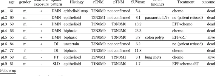
Table 1 A summary of the patients. Initial diagnosis
age gender asbestos exposure
uptake
pattern Histlogy cTNM pTNM SUVmax
other
findings Treatment outcome pt.1 61 m + DMN epithelioid susp. T2N0M0 not confirmed 5.4 chemo dead pt.2 60 m - DMN epithelioid T3N2M1 not confirmed 8.1 paraaortic LN+ no (patient refused) dead pt.3 59 m + DMN epithelioid T3N0M0 T3N0M0 13.2 EPP+chemo dead pt.4 56 m + DMN biphasic T3N2M0 T3N2M0 23.3 chemo dead pt.5 55 m + DMN biphasic T3N0M0 T3N0M0 3.7 colon polyp EPP+RT alive pt.6 84 m + DI uncertain T3N0M0 not confirmed 6.2 no (patient refused) dead pt.7 77 f - DI biphasic T4N2M0 not confirmed 11.8 chemo dead pt.8 59 m - FT epithelioid T2N0M1 T2N0M1 3.1 lung mets chemo alive pt.9 51 m - SLD epithelioid T1N0M0 T3N2M0 1.7 EPP+chemo+RT dead Follow up
Treatment time after 1st PET
uptake
pattern SUVmax pt.1 chemo 3 mo DMN local recurrence, subclavicular LN+ 7.4 pt.3 EPP+chemo 5 mo MN local recurrence 13.2 pt.5 EPP+RT 1 mo DMN progressed disease 5.2 pt.9 EPP+chemo+RT 9 mo DMN local recurrence 6.2
DMN : diffuse multi - nodular, DI : diffuse irregular, FT : focal thickening, SLD : slight diffuse, MN : multi - nodular EPP : extrapleural pneumonectomy, RT : radiation therapy
The other patient diagnosed as N0 by PET, had a one month time lag between PET/CT examination and surgery, was confirmed as N2 by extrapleural pneumonectomy. In 3 patients, hilar or mediastinal lymph nodes were difficult to distinguish from ir-regular pleural thickening. One patient had a FDG positive lymph node in the ipsilateral supraclavicu-lar region confirmed as metastasis (N3). One patient had a FDG positive lymph node in the para-aortic region. Lung metastasis was seen in one patient (M1). In another patient, two focal nodular uptakes in the colon were detected and confirmed as colon polyps (Group 3-4).
At follow up studies (1-9 months after first PET/ CT), local recurrence and disease progression was demonstrated as FDG positive. Three of 4 patients having follow up scans showed diffuse+multi-nodu-lar uptake while the other showed multi-nodudiffuse+multi-nodu-lar up-take.
CASE PRESENTATION
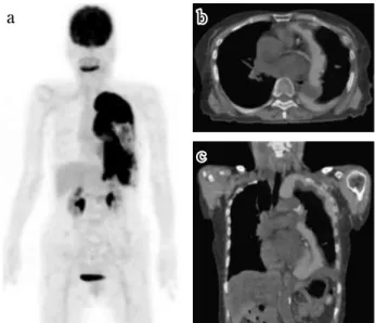
Patient 7 (Fig. 1) : A 77 year-old female with no history of direct asbestos exposure was admitted with left pleural effusion and pleural thickening. FDG-PET/CT clearly demonstrated intense diffuse thickened FDG uptake. It was difficult to differentiate a pleural lesion from hilar lymph node metastasis.
No FDG positive lymph node was seen in the con-tralateral hilar and supraclavicular region. There was no evidence of distant metastasis.
Patient 5 (Fig. 2) : A 55 year-old male with a his-tory of asbestos exposure was admitted with dysp-nea. FDG-PET/CT demonstrated diffuse and multi-nodular uptake in the right pleura. No hilar or me-diastinal lymph node abnormalities or distant metas-tasis were seen (N0 on FDG-PET/CT). He under-went an extrapleural pneumonectomy and pT3N0 was confirmed. Two focal uptakes were demon-strated in the pelvis corresponding to the rectum. Following our recommendation of colon fiberscopic examination, these uptakes confirmed as colon pol-yps (group 3-4).
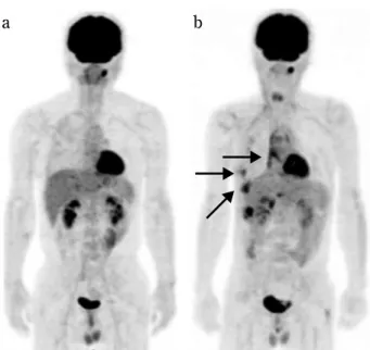
Patient 9 (Fig. 3) : A 51 year-old male with no his-tory of asbestos exposure was admitted with a pro-longed right pleural effusion. FDG-PET/CT demon-strated a slight diffuse uptake along the pleura. There was no evidence of lymph node metastasis or distant metastasis. His disease progressed very rap-idly and extrapleural pneumonectomy one month after PET/CT examination confirmed pT3N2. After 10 months, follow up PET/CT study clearly demon-strated recurrent lesions as FDG positive.
aa bb
cc
Fig. 1 Seventy seven year old female. a) MIP image of FDG -PET. b) Axial view of PET/CT. c) Coronal view of PET/CT. FDG - PET/CT clearly demonstrated intense diffuse FDG uptake in thickened pleura. It is difficult to differentiate pleural lesion from hilar lymph node metastasis. No FDG positive lymph node was seen in the contralateral hilar and supraclavicular region. There was no evidence of distant metastasis.
aa bb
cc
d d
Fig. 2 Fifty - five year - old male. a) MIP image of FDG - PET. b) Axial view of PET. c) Axial view of CT. d) Axial view of PET/ CT. FDG - PET/CT demonstrated diffuse and multi - nodular up-take in the right pleura. No hilar or mediastinal lymph node ab-normalities and no distant metastasis were seen. A focal uptake is demonstrated in the pelvis corresponding to the rectum (ar-row), confirmed as a polyp (group 4).
DISCUSSION
MPM is a very rapidly progressing disease and easily invades to adjacent structures such as the chest wall, mediastinum, and diaphragm. Lymph node metastases and/or distant metastases can oc-cur to organs such as lungs, liver, adrenal glands, and brain. Prognosis is related to the extent of the primary tumor and metastases. Stage I and II pa-tients can be candidates for surgery, while stage III and IV patients are considered unresectable. Sur-gery (extrapleural pnemonectomy, pleurectomy/ decortication), radiation therapy, chemotherapy or their combination is often performed but the over-all survival is low (3-5). Imaging techniques such as CT, MRI and PET/CT have essential pre- and post-treatment roles in detecting tumors and evalu-ating the extension of MPM (6). CT is the most widely used modality, but tends to underestimate tumor invasion and lymph node metastases (7, 8). MRI is superior for evaluating tumor involvement (9). Perfusion CT or perfusion MRI can demonstrate tumor microvasculature, while perfusion MRI is used to assess the effects of treatment (10). There are also reports concerning the utility of FDG-PET or FDG-PET/CT (6, 11, 12). PET may predict sur-vival of MPM patients, and can detect extrathoracic metastases ; thus assisting treatment planning. By
using the uptake changes of FDG, PET can monitor the response to chemotherapy (13). However, the utility of PET is limited for T and N staging with sen-sitivity reported to be 19% and 11%, respectively (12). It is sometimes difficult to evaluate MPM inva-sion to adjacent structures such as chest wall or me-diastinum. FDG accumulates not only in the tumor but also in an inflammatory lesion of asbestos related pleuritis. In one of our patient (Patient 9), a slight unilateral uptake was seen in the slightly thickened pleura. This patient had a pneumonectomy one month after the PET/CT examination. pT3N2 was confirmed at surgery, and PET/CT demonstrated a recurrence 9 months after surgery. This showed the aggressive nature of MPM and the usefulness of PET/CT to evaluate recurrent MPM. For N staging, it is sometimes difficult to distinguish hilar or me-diastinal lymph node metastases from a pleural tu-mor or irregular pleural thickening. False-negative lymph nodes and false-positive FDG uptake in me-diastinal nodes is frequently seen. In this study, hi-lar or mediastinal lymph node enhi-largement could not be differentiated from irregular pleural thicken-ing in 3 of the 9 patients. On the other hand, FDG-positive lymph nodes in the supraclavicular and para-aortic regions were clearly demonstrated. PET/CT was useful to detect lung metastasis and unsus-pected disease (colon polyps). We were also able to use intense FDG uptake as a landmark for biop-sies. In follow up studies, recurrent tumors clearly showed as FDG positive.
We classified uptake pattern in 4 types. The rela-tionship between the uptake pattern and histopa-thological type, and patient outcome could not been clarified in our small number of patients ; however, we need to know the uptake pattern and that MPM in the early stage can show a slight laterality of pleu-ral uptake. FDG-PET/CT is useful for detecting dis-tant metastasis and for evaluating activity in supracla-vicular or abdominal lymph nodes. It is also useful for identifying unsuspected diseases. It is important for clinicians to know the advantages and limitations of various imaging modalities for management of MPM patients.
REFERENCES
1. Robinson BWS, Lake RA : Advances in Malig-nant Mesothelioma. N Engl J Med 353(15) : 1591-1603, 2005
2. WHO : Histological typing of lung and pleural
aa bb
Fig. 3 Fifty - one year - old male. a) PET image at initial diagno-sis. FDG - PET/CT demonstrated a slight diffuse uptake along the pleura. There was no evidence of lymph node metastasis or dis-tant metastasis. His disease progressed very rapidly and extra-pleural pneumonectomy one month after PET/CT examination confirmed pT3N2. b) PET image at follow up (10 months after 1stPET/CT). Recurrent lesions show FDG positive (arrow).
tumors. 3rdEd, Springer, Berlin, 1999
3. Flores RM, Pass HI, Seshan VE, Dycoco J, Zakowski M, Carbone M, Bains MS, Rusch VW : VW. Extrapleural pneumonectomy versus pleurectomy/decortication in the surgical man-agement of malignant pleural mesothelioma : results in 663 patients. J Thorac Cardiovasc Surg 135(3) : 620-6, 2008
4. Flores RM, Krug LM, Rosenzweig KE,
Venkatraman E, Vincent A, Heelan R, Akhurst T, Rusch VW : Induction chemotherapy, extra-pleural pneumonectomy, and postoperative high-dose radiotherapy for locally advanced malignant pleural mesothelioma : a phase II trial. J Thorac Oncol 1(4) : 289-95, 2006 5. Yajnik S, Rosenzweig KE, Mychalczak B, Krug
L, Flores R, Hong L, Rusch VW : Hemithoracic radiation after extrapleural pneumonectomy for malignant pleural mesothelioma. Int J Radiat Oncol Biol Phys 56(5) : 1319-26, 2003
6. Yamamoto M, Gerbaudo VH, Gill RR, Jacobson FL, Sugarbaker DJ, Hatabu H : Morphologic and functional imaging of malignant pleural mesothelioma. Eur J Rad 64 : 356-366, 2007 7. Roach HD, Davies GJ, Attanoos R, Crane M,
Adams H, Phillips S : Asbestos : when the dust settles an imaging review of asbestos-related disease. Radiographics 22 : S167-84, 2002 8. Lorenzo B, Feragalli B, Sacco R, Merlino B,
Storto ML : Malignant pleural disease. Eur J Radiol 34(2) : 98-118, 2000
9. Heelan RT, Rusch VW, Begg CB, Panicek DM, Caravelli JF, Eisen C : Staging of malignant pleural mesothelioma : comparison CT and MR imaging. Am J Roentgenol 172 (4) : 1039-47, 1999
10. Giesel FL, Bischoff H, von Tengg-Kobligk H : Dynamic contrast-enhanced MRI of malignant pleural mesothelioma : a feasibility study of noninvasive assessment, therapeutic follow-up, and possible predictor of improvement out-come. Chest 129 (6) : 1570- 6, 2006
11. Flores RM, Akhurst T, Gonen M, Zakowski M, Dycoco J, Larson SM, Rusch VW : Positron emission tomography predicts survival in ma-lignant pleural mesothelioma. J Thorac Cardio-vasc Surg 132(4) : 763-8, 2006
12. Flores RM, Akhurst T, Gonen M, Larson SM, Rusch VW : Positron emission tomography de-fines metastatic disease but not locoregional disease in patients with malignant pleural meso-thelioma. J Thorac Cardiovasc Surg 126(1) : 11- 6, 2003
13. Ceresoil GL, Chiti A, Zucali PA : Early response evaluation in malignant pleural mesothelioma by positron emission tomography with [18F] fluorodeoxyglucose. J Clin Oncol 24 : 4587 -4593, 2006