Research Article
Studies on Shokyo, Kanzo, and Keihi in Kakkonto
Medicine on Prostaglandin E
2
Production in
Lipopolysaccharide-Treated Human Gingival Fibroblasts
Toshiaki Ara and Norio Sogawa
Department of Pharmacology, Matsumoto Dental University, 1780 Gobara Hirooka, Shiojiri, Nagano 399-0781, Japan Correspondence should be addressed to Toshiaki Ara; ara [email protected]
Received 31 May 2016; Revised 22 July 2016; Accepted 26 September 2016 Academic Editor: Fong-Fu Hsu
Copyright © 2016 T. Ara and N. Sogawa. This is an open access article distributed under the Creative Commons Attribution License, which permits unrestricted use, distribution, and reproduction in any medium, provided the original work is properly cited.
We previously demonstrated that a kampo medicine, kakkonto, decreases lipopolysaccharide- (LPS-) induced prostaglandin E2 (PGE2) production by human gingival fibroblasts. In this study, we examined the herbs constituting kakkonto that exhibit this effect. Shokyo strongly and concentration dependently and kanzo and keihi moderately decreased LPS-induced PGE2production. Shokyo did not alter cyclooxygenase-2 (COX-2) activity, cytosolic phospholipase A2(cPLA2), annexin 1 and COX-2 expression, and LPS-induced extracellular signal-regulated kinase (ERK) phosphorylation. Kanzo inhibited COX-2 activity but increased annexin 1 and COX-2 expression and did not alter LPS-induced ERK phosphorylation. Keihi inhibited COX-2 activity and LPS-induced ERK phosphorylation but slightly increased COX-2 expression and did not alter cPLA2and annexin 1 expression. These results suggest that the mechanism of shokyo is through the inhibition of cPLA2activity, and that of kanzo and keihi is through the inhibition of COX-2 activity and indirect inhibition of cPLA2activity. Therefore, it is possible that shokyo and kakkonto are clinically useful for the improvement of inflammatory responses.
1. Introduction
Periodontal disease is accompanied by inflammation of the gingiva and destruction of periodontal tissues, leading to alveolar bone loss in severe clinical cases. Prostaglandin E2 (PGE2), interleukin-6 (IL-6), and IL-8 are known to play important roles in inflammatory responses and tissue degradation. PGE2 has several functions in vasodilation, enhancement of vascular permeability and pain, and induc-tion of osteoclastogenesis and is believed to play important roles in inflammatory responses and alveolar bone resorption in periodontal disease [1].
A kampo medicine, kakkonto (TJ-1), has been clinically used for various diseases such as common cold, coryza, initial stage of febrile diseases, and inflammatory diseases. There are several reports showing that kakkonto possesses antiallergic [2, 3] and antiviral [4–7] effects in animal and
in vitro experimental models. Regarding anti-inflammatory
effects, kakkonto has been reported to decrease PGE2 produc-tion in cultured rabbit astrocytes [8]. Recently, we reported
that kakkonto suppresses lipopolysaccharide- (LPS-) induced PGE2production by human gingival fibroblasts (HGFs) [9], as well as shosaikoto [10], hangeshashinto [11], and orento [12].
Kakkonto is constituted with seven herbs (kakkon, taiso, mao, kanzo, keihi, shakuyaku, and shokyo). Some herbs such as keihi and shokyo are known to possess anti-inflammatory effects and are clinically used to treat inflammatory diseases [13, 14]. However, which compositions in kakkonto primarily show this effect is unclear. In this study, to elucidate the effect of kakkonto on decreasing LPS-induced PGE2 production more precisely, we examined those herbs that constitute kakkonto and their mechanisms.
2. Materials and Methods
2.1. Reagents. The ingredients of kakkonto formula are shown
in Table 1. Kakkonto was purchased from Tsumura & Co. Powders of six herbs (taiso, mao, kanzo, keihi, shakuyaku,
Volume 2016, Article ID 9351787, 9 pages http://dx.doi.org/10.1155/2016/9351787
Table 1: The ingredient of kakkonto formula.
Japanese name Latin name Amount (g) Amount (g/g of product)∗
Kakkon Puerariae Radix 4.0 0.111
Taiso Zizyphi fructus 3.0 0.083
Mao Ephedrae Herba 3.0 0.083
Kanzo Glycyrrhizae Radix 2.0 0.056
Keihi Cinnamomi Cortex 2.0 0.056
Shyakuyaku Paeoniae Radix 2.0 0.056
Shokyo Zingiberis Rhizoma 2.0 0.056
Total 18.0 0.500
∗7.5 g of kakkonto product contains 3.75 g of a dried extract of the mixed
crude drugs.
and shokyo) were provided by Tsumura & Co. (Tokyo, Japan). Kakkon (Puerariae Radix) was purchased from Tsumura & Co. A hot water extract of kakkon was prepared as reported previously [15]. In brief, 10 g of kakkon was decocted for 1 h with 100 mL of water. The decoctions were mixed, concen-trated, and lyophilized. The w/w yield of kakkon was 2.1%. Powders of herbs and kakkonto were suspended in Dulbecco’s modified Eagle’s medium (D-MEM, Sigma, St. Louis, MO) containing 10% heat-inactivated fetal calf serum, 100 U/mL penicillin, and 100 mg/mL streptomycin (culture medium) and were stored at 4∘C overnight under shaking. Then, the suspension was centrifuged and the supernatant was filtrated through a 0.45𝜇m pore membrane. Lipopolysaccha-ride (LPS) from Porphyromonas gingivalis 381 was provided by Professor Nobuhiro Hanada (School of Dental Medicine, Tsurumi University, Japan). Arachidonic acid, prostaglandin H2 (PGH2), NS-398 (cyclooxygenase-2 (COX-2) inhibitor), CAY10502 (cytosolic phospholipase A2𝛼- (cPLA2𝛼-) specific inhibitor), bromoenol lactone (calcium-independent PLA2 -(iPLA2-) specific inhibitor), and thioetheramide-PC (secre-tory PLA2- (sPLA2-) specific inhibitor) were purchased from Cayman Chemical (Ann Arbor, MI). Other reagents were purchased from Nacalai Tesque (Kyoto, Japan).
2.2. Cells. HGFs were prepared as described previously [11].
In brief, HGFs were prepared from free gingiva during the extraction of an impacted tooth, with the informed consent of the subjects who consulted Matsumoto Dental University Hospital. The free gingival tissues were cut into pieces and seeded onto 24-well plates (AGC Techno Glass Co., Chiba, Japan). HGFs were maintained in the culture medium at 37∘C in a humidified atmosphere of 5% CO2. For passage, HGFs were trypsinized, suspended, and plated into new cultures in a 1 : 3 dilution ratio. HGFs were used between the 10th and 15th passages in the assays. This study was approved by the Ethical Committee of Matsumoto Dental University (number 0063).
2.3. Measurement of Cell Viability. The numbers of cells were
measured using WST-8 (Cell Counting Kit-8; Dojindo, Kumamoto, Japan) according to the manufacturer’s instruc-tions. In brief, the media were removed by aspiration and
the cells were treated with 100𝜇L of mixture of WST-8 with culture medium for 2 h at 37∘C in CO2 incubator. Optical density was measured (measured wavelength at 450 nm and reference wavelength at 655 nm) using an iMark microplate reader (Bio-Rad, Hercules, CA), and the mean background value was subtracted from each value.
2.4. Measurement of PGE2. HGFs were seeded in 96-well
plates (10,000 cells/well) and incubated in serum-containing medium at 37∘C overnight. Then, the cells were treated with various concentrations of each herb or kakkonto in the absence or presence of LPS (10 ng/mL) for 24 h (200𝜇L each well) in triplicate or quadruplicate for each sample. After collecting the culture supernatants, viable cell numbers were measured using WST-8 as described above.
The concentrations of PGE2 in the culture supernatants were measured by enzyme-linked immunosorbent assay (ELISA), according to the manufacturer’s instructions (Cay-man Chemical), and were adjusted by the number of viable cells. Data are represented as pg per 10,000 cells (mean± SD).
2.5. Measurement of COX-2 and Prostaglandin E Synthase.
COX-2 and prostaglandin E (PGE) synthase activities were evaluated as shown previously [16] with slight modifications. In brief, to estimate COX-2 activity, HGFs were treated with LPS and herb for 8 h, washed, and incubated in culture medium containing exogenous arachidonic acid (10𝜇M). The concentrations of PGE2 in the supernatants were measured by ELISA. PGE synthase activity was determined after a 15 min incubation with exogenous PGH2 (10 nM), and the concentrations of PGE2were measured. Data are represented as pg per 10,000 cells (mean± SD).
2.6. Preparation of Cell Lysates. HGFs were cultured in
60 mm dishes and treated with combinations of LPS and herb for the indicated times. Then, cells were washed twice with Tris-buffered saline, transferred into microcentrifuge tubes, and centrifuged at 6,000×g for 5 min at 4∘C. Supernatants were aspirated and the cells were lysed on ice in lysis buffer (50 mM Tris-HCl, pH 7.4, 1% Nonidet P-40, 0.25% sodium deoxycholate, 150 mM NaCl, 1 mM ethylene glycol bis(2-aminoethyl ether)tetraacetic acid (EGTA), 1 mM sodium orthovanadate, 10 mM sodium fluoride, 1 mM phenylmethyl-sulfonyl fluoride, 10𝜇g/mL aprotinin, 5 𝜇g/mL leupeptin, and 1𝜇g/mL pepstatin) for 30 min at 4∘C. Then, the samples were centrifuged at 12,000×g for 15 min at 4∘C, and the supernatants were collected. The protein concentration was measured using a BCA Protein Assay Reagent kit (Pierce Chemical Co., Rockford, IL).
2.7. Western Blotting. The samples (10𝜇g of protein) were
fractionated in a polyacrylamide gel under reducing con-ditions and transferred onto a polyvinylidene difluoride (PVDF) membrane (Hybond-P; GE Healthcare, Uppsala, Sweden). The membranes were blocked with 5% ovalbu-min for 1 h at room temperature and incubated with pri-mary antibody for an additional 1 h. The membranes were further incubated with horseradish peroxidase-conjugated
secondary antibodies for 1 h at room temperature. Protein bands were visualized with an ECL kit (GE Healthcare).
Antibodies against COX-2 (sc-1745, 1 : 500 dilution), cPLA2(sc-438, 1 : 200 dilution), annexin 1 (sc-11387, 1 : 1,000 dilution), and actin (sc-1616, 1 : 1,000 dilution), which detects a broad range of actin isoforms, were purchased from Santa Cruz Biotechnology (Santa Cruz, CA). Antibodies against extracellular signal-regulated kinase (ERK; p44/42 MAP kinase antibody, 1 : 1,000 dilution) and phosphorylated ERK (Phospho-p44/42 MAPK (Thr202/Tyr204) (E10) monoclonal antibody, 1 : 2,000 dilution) were purchased from Cell Sig-naling Technology (Danvers, MA). Horseradish peroxidase-conjugated anti-goat IgG (sc-2020, 1 : 20,000 dilution) was procured from Santa Cruz, and anti-rabbit IgG (1 : 20,000 dilution) and anti-mouse IgG (1 : 20,000 dilution) were pur-chased from DakoCytomation (Glostrup, Denmark).
2.8. Statistical Analysis. Differences between groups were
evaluated using the two-tailed pairwise comparison test with a pooled variance, followed by correction with Holm’s method (total of 10 null hypotheses; 3 null hypotheses without herb versus with herb in the absence of LPS, 3 null hypotheses without herb versus with herb in the presence of LPS, and 4 null hypotheses without LPS versus with LPS) (Figure 1). Dif-ferences between the control group and experimental groups were evaluated using two-tailed Dunnett’s test (Figures 3 and 4).
All computations were performed with the statistical pro-gram R (http://www.r-project.org/). Dunnett’s test was per-formed using the “glht” function in the “multcomp” package. Values with𝑃 < 0.05 were considered significantly different.
3. Results
3.1. Effect of Herbs on PGE2 Production. We examined
whether the herbs affect LPS-induced PGE2 production by HGFs. The concentrations of PGE2were adjusted according to viable cell number. When HGFs cells were treated with 10 ng/mL of LPS, HGFs cells produced large amounts of PGE2. Shokyo strongly and significantly decreased LPS-induced PGE2 production in a concentration-dependent manner (Figure 1). Kanzo and keihi moderately decreased LPS-induced PGE2 production (Figure 1). Taiso and mao had no effect on LPS-induced PGE2 production. Kakkon and shakuyaku increased LPS-induced PGE2 production (Figure 1). In the absence of LPS, kakkon increased PGE2 production, but kanzo decreased PGE2production (Figure 1). Other herbs had no or little effect on PGE2 production. Therefore, we used three herbs (kanzo, keihi, and shokyo) in the following experiments.
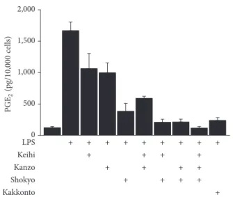
Next, we examined the synergistic effect of three herbs (shokyo, kanzo, and keihi) on PGE2 production and com-pared it with that of kakkonto. The concentrations of each herb (56𝜇g/mL) were determined based on the ingredient of kakkonto formula (Table 1). The mixture of herbs further decreased PGE2production. The combination of two herbs (shokyo + keihi and shokyo + kanzo) decreased PGE2 production to a similar level with 1 mg/mL of kakkonto.
Moreover, the mixture of three herbs decreased PGE2 pro-duction more than kakkonto (Figure 2).
3.2. PLA2 Isoform Activities in HGFs. PLA2 is the most upstream enzyme in the arachidonic acid cascade and releases arachidonic acid from the plasma membrane. PLA2s form a superfamily and are classified into cytosolic PLA2 (cPLA2), calcium-independent PLA2 (iPLA2), secre-tory PLA2(sPLA2), and others [13]. To elucidate which type of PLA2(s) contribute to arachidonic acid production in HGFs, we used selective PLA2inhibitors. cPLA2𝛼-specific inhibitor CAY10502 significantly decreased LPS-induced PGE2 pro-duction by approximately half (Figure 3). However, both iPLA2-specific inhibitor BEL and sPLA2-specific inhibitor thioetheramide-PC did not alter LPS-induced PGE2 produc-tion (Figure 3). Therefore, we examined cPLA2among these PLA2s in the following experiments.
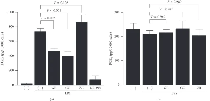
3.3. Effect of Herbs on COX-2 and PGE Synthase Activities.
Then, we examined the mechanism by which kanzo, keihi, and shokyo decreased LPS-induced PGE2production more directly. In order to bypass PLA2, we added exogenous arachidonic acid. Kanzo and keihi significantly decreased LPS-induced PGE2production to approximately half, while shokyo slightly but not significantly increased PGE2 produc-tion (Figure 4(a)). NS-398, as a positive control, decreased LPS-induced PGE2production.
The formation of PGE2 from arachidonic acid requires both COX and PGE synthase. To examine the effect of herbs on PGE synthase, we determined PGE2formation from exogenous PGH2. However, all herbs had no effect on PGE2 formation from exogenous PGH2(Figure 4(b)).
3.4. Effects of Herbs on Molecular Expression in the Arachi-donic Acid Cascade. We examined whether herbs affect the
expression of molecules in the arachidonic acid cascade. Kanzo increased cPLA2 expression, while keihi and shokyo showed no effect (Figure 6). Based on its molecular weight (approximately 90 kDa in human) [17, 18], this cPLA2 is believed to be cPLA2𝛼 subtype.
Annexin 1, also named as lipocortin, is an anti-inflam-matory mediator produced by glucocorticoids and inhibits cPLA2activity [19, 20]. Kanzo increased annexin 1 expression, while keihi and shokyo showed no effect (Figure 6).
COX-2 was not detected in the absence of LPS and LPS-induced COX-2 expression in HGFs. Kanzo and keihi increased LPS-induced COX-2 expression. However, shokyo did not alter LPS-induced COX-2 expression (Figure 5).
3.5. Effects of Herbs on ERK Phosphorylation. cPLA2 is reported to be directly phosphorylated at Ser505 by phos-phorylated ERK, resulting in cPLA2 activation [21, 22]. Therefore, we examined whether herbs suppress LPS-induced ERK phosphorylation. Keihi suppressed LPS-induced ERK phosphorylation at 30 min, while kanzo and shokyo did not (Figure 6).
Taiso 100 10 0 30 800 600 400 200 0 1,200 1,000 ### # ## # Herb (𝜇g/mL) PGE 2 (pg/ 10 ,000 cells) Mao 100 10 0 30 1,000 800 600 400 200 0 ### ### ### ### Herb (𝜇g/mL) PGE 2 (pg/ 10 ,000 cells) Keihi 100 10 0 30 800 600 400 200 0 1,000 ### ### ### ### Herb (𝜇g/mL) PGE 2 (pg/ 10 ,000 cells) ∗ ∗ ∗∗ Shakuyaku 100 10 0 30 1,200 800 600 400 200 0 1,000 ### ### ### ### Herb (𝜇g/mL) PGE 2 (pg/ 10 ,000 cells) ∗∗ Kanzo 100 10 0 30 1,000 800 600 400 200 0 ### ### ### ### Herb (𝜇g/mL) PGE 2 (pg/ 10 ,000 cells) ∗∗∗ 100 10 0 30 Kakkon 800 600 400 200 0 1,000 ### ### ### ### Herb (𝜇g/mL) PGE 2 (pg/ 10 ,000 cells) ∗∗ ∗∗ ∗∗∗ ∗∗∗ Figure 1: Continued.
Shokyo 100 10 0 30 800 600 400 200 0 1,000 ### ### ## Herb (𝜇g/mL) PGE 2 (pg/ 10 ,000 cells) ∗∗∗ ∗∗∗
Figure 1: Effects of herbs on the production of PGE2. HGFs were treated with combinations of LPS (0 and 10 ng/mL) and herb (0, 10, 30, and 100𝜇g/mL) for 24 h. Concentrations of PGE2were measured by ELISA, adjusted by cell number, and expressed as pg per 10,000 cells (mean ± SD, 𝑛 = 3). Open circles, treatment without LPS; closed circles, treatment with 10 ng/mL of LPS.∗𝑃 < 0.05,∗∗𝑃 < 0.01, and∗∗∗𝑃 < 0.001 (without herb versus with herb).#𝑃 < 0.05,##𝑃 < 0.01, and###𝑃 < 0.001 (without LPS versus with LPS). 𝑃 values were calculated by pairwise comparisons and corrected with Holm’s method (10 null hypotheses).
0 500 1,000 1,500 2,000 LPS Keihi Kanzo Shokyo Kakkonto + + + + + + + + + + + + + + + + + + + + + + PGE 2 (pg/ 10 ,000 cells)
Figure 2: Effects of mixture of keihi, kanzo, and shokyo on the production of PGE2. HGFs were treated with combinations of LPS (10 ng/mL) and herbs (56𝜇g/mL) or kakkonto (1 mg/mL) for 24 h. Concentrations of PGE2were measured by ELISA, adjusted by cell number, and expressed as pg per 10,000 cells (mean± SD, 𝑛 = 4).
4. Discussion
In the present study, we examined the effect of herbs consti-tuting kakkonto on LPS-induced PGE2production by HGFs. Shokyo, kanzo, and keihi decreased LPS-induced PGE2 pro-duction in a concentration-dependent manner. In particular, shokyo showed the most marked effect. Previously, we exam-ined the mechanisms of kakkonto [9] and shosaikoto [10] that contain shokyo and demonstrated that shosaikoto inhibited COX-2 activity and LPS-induced COX-2 expression and that
0 500 1,000 1,500
(—) CAY BEL Thio-PC ZR
(—) LPS PGE 2 (pg/ 10 ,000 cells) P = 0.539 P = 0.781 P < 0.001
Figure 3: The contribution of PLA2isoforms in HGFs. HGFs were treated with LPS (10 ng/mL) and PLA2 inhibitor for 24 h. Con-centrations of PGE2 were measured by ELISA, adjusted by cell number, and expressed as pg per 10,000 cells (mean± SD, 𝑛 = 4). CAY10502, cPLA2𝛼 inhibitor (100 nM); BEL (bromoenol lactone), iPLA2 inhibitor (20𝜇M); Thio-PC (thioetheramide-PC), sPLA2 inhibitor (20𝜇M); and ZR (shokyo, 100 𝜇g/mL) a positive control.
kakkonto suppressed ERK phosphorylation. Based on our findings in the present study, shokyo is believed to play an important role in decreasing LPS-induced PGE2production by HGFs in kakkonto and shosaikoto. In addition, the mix-ture of three herbs (shokyo, kanzo, and keihi) synergistically
GR CC ZR NS-398 LPS 1,000 800 600 400 200 0 PGE 2 (pg/ 10 ,000 cells) (—) (—) P = 0.106 P < 0.001 P = 0.002 (a) GR CC ZR LPS 200 100 0 300 PGE 2 (pg/ 10 ,000 cells) (—) (—) P = 0.980 P = 0.495 P = 0.969 (b)
Figure 4: Effects of herbs on COX and PGE synthase activities. HGFs were treated with LPS (10 ng/mL) and herb (100𝜇g/mL) for 8 h, washed, and then treated with (a) 10𝜇M arachidonic acid or (b) 10 nM PGH2for (a) 30 min or (b) 15 min. Concentrations of PGE2were measured by ELISA, adjusted by cell number, and expressed as pg per 10,000 cells (mean± SD, 𝑛 = 3). 𝑃 values by Dunnett’s test are indicated. GR, kanzo; CC, keihi; ZR, shokyo; and NS-398, COX-2 inhibitor (20𝜇M) as a positive control.
Actin Annexin 1 COX-2 GR CC ZR LPS (—) (—) cPLA2
Figure 5: Effects of herbs on cPLA2, annexin 1, and COX-2 expres-sions. HGFs were treated with a combination of LPS (10 ng/mL) and herb (100𝜇g/mL) for 8 h, and protein levels were examined by Western blotting. GR, kanzo; CC, keihi; and ZR, shokyo.
decreased PGE2 production (Figure 2). The effect of two herbs mixture including shokyo was comparable to that of kakkonto. Moreover, the effect of the three herbs mixture was stronger than that of kakkonto because mao and shakuyaku, which increase LPS-induced PGE2 productions (Figure 1), are not included. These results suggest that the combination of these herbs in kakkonto is sufficient to decrease PGE2 production.
Shokyo (Zingiberis Rhizoma) is the powdered rhizome of ginger (Zingiber officinale Roscoe). Several reports have shown that ginger has anti-inflammatory effects in humans,
ERK pERK
GR CC ZR
LPS
(—) (—)
Figure 6: Effects of herbs on LPS-induced ERK phosphorylation. HGFs were untreated (0 h), treated with LPS (10 ng/mL), or treated with both LPS and herb (100𝜇g/mL) for 30 min. Western blotting was performed using antiphosphorylated ERK or anti-ERK antibod-ies. pERK, phosphorylated ERK. Upper band indicates ERK1 (p44 MAPK) and lower band ERK2 (p42 MAPK). GR, kanzo; CC, keihi; and ZR, shokyo.
animal models, and in vitro models. Ginger has been widely used in diet and also as a treatment for rheumatoid arthritis, fever, emesis, nausea, and migraine headache [14]. Recently, a systematic review and meta-analysis reported that the extracts of Zingiberaceae including turmeric, ginger, Javanese ginger, and galangal are clinically effective as hypoanalgesic agents [23]. In an animal model, the aqueous extract of ginger significantly decreased serum PGE2 level by oral or intraperitoneal administration by the rat [24]. Moreover, crude hydroalcoholic extract of ginger reduced the serum level of PGE2and improved tracheal hyperreactivity and lung inflammation induced by LPS in rat [25]. Ethanol extract of ginger reduced the tissue level of PGE2 and improved acetic acid-induced ulcerative colitis in the rat [26]. In in
are reported to decrease PGE2production by several mech-anisms. 10-Gingerol, 8, 10-shogaol [27], and 8-shogaol and 8-paradol [28] inhibit COX-2 activity. Moreover, gingerols, but not 6-shogaol, suppress COX-2 expression in LPS-treated human leukemic monocyte lymphoma U937 cells [29].
Our data showed that shokyo did not suppress COX-2 expression and that shokyo did not alter PGE2 production when arachidonic acid or PGH2 is added to bypass their upstream pathway. These data suggest that shokyo did not affect the downstream pathway of arachidonic acid, which includes COX-2 and PGE synthase. Therefore, shokyo is considered to inhibit PLA2, which is the upstream pathway of arachidonic acid. PLA2hydrolyses the sn-2 ester bond of glyc-erophospholipids. Although PLA2s are classified into cPLA2, iPLA2, and sPLA2[13], shokyo is suggested to act on cPLA2 because cPLA2 is the primary isoform in HGFs (Figure 3). Our data showed that shokyo only slightly decreased cPLA2 expression but did not alter annexin 1 expression, which suppresses PLA2 activity. Therefore, shokyo may primarily inhibit cPLA2 activity. Although we have no direct data to show that shokyo inhibits cPLA2 activity, this assumption is consistent with the fact that gingerols in ginger inhibit i/cPLA2activities [30].
There are six molecules in cPLA2: cPLA2𝛼, cPLA2𝛽, cPLA2𝛾, cPLA2𝛿, cPLA2𝜖, and cPLA2𝜁 [31]. cPLA2𝛼 was first identified and characterized by Ca2+-dependence and substrate preference for arachidonoyl phospholipids [32]. We detected cPLA2 at approximately 90 kDa as well as in human platelets and erythrocytes [17, 18], although the molecular weight of cPLA2𝛼 protein on the basis of amino acid sequence is 85 kDa. Therefore, cPLA2 that we detected in HGFs is believed to be cPLA2𝛼. In contrast, cPLA2 𝛼-specific inhibitor CAY10502 decreased LPS-induced PGE2 production to approximately half (Figure 3), suggesting that other cPLA2s such as cPLA2𝛽 and cPLA2𝛾 may contribute to producing arachidonic acid, and shokyo may inhibit these cPLA2s. However, we could not detect cPLA2𝛽 (114 kDa in humans), cPLA2𝛾 (61 kDa in humans), cPLA2𝜖 (100 kDa in murine), and cPLA2𝜁 (96 kDa in murine) [13]. Although the molecular weight of cPLA2𝛿 from human/murine is 92-93 kDa [13], cPLA2𝛿 is distributed in the placenta [31]. These results suggest that there is no or very little contribution of cPLA2s other than cPLA2𝛼 in HGFs; therefore, the remaining mechanisms remain to be elucidated.
As described above, our data that shokyo did not alter COX-2 activity and COX-2 expression are different from those of gingerols and shogaols. Although there is no obvious evidence, the reason may be the preparation method of shokyo. Gingerols and shogaols are extremely hydrophobic by their structures. Indeed, these compositions were extracted from hydrophobic phase in previous studies. However, the powders of herbs used in this study are prepared by decoc-tion; therefore, hydrophilic compositions are likely to be extracted but hydrophobic compositions are unlikely to be extracted.
Kanzo (Glycyrrhizae Radix) is the powdered root or stolon of Glycyrrhiza uralensis Fischer. Kanzo is also known to have anti-inflammatory effects [33]. We demonstrated that
kanzo decreased LPS-induced PGE2 production (Figure 1) and further demonstrated that kanzo increased annexin 1 expression (Figure 5), regardless of the increase of cPLA2 expression, suggesting that kanzo decreases LPS-induced PGE2 production by enhancement of annexin 1 expression and following inhibition of cPLA2activity. However, the com-positions that increase annexin 1 expression have not been reported. Moreover, we demonstrated that kanzo increased LPS-induced COX-2 expression (Figure 5). These findings are similar to those obtained using kampo medicines orento [12] and saireito [34], which contain kanzo. In contrast, kanzo decreased LPS-induced PGE2production when arachidonic acid is added, while kanzo did not decrease when PGH2was added (Figure 4). These results suggest that kanzo inhibits COX-2 activity but not PGE synthase. Indeed, kanzo inhibits COX-2 activity [35]. Therefore, kanzo inhibits arachidonic acid cascade in multiple points and cPLA2 and COX-2 activities. However, because the contribution of kanzo in kakkonto may be little, the ability of kanzo to decrease LPS-induced PGE2production is weak. Kanzo contains the com-positions such as glycyrrhizin, glycyrrhizic acid, liquiritin, and isoliquiritigenin. Nonetheless, the contributions of these compositions are unlikely in this study because they sup-pressed LPS-induced COX-2 expression [36–39]. Moreover, the compositions that inhibit COX-2 activity have not been reported. Therefore, other compositions may contribute to our findings.
Keihi (Cinnamomi Cortex) is the powdered bark of
Cinnamomum cassia. Cinnamon has been widely used for
the treatment of fever and inflammation [13]. Cinnamon improves nephritis, purulent dermatitis, and hypertension and enhances wound healing. Cinnamon extracts have been used for the improvement of or protection against com-mon cold, diarrhea, and pain [13]. In a previous study, we demonstrated that ERK phosphorylation was suppressed by kakkonto [23] and orento [12], which also contains keihi. In this study, we demonstrated that this effect is responsible for keihi (Figure 6). Moreover, we demonstrated that keihi increased LPS-induced COX-2 expression (Figure 5) and that keihi decreased LPS-induced PGE2 production when arachidonic acid is added while keihi did not decrease when PGH2 was added. These results suggest that keihi inhibits COX-2 activity but not PGE synthase. Therefore, keihi inhibits arachidonic acid cascade in multiple points, cPLA2 activation, and COX-2 activity. However, the contribution of keihi in kakkonto may be little because the ability of keihi to decrease LPS-induced PGE2production is weak. Keihi con-tains the compositions such as cinnamic aldehyde, cinnamic alcohol, cinnamic acid, and coumarin. Cinnamic aldehyde, but not others, suppressed LPS-induced COX-2 expression and decreased PGE2 production by RAW264.7 cells [40, 41]. Moreover, cinnamic aldehyde suppressed carrageenan-induced COX-2 expression and improved footpad edema in mouse [40]. However, the contribution of cinnamic aldehyde is unlikely in this study.
Aspirin-induced asthma (AIA) occurs after ingestion of acid nonsteroidal anti-inflammatory drugs (NSAIDs) such as aspirin and indomethacin [42, 43]. It is believed that AIA is caused by leukotorienes (LTs), in which contract
bronchus are increased by acid NSAIDs [42, 43]. Similarly, acid NSAIDs are known to exacerbate a usual asthma. In this study, we speculate that shokyo inhibits cPLA2activity. Therefore, the production of LTs is believed to be decreased because shokyo blocks arachidonic acid cascade at cPLA2 level. In this case, shokyo may be safely used for patients with asthma, including AIA, instead of conventional anti-inflammatory drugs. Moreover, oral administration of ginger protects against aspirin-induced gastric ulcers in rats [44]. Therefore, shokyo is possible to be available as an anti-inflammatory drug instead of NSAIDs.
5. Conclusion
We demonstrated that shokyo strongly and kanzo and keihi moderately decreased LPS-induced PGE2production. More-over, shokyo may inhibit cPLA2activity and kanzo and keihi inhibit COX-2 activity directly and cPLA2activity indirectly. These results suggest that shokyo, and kakkonto, is clinically useful for the improvement of inflammatory responses in periodontal disease and other diseases.
Ethical Approval
This study was approved by the Ethical Committee of Mat-sumoto Dental University (no. 0063).
Competing Interests
The authors have no conflict of interests to disclose.
Acknowledgments
The authors thank Professor Nobuo Yoshinari (Department of Periodontology) for HGFs preparation. The study was aided by funding from the Nagano Society for the Promotion of Science and a Scientific Research Special Grant from Matsumoto Dental University.
References
[1] K. Noguchi and I. Ishikawa, “The roles of cyclooxygenase-2 and prostaglandin E2in periodontal disease,” Periodontology 2000, vol. 43, no. 1, pp. 85–101, 2007.
[2] Y. Ozaki, “Studies on antiinflammatory effect of Japanese ori-ental medicines (kampo medicines) used to treat inflammatory diseases,” Biological and Pharmaceutical Bulletin, vol. 18, no. 4, pp. 559–562, 1995.
[3] T. Yamamoto, K. Fujiwara, M. Yoshida et al., “Therapeutic effect of kakkonto in a mouse model of food allergy with gas-trointestinal symptoms,” International Archives of Allergy and Immunology, vol. 148, no. 3, pp. 175–185, 2009.
[4] K. Nagasaka, M. Kurokawa, M. Imakita, K. Terasawa, and K. Shiraki, “Efficacy of Kakkon-to, a traditional herb medicine, in herpes simplex virus type 1 infection in mice,” Journal of Medical Virology, vol. 46, no. 1, pp. 28–34, 1995.
[5] M. Kurokawa, M. Tsurita, J. Brown, Y. Fukuda, and K. Shiraki, “Effect of interleukin-12 level augmented by Kakkon-to, a herbal medicine, on the early stage of influenza infection in mice,” Antiviral Research, vol. 56, no. 2, pp. 183–188, 2002.
[6] M.-S. Wu, H.-R. Yen, C.-W. Chang et al., “Mechanism of action of the suppression of influenza virus replication by Ko-Ken Tang through inhibition of the phosphatidylinositol 3-kinase/Akt signaling pathway and viral RNP nuclear export,” Journal of Ethnopharmacology, vol. 134, no. 3, pp. 614–623, 2011.
[7] J. S. Chang, K. C. Wang, D. E. Shieh, F. F. Hsu, and L. C. Chiang, “Ge-Gen-Tang has anti-viral activity against human respiratory syncytial virus in human respiratory tract cell lines,” Journal of Ethnopharmacology, vol. 139, no. 1, pp. 305–310, 2012.
[8] M. Kutsuwa, N. Nakahata, M. Kubo, K. Hayashi, and Y. Ohizumi, “A comparative study of Kakkon-to and Keishi-to on prostaglandin E2release from rabbit astrocytes,” Phytomedicine, vol. 5, no. 4, pp. 275–282, 1998.
[9] H. Kitamura, H. Urano, and T. Ara, “Preventive effects of a kampo medicine, kakkonto, on inflammatory responses via the suppression of extracellular signal-regulated kinase phospho-rylation in lipopolysaccharide-treated human gingival fibrob-lasts,” ISRN Pharmacology, vol. 2014, Article ID 784019, 7 pages, 2014.
[10] N. Horie, K. Hashimoto, T. Kato et al., “COX-2 as possible target for the inhibition of PGE2 production by Rikko-san activated macrophage,” In Vivo, vol. 22, no. 3, pp. 333–336, 2008. [11] Y. Nakazono, T. Ara, Y. Fujinami, T. Hattori, and P.-L. Wang,
“Preventive effects of a kampo medicine, hangeshashinto on inflammatory responses in lipopolysaccharide-treated human gingival fibroblasts,” Journal of Hard Tissue Biology, vol. 19, no. 1, pp. 43–50, 2010.
[12] T. Ara, K.-I. Honjo, Y. Fujinami, T. Hattori, Y. Imamura, and P.-L. Wang, “Preventive effects of a kampo medicine, orento on inflammatory responses in lipopolysaccharide treated human gingival fibroblasts,” Biological and Pharmaceutical Bulletin, vol. 33, no. 4, pp. 611–616, 2010.
[13] J. E. Burke and E. A. Dennis, “Phospholipase A2biochemistry,” Cardiovascular Drugs and Therapy, vol. 23, no. 1, pp. 49–59, 2009.
[14] M. Afzal, D. Al-Hadidi, M. Menon, J. Pesek, and M. S. I. Dhami, “Ginger: an ethnomedical, chemical and pharmacolog-ical review,” Drug Metabolism and Drug Interactions, vol. 18, no. 3-4, pp. 159–190, 2001.
[15] T.-J. Lin, C.-F. Yeh, K.-C. Wang, L.-C. Chiang, J.-J. Tsai, and J.-S. Chang, “Water extract of Pueraria lobata Ohwi has anti-viral activity against human respiratory syncytial virus in human respiratory tract cell lines,” Kaohsiung Journal of Medical Sciences, vol. 29, no. 12, pp. 651–657, 2013.
[16] J. Wilborn, L. J. Crofford, M. O. Burdick, S. L. Kunkel, R. M. Strieter, and M. Peters- Golden, “Cultured lung fibroblasts isolated from patients with idiopathic pulmonary fibrosis have a diminished capacity to synthesize prostaglandin E2 and to express cyclooxygenase-2,” The Journal of Clinical Investigation, vol. 95, no. 4, pp. 1861–1868, 1995.
[17] K. Takayama, I. Kudo, D. K. Kim, K. Nagata, Y. Nozawa, and K. Inoue, “Purification and characterization of human platelet phospholipase A2 which preferentially hydrolyzes an arachi-donoyl residue,” FEBS Letters, vol. 282, no. 2, pp. 326–330, 1991. [18] D. J. Macdonald, R. M. Boyle, A. C. A. Glen, and D. F. Horrobin, “Cytosolic phospholipase A2type IVA is present in human red cells,” Blood, vol. 103, no. 9, pp. 3562–3564, 2004.
[19] C. Gupta, M. Katsumata, A. S. Goldman, R. Herold, and R. Piddington, “Glucocorticoid-induced phospholipase A2 -inhib-itory proteins mediate glucocorticoid teratogenicity in vitro,” Proceedings of the National Academy of Sciences of the United States of America, vol. 81, no. 4, pp. 1140–1143, 1984.
[20] B. P. Wallner, R. J. Mattaliano, C. Hession et al., “Cloning and expression of human lipocortin, a phospholipase A2inhibitor with potential anti-inflammatory activity,” Nature, vol. 320, no. 6057, pp. 77–81, 1986.
[21] L.-L. Lin, M. Wartmann, A. Y. Lin, J. L. Knopf, A. Seth, and R. J. Davis, “cPLA2 is phosphorylated and activated by MAP kinase,” Cell, vol. 72, no. 2, pp. 269–278, 1993.
[22] M. A. Gij´on, D. M. Spencer, A. L. Kaiser, and C. C. Leslie, “Role of phosphorylation sites and the C2 domain in regulation of cytosolic phospholipase A2,” The Journal of Cell Biology, vol. 145, no. 6, pp. 1219–1232, 1999.
[23] S. E. Lakhan, C. T. Ford, and D. Tepper, “Zingiberaceae extracts for pain: a systematic review and meta-analysis,” Nutrition Journal, vol. 14, no. 1, article 50, 2015.
[24] M. Thomson, K. K. Al-Qattan, S. M. Al-Sawan, M. A. Alnaqeeb, I. Khan, and M. Ali, “The use of ginger (Zingiber officinale Rosc.) as a potential anti-inflammatory and antithrombotic agent,” Prostaglandins Leukotrienes and Essential Fatty Acids, vol. 67, no. 6, pp. 475–478, 2002.
[25] F. Aimbire, S. C. Penna, M. Rodrigues, K. C. Rodrigues, R. A. B. Lopes-Martins, and J. A. A. Serti´e, “Effect of hydroalcoholic extract of Zingiber officinalis rhizomes on LPS-induced rat airway hyperreactivity and lung inflammation,” Prostaglandins Leukotrienes and Essential Fatty Acids, vol. 77, no. 3-4, pp. 129– 138, 2007.
[26] H. S. El-Abhar, L. N. A. Hammad, and H. S. A. Gawad, “Mod-ulating effect of ginger extract on rats with ulcerative colitis,” Journal of Ethnopharmacology, vol. 118, no. 3, pp. 367–372, 2008. [27] R. B. van Breemen, Y. Tao, and W. Li, “Cyclooxygenase-2 inhibitors in ginger (Zingiber officinale),” Fitoterapia, vol. 82, no. 1, pp. 38–43, 2011.
[28] E. Tjendraputra, V. H. Tran, D. Liu-Brennan, B. D. Roufogalis, and C. C. Duke, “Effect of ginger constituents and synthetic ana-logues on cyclooxygenase-2 enzyme in intact cells,” Bioorganic Chemistry, vol. 29, no. 3, pp. 156–163, 2001.
[29] R. C. Lantz, G. J. Chen, M. Sarihan, A. M. S´olyom, S. D. Jolad, and B. N. Timmermann, “The effect of extracts from ginger rhi-zome on inflammatory mediator production,” Phytomedicine, vol. 14, no. 2-3, pp. 123–128, 2007.
[30] A. Nievergelt, J. Marazzi, R. Schoop, K.-H. Altmann, and J. Gertsch, “Ginger phenylpropanoids inhibit IL-1𝛽 and prostan-oid secretion and disrupt arachidonate-phospholipid remodel-ing by targetremodel-ing phospholipases A2,” Journal of Immunology, vol. 187, no. 8, pp. 4140–4150, 2011.
[31] T. Ohto, N. Uozumi, T. Hirabayashi, and T. Shimizu, “Identifi-cation of novel cytosolic phospholipase A2s, murine cPLA2𝛿, 𝜀, and𝜁, which form a gene cluster with cPLA2𝛽,” The Journal of Biological Chemistry, vol. 280, no. 26, pp. 24576–24583, 2005. [32] C. C. Leslie, “Properties and regulation of cytosolic
phospholi-pase A2,” The Journal of Biological Chemistry, vol. 272, no. 27, pp. 16709–16712, 1997.
[33] S. Shibata, “A drug over the millennia: pharmacognosy, chem-istry, and pharmacology of licorice,” Yakugaku Zasshi, vol. 120, no. 10, pp. 849–862, 2000.
[34] T. Kaneko, H. Chiba, N. Horie et al., “Effect of Sairei-to and its ingredients on prostaglandin E2 production by mouse macrophage-like cells,” In Vivo, vol. 22, no. 5, pp. 571–575, 2008. [35] Y. Kase, K. Saitoh, A. Ishige, and Y. Komatsu, “Mechanisms by which Hange-shashin-to reduces prostaglandin E2 levels,” Biological and Pharmaceutical Bulletin, vol. 21, no. 12, pp. 1277– 1281, 1998.
[36] T. Takahashi, N. Takasuka, M. Iigo et al., “Isoliquiritigenin, a flavonoid from licorice, reduces prostaglandin E2 and nitric oxide, causes apoptosis, and suppresses aberrant crypt foci development,” Cancer Science, vol. 95, no. 5, pp. 448–453, 2004. [37] J.-Y. Kim, S. J. Park, J. Yun, Y.-W. Cho, H.-J. Park, and K.-T. Lee, “Isoliquiritigenin isolated from the roots of Glycyrrhiza uralensis inhibits LPS-induced iNOS and COX-2 expression via the attenuation of NF-𝜅B in RAW 264.7 macrophages,” European Journal of Pharmacology, vol. 584, no. 1, pp. 175–184, 2008.
[38] J.-H. Song, J.-W. Lee, B. Shim et al., “Glycyrrhizin alleviates neuroinflammation and memory deficit induced by systemic lipopolysaccharide treatment in mice,” Molecules, vol. 18, no. 12, pp. 15788–15803, 2013.
[39] J.-Y. Yu, J. Y. Ha, K.-M. Kim, Y.-S. Jung, J.-C. Jung, and S. Oh, “Anti-inflammatory activities of licorice extract and its active compounds, glycyrrhizic acid, liquiritin and liquiritigenin, in BV2 cells and mice liver,” Molecules, vol. 20, no. 7, pp. 13041– 13054, 2015.
[40] J.-C. Liao, J.-S. Deng, C.-S. Chiu et al., “Anti-inflammatory activities of Cinnamomum cassia constituents in vitro and in vivo,” Evidence-Based Complementary and Alternative Medicine, vol. 2012, Article ID 429320, 12 pages, 2012.
[41] T. Yu, S. Lee, W. S. Yang et al., “The ability of an ethanol extract of Cinnamomum cassia to inhibit Src and spleen tyrosine kinase activity contributes to its anti-inflammatory action,” Journal of Ethnopharmacology, vol. 139, no. 2, pp. 566–573, 2012. [42] L. T. Vaszar and D. D. Stevenson, “Aspirin-induced asthma,”
Clinical Reviews in Allergy and Immunology, vol. 21, no. 1, pp. 71–87, 2001.
[43] G. Bochenek, K. B´anska, Z. Szab´o, E. Nizankowska, and A. Szczeklik, “Diagnosis, prevention and treatment of aspirin-in-duced asthma and rhinitis,” Current Drug Target—Inflamma-tion & Allergy, vol. 1, no. 1, pp. 1–11, 2002.
[44] Z. Wang, J. Hasegawa, X. Wang et al., “Protective effects of ginger against aspirin-induced gastric ulcers in rats,” Yonago Acta Medica, vol. 54, no. 1, pp. 11–19, 2011.