Studies on Ion Transportation System by Small-Ring
Cyclic Peptides
March 2013
Department of Science and Advanced Technology
Graduate School of Science and Engineering
Saga University
Contents
Abstract 4
1. Introduction 1.1. Chemistry for Life 5
1.2. Peptide Chemistry for Life 6
1.3. Ion Channel Peptides Make It Possible to Transport Ions 7
1.4. Formation of Ion Channel by Cyclic Peptides 8
1.5. Main Purpose in This Study 11
2. Ion Channel Formation by Cyclic Tetrapeptides 2.1. Results 14
2.1.1. Chemical Structures of Cyclic Tetrapeptides 14
2.1.2. Exploration of The Most Appropriate Peptide Sequence for Ion Channel Formation 16
2.1.3. Ion Selectivity of Ion Channel by cyclo(D-Ala-Dap)2 and cyclo(D-Ala-Glu)2 19
2.1.4. Evaluation of Secondary Structure of cyclo(D-Ala-Dap)2 and cyclo(D-Ala-Glu)2 23
2.1.5. Ion Channel Formation in Negative Charged Acid Membrane 24
2.1.6. Disruption of Hydrogen Bonding by Urea 26
2.2. Discussion 29
2.2.1. Favorable Condition for Ion Channel Formation by Cyclic Tetrapeptides 29
2.2.2. Ion Selectivity for Ion Channel Formation by Cyclic
Tetrapeptides 31
2.2.3. Conformation Data for Cyclic Tetrapeptides 32
2.2.4. Effect of Charge State of Membrane on Ion Channel Formation by Cyclic Tetrapeptides 33
2.2.5. Effect of Hydrogen-Bonding Denaturant on Ion Channel Formation by Cyclic Tetrapeptides 34
3. Ion Channel Formation by Cyclic Pentapeptides 3.1. Results 37
3.1.1. Chemical Structures of Cyclic Pentapeptides 37
3.1.2. Effect of Peptide Concentration on Ion Channel Formation 39
3.1.3. Effect of Side-Chain of Cyclic Pentapeptides on Ion Channel Formation 40
3.1.4. Ion Selectivity of Cyclic Pentapeptides for Ion Channel Formation 42
3.1.5. Effect of Low Concentration Urea on Ion Channel Formation by Cyclic Pentapeptides 47
3.2. Discussion 51
3.2.1. Effect of Peptide Concentration on Ion Channel Formation 51
3.2.2. Effect of Side-Chains on Ion Channel Formation 52
3.2.3. Factor of Ion Selectivity by Cyclic Pentapeptides 53
3.2.4. Appropriate Ion Channel Formation by Urea Additive 54
4. Conclusion 57 5. Experimental Procedures
5.1. Materials 62
5.2. Peptide Synthesis 62
5.3. Circular Dichroism Measurements 64
5.4. Single-Channel Measurements 65
6. Abbreviations 67
7. References 68
Abstract
Cyclic ion channel peptides interact with a lipid membrane and stack each other through inter-molecular hydrogen bondings, followed by forming “peptide nanotube” perpendicular to the membrane. This nanotube has a pore structure which is counterpart with the hollow of nanotube, that is, nanotube pore”. In addition to “intra-nanotube pore”, it is also suggested that “inter-“intra-nanotube pore” is formed by aggregation and association of peptide nanotubes in the membrane. This supramolecular-like behavior is unstable and difficult to control its pore formation though inherent advantages to employ cyclic peptides for ion channel formation compared to linear ones with the respect to their rigid conformation and molecular stability. Main purpose in this study is to regulate ion channel formation by cyclic peptides. To accomplish this mission, we employed small-ring cyclic peptides whose the number of amino acid were 4 and 5 aiming for forming inter-nanotube pore mainly as much as possible. Some cyclic tetra- and penta- peptides were synthesized. From the results of cyclic tetrapeptides, it may be preferable for cyclic tetrapeptides that contain less interactive factors such as low hydrophobic residues which have little bulky side-chain groups. From the results of cyclic pentapeptides, charged peptides were less likely to form ion channel compared to the neutral one. By polarizing the charge of both cyclic peptides, specific ion selectivity was observed meaning the charge state of peptides might lead to the ion filter against the passing ions. Interestingly, the cyclic pentapeptide with appropriate urea concentration led to the stable pore formation. In this study, the structure-activity-relationship study gave the specific insight to achieve the regulation of ion channel formation by cyclic peptides. This study may shed the light into the field of the ion channel formation by cyclic peptides.
1. Introduction
1.1. Chemistry for Life
What is a main purpose of chemistry? It is quite a simple question but important one for all chemists. Making useful materials and compounds are crucial for our daily life. As chemists, they have been making a lot of efforts for designing, synthesizing, analyzing, and applying matters for all organism especially human being using chemical methodologies. In the Old Testament reveals, “Then God said, “Let there be light”; and
there was light.” (Genesis 1:3)1 Of course, we chemists never dare do things like that.
But we have tried doing a lot of challenges and accumulating much work tenaciously. For instance, in 17th century, alchemists who have seemed origin of chemists tried to transmute of common metals into the gold, actually they failed at then because they didn’t have much understandings about the law of nature about atomic and molecular regions.2
However, now that many chemists are there who work in various regions and fields to harness chemistry for material, agricultural, physical, biomedical, and various subjects.
One of the typical approaches for chemists is to design the chemicals which have some specific functions and additional values. There are lots of targets which could be useful chemical compounds on the earth. Some chemists design compounds from nearly zero state called “de novo design”, others utilize natural complexed compounds and get a hint from them. Generally this process is “two-way street”, which means chemists design materials from natural compounds and natural compounds also lead chemists to develop novel materials vice versa. In biochemistry fields, many biochemists often get tips from protein.3 Protein is a typical natural compound that exists everywhere around us. Protein
complexed ones and have multi-functions for life activity. For example, enzymes work as catalysts in human body to promote some chemical reaction in many metabolic systems. Hemoglobin is a carrier protein containing in a red blood corpuscle to convey oxygen to many organs. Moreover, proteins break down into association of simpler compounds, that is, “peptides”. Function and variety of peptides are limited compared to proteins because of structural simplicity of peptides. Although peptides are simpler than proteins, many reports have been published that peptides could be targets of useful materials, engineering application, biological tools, medicinal reagents, and pharmaceutical drugs.4-6
1.2. Peptide Chemistry for Life
Peptide is a kind of general name of the compound composed by amino acids. Amino acid is an unit including amino group (-NH2) and carboxyl group (-COOH).
Amino acids are bonded with each other through amide bond (-NHCO-) to become the peptide. In fact, a lot of peptides exist in the world and many of peptides are produced in organism needless to say human being. For example, insulin is a peptide hormone secreted from pancreas to adjust the blood sugar level. This insulin is composed of 51 amino acids and binds to the specific receptor protein on the cell surface to transmit a signal to activate to regulate the metabolic system. Many studies about insulin allow people who suffer from diabetes to alleviate their symptoms.7, 8 The other famous
example is an artificial sweetener known as the aspartame which has about 100-200 times sweeter but much lower calories than sucrose.9 The aspartame is a dipeptide which
means it is composed of only 2 amino acids and synthesized artificially. This totally artificial sugar is useful sweetener and literally “sweet” for people who are conscious about food calories today.
Figure 1-1: Chemical structure of alamethicin and mechanism of ion channel formation by alamethicin peptide. Conformaiton of alamethicin is α-helix structure depicted as a
coil and rod. The peptide interacts with the lipid membrane and makes a pore to pass through ions.10
1.3. Ion Channel Peptides Make It Possible to Transport Ions
In addition to the insulin and the aspartame, there are many useful peptides that could be marvelous technology and fantastic medicinal approach. The alamethicin is known as famous ion channel peptide derived from fungus “Trichoderma viride” and composed of 20 amino acids.10 In molecular level, alamethicin looks like a coil called as
α-helix structure. Ion channel peptide is a general term of peptide which makes it possible to transport ions in the lipid membrane system whose atmosphere is similar to a cell membrane. The mechanism of ion channel for alamethicin is following as shown in Fig. 1-1; Firstly, a few alamethicin peptides interact with the membrane and insert into that. Secondly, alamethicin aggregates and associates with each other perpendicular to the membrane resulting formation of pore structure which is counterpart with the opening
space within the bundle structure. Finally, many ions which exist in inside and outside of the membrane are influx to and efflux from the compartment surrounded by the lipid membrane.10-12
The pore structure of ion channel formed by the ion channel peptide is a dynamic construction and the pore experiences open-close transition depending on aggregation and association of the peptides. The alamethicin makes it possible to transport ions and change amount of ions in a specific area. This phenomenon is applied to anti-biotic reagents. Because keeping constant ion concentration and ion gradient are so important for organism. If adequate ion maintenance is disrupted, many species will not be able to keep on their life activities. This concept allows alamethicin peptide to work as anti-biotic reagents for some bacteria through disrupting ion gradient by interaction with and destruction of bacteria’s cell wall and their membrane. Ion channel peptides have much attraction for many chemists. Actually many studies have been reported about ion channel peptides for their structure-activity relationships to understand a principle and mechanism on control of ion concentration and gradient in various circumstances.13-15
1.4. Formation of Ion Channel by Cyclic Peptides
Ion channel peptides break down to two types from their structural aspects; linear ion channel peptides and cyclic ones. The alamethicin is a typical linear channel peptide whose N-terminal and C-terminal amino acids are open-ended.10 On the other hand,
cyclic ion channel peptides have been reported by Ghadiri et al., whose N- and C- terminal amino acids are bonded as head-tail motif as shown in Fig. 1-2.16 The
mechanism of ion channel for cyclic octapeptide cyclo(Trp-D-Leu-Trp-D-Leu-Trp-D -Leu-Gln-D-Leu) is following as shown in Fig. 1-2; Firstly, cyclic peptides interact with
membrane and stack each other through the hydrogen bondings by inter-molecular amide skeletons (-NHCO-) and hydrophobic interaction between the side-chains of amino acid residues and lipid molecules of the membrane, followed by forming “peptide nanotube” perpendicular to the membrane. Secondly, this nanotube has a pore structure which is counterpart with the hollow of nanotube, that is, “intra-nanotube pore”. Finally, many ions which exist in inside and outside of the membrane are influx to and efflux from the compartment through this intra-nanotube pore. A size of intra-nanotube pore depends on the number of amino acid residue because size of intra-nanotube pore is proportion to the ring size of the cyclic peptide. In addition to “intra-nanotube pore”, it is also suggested that “inter-nanotube pore” is formed by aggregation and association of peptide nanotubes similar to the bundle of linear ion channel peptides when they form pore structure as shown in Fig. 1-2.17 As shown in Fig. 1-2, inter-nanotube pore is a supramolecular-like
architecture. Ions pass through the opening space within nanotube-bundle structure. A size of inter-nanotube pore depends on a degree of aggregation by nanotubes. So the
Figure 1-2: Chemical structure of cyclic ion channel peptide and proposed mechanism of ion channel formation by the peptide nanotube formed by the cyclic peptide. Intra-nanotube pore and inter-Intra-nanotube pore are depicted, respectively.16, 35
number of amino acid residue does not have correlation with the pore size compared to the intra-nanotube one.
As a result, the study of ion channel peptide formed by cyclo(Trp-D-Leu-Trp-D -Leu-Trp-D-Leu-Gln-D-Leu) has stimulated and induced other studies as shown in Table 1-1.18-21 There are some advantages to employ cyclic peptides for ion channel formation
compared to linear peptides. Firstly, it is a rigid conformation because of cyclized conformation. Ion channel formation by peptides needs to interact, aggregate, and associate between own molecules. These processes lead to reduce entropy that is an unfavorable phenomenon from the chemical thermodynamics aspect but cyclized peptide is less flexible and pre-organized to lose less entropy about interaction between cyclic peptides compared to linear ones.22 Secondly, it is a molecular stability which comes
from N- and C- terminal amino acids blocked by the head-to-tail cyclization.23 In
bio-organism system, many digestive and decomposable proteins recognize N-terminal amino acid to decompose the substrates. It can be certain merits when the cyclic ion channel peptides are applied to experiments in living body (in vivo). Besides cyclic peptides tend to be stable against heat and pH change compared to linear ones because of a thermodynamic stabilization by the intra-molecular interaction such as hydrogen
bondings. These advantages for employing cyclic ion channel peptides can lead to bio-availability. If we can take advantage of these unique characteristics of cyclic peptides for ion channel formation, many useful materials will be achieved in the future.
However, a problem of employing cyclic peptides for ion channel formation is a regulation. There are many challenges about regulation; ion-selectivity, charge selectivity, passing amount of ions, pore size, stable channel formation, and long life-time pore formation. As shown in Fig. 1-2, cyclic peptides behave in the dynamic atmosphere which is aqueous condition in the lipid membrane system. This supramolecular-like behavior is unstable and difficult to control. Cyclic ion channel peptides need to proceed a few steps to form ion channel such as interaction with the lipid membrane, association among peptides, forming the peptide nanotubes, and keeping the nanotube structure for ion passing in the membrane. Achieving stable and functional pore structure is an important agenda. Actually there are scarcely reports about cyclic ion channel peptides, especially small ring size composed of 4 and 5 amino acids which are cyclic tetra- and penta- peptides. These small ring size peptides have difficulties of synthesis and handling because of their high mobility.24 Discovery of rule for cyclic ion channel peptides is
important to achieve useful bio-available materials for various regions.
1.5. Main Purpose in This Study
Main purpose in this study is to regulate ion channel formation by using cyclic peptides. Achievement of stable, functional ion channel formation is not easy process. So steady structure-activity-relationships study must be needed. The differences of side-chains were evaluated systematically. The strategy was employing small ring size which meant short diameter of cyclic peptides. Cyclic tetra- and pentapeptides were employed
that has not been much synthesized and evaluated. This aiming allows cyclic peptides to form the inter-nanotube pore mainly as much as possible. The small ring size whose diameter is about 0.3-0.4 nm is as same diameter as compared to general passing ions K+
and Cl-. So this size restriction probably enables ions to pass through the inter-nanotube
pore mainly not the intra-nanotube pore. This attempts leads to simplify the experiment system that is helpful to understand the complexed problem such as supramolecular behavior of aggregation of cyclic peptides in the membrane. It is a part of our main scheme CYBAR (Cyclic peptides BAsed Regulation) system. The concept of this system is to regulate various biological activity by a combination of various peptides as much as possible using comparatively simple structure cyclic peptides. Cyclic peptides function not only ion channel formation but also other biological activities such as protein inhibitors and signaling ligand molecules. The combination of these cyclic peptides is attractive for medicinal fields because of some advantages of cyclic peptide per se. Some tetra- (4 amino acids) and penta- (5 amino acids) cyclic peptides were employed for this purpose. Many reports of cyclic ion channel peptides are about over hexa- (6 amino acids) cyclic peptides.16, 19 Achievement of simple molecular regulation of ion channel
formation, it may accelerate and stimulate other studies and developments. This study may shed the light into the field of the ion channel formation by cyclic peptides.
In this study, some cyclic peptides were synthesized. The following chapters are written separately about cyclic tetra- (chapter 2) and penta- peptide (chapter 3). From the results of cyclic tetrapeptides, the difference of stability for ion channel formation is dependent on the amino acid sequence. It may be preferable for cyclic tetrapeptides that contain less interactive factors such as low hydrophobic molecules which have little bulky side-chain groups. The less interactive factor enable cyclic peptides to move and
arrange the most appropriate conformation to achieve supramolecular-like structure such as “nanotube-bundle” in the dynamic lipid membrane atmosphere. Therefore, it is possible that the use of the low-hydrophobic cyclic tetrapeptide has the advantage for energetically stable and having constant pore-size for the ion channel formation compared to the high-hydrophobic cyclic tetrapeptide in the peptide library. From the results of cyclic pentapeptides, charged peptides were less likely to form ion channel compared to the neutral one. The decrease of the reproducibility for ion channel formation is caused by electrostatic repulsion between the charged cyclic peptide and the DPhPC lipid membrane.
Ion selectivity and effect of denaturant of hydrogen-bonding on ion channel formation were evaluated about both cyclic tetra- and pentapeptides. By polarizing the charge of cyclic peptides, specific ion selectivity was observed that meant the charge state of peptides might lead to the ion filter against the passing ions. The urea was used as inhibitor of inter-molecular hydrogen-bondings. In the high urea concentration, the specific inhibition of ion channel formation was observed. However, the urea was favorable additive for the cyclic pentapeptide to form ion channel. An appropriate urea concentration led to the stable pore formation. The detail of experiments and insights are mentioned in this dissertation.
2. Ion Channel Formation by Cyclic Tetrapeptides
2.1. Results
2.1.1. Chemical Structures of Cyclic Tetrapeptides
To prevent cyclic peptides from forming the intra-nanotube pore and to focus on the inter-nanotube pore formation, a search for minimum requirement of amino acid residues was attempted. From a calculation based on CPK molecular model, a diameter of cyclic tetrapeptide is ~0.2 nm and that of general evaluated ions that employed are ~0.2-0.3 nm such as dehydrated K+ and Cl- ions.25 When assuming the hollow peptide
nanotube, ion channel formed by cyclic tetrapeptides is supposed to form the inter-nanotube pore mainly in the lipid membrane because of size-restriction between diameter of nanotube and passing ions. Tentoxin is a cyclic tetrapeptide whose peptide sequence is cyclo(N-Me-Ala-Leu-N-Me-ΔPhe-Gly) (ΔPhe is dehydrophenylalanine).24, 26 This cyclic
tetrapeptide causes ion channel formation and channel current is ~2 pA under +150 mV potential. This report has been published in 1986 but there are scarcely reports for ion channel formation by cyclic tetrapeptides. The report by Ghadri et al. was referred that cyclic ion channel peptides designed to contain even numbers of alternating D- and L -amino acid residues can adopt a flat-ring conformation to promote stacking cyclic peptides, followed by forming a contiguous amide-skeleton hydrogen-bonded hollow structure of peptide nanotube.27 This even number of alternating D- and L-amino acid
sequence was employed as the peptide design strategy. In addition to alternating D- and
L-amino acid sequences, peptides were designed considering as follows. Evaluation of ion channel formation is tested in aqueous solution so that the synthesized peptides needed to have some solubility for water by introducing charged residues. But at the
same time, cyclic peptides were also designed having hydrophobic structure because they needed to interact with the lipid membrane. Given the supramolecular-like structure “inter-nanotube pore”, it was necessary for cyclic tetrapeptides to experience multi-step complicated interaction. For promotion of interaction, various functional groups were introduced into the cyclic tetrapeptides to stimulate molecular aggregation. As shown in Fig. 2-1, we designed seven peptides based on the strategy above mentioned. For evaluation of differences of side-chain groups, aromatic; D-Phe, non-aromatic; D-Ala, long alkyl chain; Lys, short alkyl chain; Dap (L-2,3-diaminopropionic acid), positive charge; Lys and Dap, negative charge; Glu, and highly hydrophobic; Ile and Aib
amino-2-methylpropanoic acid) were introduced into the cyclic tetrapeptides designed in this study.
2.1.2. Exploration of The Most Appropriate Peptide Sequence for Ion Channel Formation
Evaluation of ion channel formation was performed by the tip-dipping method.28, 29 Detail of experimental procedure is written in experimental session (see in chapter 5).
This electrophysiological approach allows us to get information about ion channel formation. Specifically, x-axis is a time-scale which shows from milli-second (msec.) to second (sec.) real time-scale observation and y-axis is an ion passing current called the single-channel current which is a barometer of ion amounts. A unit of the current is pA which stands for C/sec. C is a clone unit and 1 C stands for ~6×10¹⁸ charged particles.
So 1 pA stands for 10-12 C/sec that is equivalent of ~6×10⁶ charged particles/sec. For
example, 10 pA scale current keeps its signal 1 sec in the KCl evaluating system, it is estimated that ~6×10⁷ K+ and Cl- ions are passing through the lipid membrane within 1
sec observation time. And electrical potential value is a membrane potential. This
potential causes electrochemical gradient which is a main driving force for ion passing when the pore is formed.
The single-channel current recording was measured to evaluate the difference of molecular-hydrophobic effects on ion channel formation for cyclo(D-Phe-Lys)2 (3),
cyclo(D-Phe-Dap)2 (4), cyclo(D-Ala-Lys)2 (5) and cyclo(D-Ala-Dap)2 (1) with the peptide
concentration 10 nM in 500 mM KCl. As shown in Fig. 2-2a and b (left panels), the recording for the peptide 3 showed fluctuated and discrete current values in the range up to 60 pA. To clarify the distribution of current value with its frequency for each
single-current recordings, the recording data were converted to the histogram analysis data. From the histogram analysis of the peptide 3 shown in Fig. 2-2a and b (right panels), some broad peaks were observed which meant the distribution of current values was dispersed. As shown in Fig. 2-2c and d (left panels), the recording for the peptide 4–5
Figure 2-2: Ion channel recording of cyclo(D-Phe-Lys)2 (3), cyclo(D-Phe-Dap)2 (4),
cyclo(D-Ala-Lys)2 (5) and cyclo(D-Ala-Dap)2 (1) in 500 mM KCl solution under specific
electrical potential. Left panel is a single ion channel conductance and right one is a histogram analysis for each current recording. Artificial lipid membrane was prepared using neutral DPhPC lipid. Measurements were carried out under neutral (pH 7.4) conditions buffered with 5 mM HEPES. All samples were prepared for 10 nM peptide concentration.
showed also fluctuated and discrete current values in the range up to 30 pA. From the histogram analysis of the peptide 4–5 shown in Fig. 2-2c and d (right panels), some broad peaks were observed which meant the distribution of current values was dispersed. On the contrary, as shown in Fig. 2-2e and f (left panels), the recording for the peptide 1 showed stable and continuous current values in the range up to 50 pA which meant open-close pore transition states of the ion channel formation. From the histogram analysis of the peptide 1 shown in Fig. 2-2e and f (right panels), some sharp peaks were observed which meant distribution of current values was constantly focused. In Fig. 2-2e (left panel), the step-wise current changes were observed indicating multi-pore formation by the peptide 1. From these results, it was preferable for energetically stable ion channel formation to use the low-hydrophobic cyclic tetrapeptide 1 compared to the comparatively high-hydrophobic cyclic tetrapeptide 3-5.
To evaluate more hydrophobic cyclic peptide for the ion channel formation, D-Ala was fixed as a D-amino acid and the another amino acid was substituted with Aib and Leu. Aib is an unusual amino acid which is not coded as a standard amino acid in human amino-acid components for the protein bio-synthesis. Hydrophobicity of Leu is larger than that of Aib. Both synthesized peptides cyclo(D-Ala-Aib)2 (7) and cyclo(D-Ala-Ile)2
(6) were insoluble in 100% water solution. So the sample was dissolved in 5 vol% EtOH with HEPES buffer. As shown in Fig. 2-3a and b (left panels), the recording for the peptide 7 showed stable and continuous current values in the range up 90 pA which meant open-close pore transition state of ion channel formation. On the contrary, as shown in Fig. 2-3c and d (left panels), the recording for the peptide 6 showed fluctuated discrete current values in the range up 40 pA. The stable ion channel formation was observed in cyclo(D-Ala-Aib)2 (7) than cyclo(D-Ala-Ile)2 (6). The results of the peptide 6-7 were
re-validated the tendency that low-molecular hydrophobic peptide tended to form more stable ion channel than high-hydrophobic one.
2.1.3. Ion Selectivity of Ion Channel by cyclo(D-Ala-Dap)2 and cyclo(D-Ala-Glu)2
In the peptide library shown in Fig. 2-1, the most preferable peptide for ion channel formation was cyclo(D-Ala-Dap)2 (1) because of its energetic stability of channel
formation, the long life-time of pore structure, and the good solubility in water as shown in Fig. 2-2. Then ion selectivity for this cyclo(D-Ala-Dap)2 (1) and cyclo(D-Ala-Glu)2 (2)
Figure 2-3: Ion channel recording of cyclo(D-Ala-Aib)2 (7) and cyclo(D-Ala-Ile)2 (6) in
500 mM KCl solution under specific electrical potential. Left panel is a single ion channel current and right one is a histogram analysis for each current recording. Artificial lipid membrane was prepared using neutral DPhPC lipid. Measurements were carried out under neutral (pH 7.4) conditions buffered with 5 mM HEPES 5 vol% EtOH. All samples were prepared for 10 nM peptide concentration.
were evaluated to get wider possibility for useful materials. The anionic cyclic tetrapeptide cyclo(D-Ala-Glu)2 (2) was synthesized as inverse charged one against the
cationic cyclic tetrapeptide cyclo(D-Ala-Dap)2 (1).
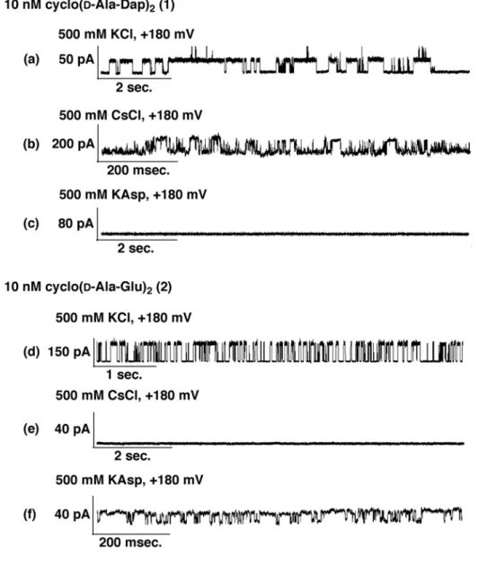
Figure 2-4: Ion channel recording of cyclo(D-Ala-Dap)2 (1) and cyclo(D-Ala-Glu)2 (2) in
500 mM KCl, CsCl, and KAsp solution under specific electrical potential. Artificial lipid membrane was prepared using neutral DPhPC lipid. Measurements were carried out under neutral (pH 7.4) conditions buffered with 5 mM HEPES. All samples were prepared for 10 nM peptide concentration.
Evaluation of the difference of ion selectivity for the cationic cyclic tetrapeptide cyclo(D-Ala-Dap)2 (1) with peptide concentration 10 nM in 500 mM three kinds of
electrolytes which were KCl, CsCl and KAsp (potassium aspartate), independently. As shown in Fig. 2-4a and b, the current recording at KCl and CsCl measurements showed the stable and continuous current values in the range up to 100 pA which meant open-close pore transition states of the ion channel formation. The reproducibility of recording for ion channel formation was two times out of five trials (2/5) about both KCl and CsCl measurements. However, it was not observed at KAsp measurement (reproducibility; 0/5) as shown in Fig. 2-4c in this measurement.
The single-channel current recording was also measured for anionic cyclic tetrapeptide cyclo(D-Ala-Glu)2 (2) with peptide concentration 10 nM in 500 mM three
kinds of electrolytes which were KCl, CsCl and KAsp, independently. As shown in Fig. 2-4d and f, the current recording at KCl and KAsp measurements showed the stable and continuous current values in the range up to 100 pA which meant open-close pore transition states of the ion channel formation. The reproducibility of recording for the ion channel formation was 2/5 about both KCl and KAsp measurements. However, it was not observed current values that meant ion channel formation at CsCl measurement (reproducibility; 0/5) shown in Fig. 2-4e in this measurement.
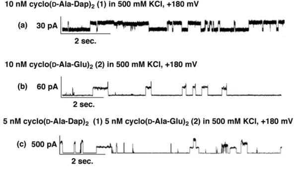
The single-channel current recording was also measured for the mixing sample with cationic cyclic tetrapeptide cyclo(D-Ala-Dap)2 (1) and anionic one cyclo(D
-Ala-Glu)2 (2) with molar ratio, 1:1. The mixing sample was prepared to add both 10 nM
peptide solutions into a same tube with molar ratio, 1:1. It was also employed three kinds of electrolytes which were KCl, CsCl and KAsp, independently. As shown in Fig. 2-5a and c, the current recording at KCl and KAsp measurements showed the stable and
continuous current values in the range up to 500 pA which meant open-close pore transition states of the ion channel formation. The reproducibility of recording for the ion channel formation was 1/5 about both KCl and KAsp measurements. However, it was not observed current values that meant the ion channel formation at CsCl measurement (reproducibility; 0/5) in this measurement. The current values of at KCl and KAsp measurements using mixing sample (100-500 pA see in Fig. 2-5) were higher than that of at the measurements using each cyclic tetrapeptides separately (20-100 pA see in Fig. 2-4). The specific ion selectivity was observed by using simple charged cyclic tetrapeptides 1, 2, and both mixing sample. The results of ion channel formation by two cyclic peptides and both mixing sample were summarized in Table. 2-1.
Figure 2-5: Ion channel recording of mixing sample with cyclo(D-Ala-Dap)2 (1) and
cyclo(D-Ala-Glu)2 (2) in 500 mM KCl, CsCl, and KAsp solution under specific electrical
potential. Artificial lipid membrane was prepared using neutral DPhPC lipid. Measurements were carried out under neutral (pH 7.4) conditions buffered with 5 mM HEPES. The sample was prepared to mix two peptides; peptide 1 and 2 with 1:1 molar ratio.
2.1.4. Evaluation of Secondary Structure of cyclo(D-Ala-Dap)2 and cyclo(D
-Ala-Glu)2
To evaluate conformation of cyclic tetrapeptide, circular dichroism (CD) spectra were measured about cyclo(D-Ala-Dap)2 (1), cyclo(D-Ala-Glu)2 (2), and both mixing
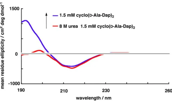
sample. As shown in Fig. 2-6, 1.5 mM peptide samples dissolved in 2,2,2-trifluoroethanol (TFE) were measured. There were no specific spectra observed that meant secondary structure about three samples. Around 210 nm of ellipticity decreased in stepwise when both peptide was mixed. The value of ellipticity was 103 cm2 deg
dmol-1 that was lower than other specific secondary structure such as α-helix and β-sheet
structure whose ellipticity values are often observed over 104 cm2 deg dmol-1.30, 31
Table 2-1: Summary of ion channel formation by various electrolytes. +; ion channel formation observed. -; not observed. Observed average current value noted under + sign and common counter ion species are described, respectively.
2.1.5. Ion Channel Formation in Negative Charged Acid Membrane
To evaluate the ion channel formation in a negative-charged lipid membrane, the single channel current recording was measured using an acid membrane. In bacteria cell surface has negative charge derived from acid amino acid and glycosaminoglycans.32 To
evaluate ion channel formation in a different membrane, it was employed that not neutral lipid membrane but acid membrane which was negative-charged similar to the cell membrane of micro-organisms. The acid membrane was composed of two lipids; DPhPC/DPhPG, 3/1, w/w. DPhPG has negative charge because of its glycerol moiety and phosphate ion. So, this mixture of lipids was negative charged membrane in this measurement system.
Figure 2-6: CD spectra of cyclic tetrapeptides cyclo(D-Ala-Dap)2 (1) and cyclo(D
-Ala-Glu)2 (2) and both mixing sample. The peptide 1 and 2 were dissolved in TFE to 1.5 mM
peptide concentration. Mixing sample was prepared by mixing both 1.5 mM peptide solution with 1:1 molar ratio.
As shown in Fig. 2-7, cyclo(D-Ala-Dap)2 (1), cyclo(D-Ala-Glu)2 (2) and mixing
sample were measured their ion channel formation. From Fig. 2-7a and b, cyclo(D -Ala-Dap)2 (1) and cyclo(D-Ala-Glu)2 (2) formed specific ion channel formation whose current
value was 20-50 pA scale. It was similar to the measurement in the neutral lipid membrane shown in Fig. 2-4a and d. From Fig. 2-7c, the current value was 200-300 pA which was about 10 times higher than that of the measurement using cationic cyclo(D -Ala-Dap)2 (1) and anionic cyclo(D-Ala-Glu)2 (2), independently. That was similar
tendency of the measurement at the neutral membrane shown in Fig. 2-5a. This was clearly indicated mixing cationic cyclic tetrapeptide 1 and anionic peptide 2 led to
Figure 2-7: Ion channel recording of mixing sample cyclo(D-Ala-Dap)2 (1) and cyclo(D
-Ala-Glu)2 (2) and mixing sample with the peptide 1 and 2 in 500 mM KCl solution under
specific electrical potential. Artificial lipid membrane was prepared using mixture of neutral DPhPC and acidic DPhPG (3/1, w/w) lipid. Measurements were carried out under neutral (pH 7.4) conditions buffered with 5 mM HEPES. The 10 nM peptide concentration was configured. The mixing sample was prepared to mix two pepitdes with 1:1 molar ratio.
observation of the large current values regardless of the charge state of the lipid membrane.
2.1.6. Disruption of Hydrogen Bonding by Urea
One of the important driving forces about the ion channel formation by cyclic ion channel peptide is aggregation through hydrogen bondings between -NHCO- amide skeletons.16, 27 To evaluate the importance of this intermolecular interaction to form the
nanotube and the ion channel, the inhibitor of hydrogen bonding, that is, a denaturant of hydrogen bonding was co-exited with cyclic peptides and evaluated their ion channel formation. In general, denaturation of hydrogen bonding of protein and peptides is carried out using 6-8 M urea.33 The urea (NH
2CONH2) interrupts and intervenes with
hydrogen bondings, followed by denaturing molecular hydrogen bondings. The measuring sample was prepared by mixing with urea and peptide 1. As shown in Fig.
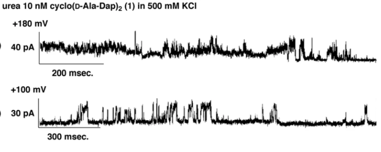
Figure 2-8: Ion channel recording of 8 M urea 10 nM cyclo(D-Ala-Dap)2 (1) in 500 mM
KCl solution under specific electrical potential. Artificial lipid membrane was prepared using neutral DPhPC lipid. Measurements were carried out under neutral (pH 7.4) conditions buffered with 5 mM HEPES.
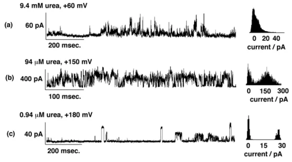
2-8a and b, fluctuated and discrete current values were observed. The spike signal which meant transient ion passing because of unstable pore formation. It was not clear open-close pore transition state indicating disturbance of pore formation by 8 M urea. To obtain more detail information about urea denaturation, less urea concentration was tested. As shown in Fig. 2-9, 9.4 mM and 94 µM urea concentration led to disrupt the ion
channel formation similar to 8 M urea denaturation test shown in Fig. 2-8. However, in the 94 µM urea evaluation, the specific open-close transition of pore formation was restored. The lower the urea concentration was, the more stable ion channel was observed in this evaluation.
Figure 2-9: Ion channel recording of 0.94 µM, 94 µM, and 9.4 mM urea 10 nM cyclo(D -Ala-Dap)2 (1) in 500 mM KCl solution under specific electrical potential. Left panel is a
single ion channel current and right one is a histogram analysis for each current recording. Artificial lipid membrane was prepared using neutral DPhPC lipid. Measurements were carried out under neutral (pH 7.4) conditions buffered with 5 mM HEPES.
CD spectra were measured to evaluate effect of urea on the peptide conformation. As show in Fig. 2-10, the specific changes of spectra were observed when 8 M urea was co-existed with 1.5 mM cyclo(D-Ala-Dap)2 (1) in 50 mM phosphate buffer. It indicated
clearly that the urea effected the peptide conformation. Particularly, around 195 nm ellipticity from π-π* transition of the amide was decreased by adding urea. This result showed that the urea effected on -NHCO- amide of the peptide 1.
To evaluate urea effect for highly hydrophobic peptide cyclo(D-Ala-Aib)2 (7) and
cyclo(D-Ala-Ile)2 (6), the ion channel formation co-existed with 8 M urea was also
measured. As shown in Fig. 2-11a, fluctuated and discrete current values were observed. The spike signal which meant transient ion passing because of unstable pore formation compared to no urea condition shown in Fig. 2-3a and b. It was not clear open-close pore transition state indicating disturbance of pore formation by 8 M urea for the peptide 6.
Figure 2-10: CD spectra of 1.5 mM cyclo(D-Ala-Dap)2 (1) co-existed 8 M urea in 50
On the contrary, as shown in Fig. 2-11b, the specific open-close transition of pore formation was restored about the peptide 6 compared to the no urea condition shown in Fig. 2-3c and d. In the range up to 20 pA channel current and stable open-close pore transition state were surely observed.
2.2. Discussion
2.2.1. Favorable Condition for Ion Channel Formation by Cyclic Tetrapeptides To get the most available and appropriate peptide sequences for the ion channel formation by cyclic tetrapeptides, 7 species cyclic peptides were synthesized and evaluated their ion channel activity. In this library shown in Fig. 2-1, it is surely limited the variety of amino acids components and their side-chain varieties. But some tendencies and rules are found out in the experiments.
Figure 2-11: Ion channel recording of 8 M urea 10 nM cyclo(D-Ala-Aib)2 (7) and
cyclo(D-Ala-Ile)2 (6) in 500 mM KCl solution under specific electrical potential.
Artificial lipid membrane was prepared using neutral DPhPC lipid. Measurements were carried out under neutral (pH 7.4) conditions buffered with 5 mM HEPES.
From the results of single ion channel recording of the peptide 3-5 and the peptide 1, the recording of the peptide 1 showed energetically stable ion channel formation compared to that of the peptide 3-5 shown in Fig. 2-2. The open-time of the pore for the peptide 1 showed continuously about 10 sec compared to that of the peptide 3-5 whose conductance recording showed the fluctuated and discrete current values change. The difference of stability for the ion channel formation is dependent on the amino acid sequence. It may be preferable for cyclic peptides that contain less interactive factors such as low hydrophobic molecules that have little bulky side-chain groups. Because the less interactive factor enable cyclic peptides to move and arrange the most appropriate conformation to achieve supramolecular-like structure such as “nanotube-bundle” in the dynamic lipid membrane atmosphere. This nanotube-bundle structure leads to form the inter-nanotube pore for the ion channel formation. Therefore, it is possible that the use of the low-hydrophobic cyclic tetrapeptide 1 has the advantage for energetically stable and having constant pore-size ion channel formation compared to the high-hydrophobic cyclic tetrapeptide 3-5. This tendency for the ion channel formation was also observed about the peptide 6 and 7. The stable ion channel formation was observed in cyclo(D-Ala-Aib)2
(7) than cyclo(D-Ala-Ile)2 (6). As shown in Fig. 2-3, the results of the peptide 6-7 were
re-validated the tendency that low-molecular hydrophobic peptide tended to form more stable ion channel than high-hydrophobic one. The hydrophobicity of Ile is larger than that of Aib.
Many studies have been reported that the evaluation of the ion channel formation using cyclic peptides.16, 18, 19, 34 For example, Ghadiri et al. have suggested that the cyclic
ocatapeptide forming the self-assembled cylindrical β-sheet peptide structure called “nanotube” by stacking the cyclic peptides in the membrane.16 As a result, ions pass
through this hollow of nanotubes, and the ion channel formation is achieved. According to the “nanotube” ion channel model, it is necessary that a diameter of cyclic peptides should be larger than that of passing ions such as K+ and Cl- (dehydrated diameter of
these ions are ~0.3 nm). However, a few reports about ion channel formation of cyclic tetrapeptide such as tentoxin have been published though a diameter of cyclic tetrapeptide (~0.2 nm) is smaller than that of K+ and Cl-.25 The mechanism of the ion channel
formation by cyclic tetrapeptides is unclear for now. In the present study, the specific ion channel formation was also observed using the cyclic tetrapeptides such as cyclo(D -Ala-Dap)2 (1) and cyclo(D-Ala-Glu)2 (2). This might indicate the nanotube-bundle model. It
is a kind of self-organizing structure that nanotubes aggregate each other in the lipid membrane, followed by forming the inter-nanotube pore between nanotubes, which is similar to the bundle structure formed by the linear ion channel peptides. The possibility for the nanotube-bundle model by cyclic peptides has been suggested before.35 By
employing a small-ring-sized cyclic peptide, cyclic tetrapeptides, that allowed peptides to form the inter-nanotube pore mainly not the intra-nanotube pore. In this condition, it is important for the ion channel formation to aggregate smoothly in the lipid membrane. The hydrophobicity and interactive factors such as alkyl-chain length and aromatic moiety might be obstacle when it comes to aggregate nanotubes each other to form a stable inter-nanotube pore. It is necessary to clarify the mechanism of the ion channel formation by the cyclic tetrapeptides in the future.
2.2.2. Ion Selectivity for Ion Channel Formation by Cyclic Tetrapeptides
From the results of ion selectivity evaluation, the ion channel formation was observed that the recording of the peptide 1 showed the ion channel formation at KCl and
CsCl, not at KAsp evaluating system (Fig. 2-4a, b, and c). On the other hand, that of the peptide 2 showed the ion channel formation at KCl and KAsp, not at CsCl system (Fig. 2-4d, e, and f). Similarly, that of the mixing sample showed the ion channel formation at KCl and KAsp, not at CsCl system (Fig. 2-5). Given the common ion species from these results, the use of the cationic peptide 1 was resulted in Cl- anion-selective and the
anionic peptide 2 led to K+ cation-selective ion channel formation, respectively shown in
Table 2-1. This ion selectivity might be attributed to the charge state of peptides, which means that a cationic cyclic peptide attract an anion and anionic one attract a cation. It has been reported that ion selectivity has changed cation-selective into anion-selective by altering the neutral amino acid residue; Gln to cationic one; Lys.36 This might indicate
that the charge state of residue function as an ion-filter. The ion selectivity of the mixing sample was similar to that of anionic cyclic peptide which showed K+ cation-selective ion
channels. Given electrostatic interaction between the cationic peptide 1 and the anionic peptide 2, the bias of charge for each peptides may be canceled and approach to the neutral charge state. This ion-selectivity for mixing sample is interesting. But it is still uncertain about the detailed mechanism of ion-selectivity for the mixing sample. The current value in the recording of the mixing sample is lager than that of each cyclic peptide employed separately. It is often said that the current value is counterpart of the pore-size of ion channel. The electrostatic interaction between cationic and anionic cyclic tetrapeptides in the mixing sample may lead to stability of pore-structure for formation of more large-size pore in the membrane.
From the results of CD spectra of the peptide 1 and 2 and both mixing sample, there were no specific spectra observed that meant the specific secondary structure about three samples shown in Fig. 2-6. The value of ellipticity was 103 cm2 deg dmol-1 that was
lower than other specific secondary structure such as α-helix and β-sheet structure whose ellipticity values are often observed over 104 cm2 deg dmol-1.30 This observation for
cyclic tetrapeptide 1 and 2 has no any β-sheet nor β-turn structure in their conformation. That is reasonable observation given the structure of the peptide 1 and 2 because their peptide sequence is totally designed artificially and a few number of amino acids; D -Ala-AA-D-Ala-AA (AA means arbitrary amino acid) are not enough to have the specific secondary structure.
2.2.4. Effect of Charge State of Membrane on Ion Channel Formation by Cyclic Tetrapeptides
To evaluate ion channel formation in negative-charged lipid membrane, the single channel current recording was measured using the acid membrane which had negative charged membrane in this measurement system.
As shown in Fig. 2-7, cyclo(D-Ala-Dap)2 (1), cyclo(D-Ala-Glu)2 (2) and mixing
sample were measured their ion channel formation. From Fig. 2-7a and b, cyclo(D -Ala-Dap)2 (1) and cyclo(D-Ala-Glu)2 (2) formed specific ion channel formation whose current
value was 20-50 pA scale. It was similar to the measurement in the neutral lipid membrane shown in Fig. 2-4a and d. This observation might suggest that the charge state of the membrane is irrelevant to the ion channel formation. Especially, the peptide 2 is anionic cyclic tetrapeptide so that the peptide 2 has a possibility to repel with negative charge membrane. But it is surely observation of the ion channel formation by the
peptide 2 with the acid membrane. The solubility is more important than hydrophobicity in the interaction with lipid membrane in this case.
From Fig. 2-7c, the current value was 200-300 pA which was about 10 times higher than that of the measurement using cationic cyclo(D-Ala-Dap)2 (1) and anionic
cyclo(D-Ala-Glu)2 (2), independently. That was similar tendency of the measurement at
the neutral membrane shown in Fig. 2-5a. This was clearly indicated mixing cationic cyclic tetrapeptide 1 and anionic peptide 2 led to the observation of the large current values regardless of the charge state of the lipid membrane. The stabilization by interaction the cationic 1 with the anionic 2 happens independent on the charge state of the membrane. The contribution of charge of cyclic peptides is larger than that of the membrane because the cyclic peptides are dispersed and scattered in the solution comparing the lipid membrane localized in the tip of glass pipette. The flexibility and degree of freedom are the main reason why ion channel formation is observed constantly independent on charge state of the membrane. This observation is simple but convenient way to make a large pore for other cyclic ion channel peptides.
2.2.5. Effect of Hydrogen-Bonding Denaturant on Ion Channel Formation by Cyclic Tetrapeptides
To evaluate importance of this intermolecular interaction to form nanotube and ion channel, the measuring sample was prepared by mixing with urea as hydrogen-bonding denaturant and peptide 1. As shown in Fig. 2-8a and b, fluctuated and discrete current values were observed. The spike signal means transient ion passing because of unstable pore formation. It was not clear open-close pore transition state indicating disturbance of the pore formation by 8 M urea. As shown in Fig. 2-9, 9.4 mM and 94 µM
urea concentration led to disrupt ion channel formation similar to 8 M urea denaturation test shown in Fig. 2-8. However, in the 94 µM urea evaluation, the specific open-close transition of pore formation was restored. The lower the urea concentration was, the more stable ion channel was observed in this evaluation. This is clearly indicated that urea effects on the ion channel formation by the cyclic tetrapeptide cyclo(D-Ala-Dap)2 (1)
depending on urea concentration. The inter-molecular hydrogen bonding for ion channel formation can be many various models. The most important one is amide-amide hydrogen bonding to from the peptide nanotube. If the peptide nanotube formation is disrupted, the ion channel formation will be difficult because aggregation between nanotubes is important for inter-nanotube pore formation.
CD spectra were measured to evaluate effect of urea on peptide conformation. As shown in Fig. 2-10, specific changes of spectra were observed when 8 M urea was co-existed with 1.5 mM cyclo(D-Ala-Dap)2 (1) in 50 mM phosphate buffer. It indicated
clearly that urea effected the peptide conformation. Particularly, around 195 nm ellipticity from π-π* transition of amide was decreased by adding urea. This result showed that urea effected on -NHCO- amide of the peptide 1. The results of ion channel recording and CD measurement suggest the inter-molecular hydrogen bonding between peptide bonds is significant for the ion channel formation by the peptide 1.
To evaluate urea effect on highly hydrophobic peptide cyclo(D-Ala-Aib)2 (7) and
cyclo(D-Ala-Ile)2 (6), the ion channel formation co-existed with 8 M urea was also
measured. Interestingly, as show in Fig. 2-11b, the specific open-close transition of pore formation was restored about the peptide 6 compared to the no urea condition shown in Fig. 2-3c and d. In the range up to 20 pA channel current and subtle open-close pore transition state were surely observed. These are totally inverse results and tendencies
compared to the peptide 1 shown in Fig. 2-8. Generally, 8 M urea is enough high concentration to inhibit the inter-molecular hydrogen bondings. The result of cyclo(D -Ala-Aib)2 (7) shows surely unstable pore formation but the result of cyclo(D-Ala-Ile)2 (6)
shows more stable ion channel formation compared to the peptide 7 with 8 M urea. It might alleviate the influence of inter-molecular hydrophobic interaction which is general van der Waals interaction by disrupting inter-molecular hydrogen bonding. Ile is a highly hydrophobic amino acid residue that causes and promotes interaction between side-alkyl chains which leads to congestion of cyclic peptides. For nanotube formation, it is important of appropriate interaction between molecules and nanotubes. Although disruption of inter-molecular hydrogen bonding for the peptide 6, the side-chain hydrophobicity covers it compensation for earning interactive driving force for the nanotube formation and the ion channel formation even if inter-molecular hydrogen bonding is totally disturbed by 8M urea.
3. Ion Channel Formation by Cyclic Pentapeptides
3.1. Results
3.1.1. Chemical Structures of Cyclic Pentapeptides
Containing even number of alternating D- and L-amino acid residue is one of the important factors of cyclic peptides to form the ion channel formation. Because this peptide sequence adopts a flat-ring conformation to promote stacking cyclic peptides, followed by forming a contiguous amide-skeleton hydrogen-bonded hollow structure of the peptide nanotube.16 It is preferable for cyclic peptides to have an inherent tendency of
the nanotube formation. In 2005, however, it has been reported that the odd numbers of only L-amino acid residues peptides cyclo(L-Gln)5 (8) forms entwined and branching
nanotube structure by using Atomic Force Microscopic (AFM) imaging.37 From the
molecular dynamics and quantum chemistry simulation, the conformation of cyclo(L -Gln)5 (8) is not flat-ring structure because of the composition of all five L-amino acids so
that stacking the cyclo(L-Gln)5 (8) each other leading to the entwined nanotube. And
entangling with each entwined nanotubes leads to apparently the branching nanotube. Ion channel formation by the cyclic pentapeptide cyclo(L-Gln)5 (8) hasn’t been evaluated.
Given ion channel formation by this cyclic pentapeptide, ion passing through the intra-nanotube pore is difficult for cyclo(L-Gln)5 (8) because ion mobility of the entwined
nanotube by cyclic homo-L-peptide can be poorer than that of the straight nanotube by cyclic hetero-D-L-peptide. On the other hand, the inter-nanotube pore might be possible to form than intra-nanotube pore because a pore size of the inter-nanotube one is variable and flexible depending on degree of aggregation of nanotubes in the membrane. The problem is a difficulty in the pore formation by aggregation of entwined nanotubes that
are congested and formation of the inter-nanotube pore may be difficult to form the pore structure even if it’s possible in theory. But cyclic tetrapeptide is composed by 4 amino acids and cyclic pentapeptide is composed by 5 amino acids. It is important and valuable to evaluate these simple and small ring size cyclic peptides for the ion channel formation in the respect of the regulation. To focus on inter-nanotube pore is preferable for the restriction of pore formation that is similar strategy of the cyclic terapeptide one in aspect of the ring-size restriction policy. In addition to the cyclo(L-Gln)5 (8), two other peptide
sequences were prepared for the evaluation of differences of side-chain groups, positive
charge including Lys; cyclo(L-Lys)5 (9) and negative charge including Glu; cyclo(L-Glu)5
(10).
3.1.2. Effect of Peptide Concentration on Ion Channel Formation
To investigate the influence of peptide concentration on the ion channel formation, two peptide concentration configured 1 µM and 10 nM were evaluated, respectively. As shown in Fig. 3-2a and b (left panels), the recording for the peptide 8 showed fluctuated and discrete current values in the range up to 10 pA. To clarify the distribution of current value with its frequency for each single-conductance recordings, the recording data were converted to the histogram analysis data. As shown in Fig. 3-2a and b (right panels), some broad peaks were observed which meant the distribution of current values were dispersed. The reproducibility of recording for the ion channel formation was one time
Figure 3-2: Ion channel recording of cyclo(L-Gln)5 (8) in 500 mM KCl solution under
electrical potential. Left panel is a single ion channel conductance and right one is a histogram analysis for each conductance. Artificial lipid membrane was prepared using neutral DPhPC lipid. Measurements were carried out under neutral (pH 7.4) conditions buffered with 5 mM HEPES. The samples were prepared for 1 µM (a) and 10 nM (b) peptide concentration.
out of five trials (1/5) about 1 µM cyclo(L-Gln)5 (8). However, the reproducibility was
increased up to 4/5 at 10 nM cyclo(L-Gln)5 (8) measurement.
3.1.3. Effect of Side-Chain of Cyclic Pentapeptides on Ion Channel Formation
The other two charged peptides cyclo(L-Lys)5 (9) and cyclo(L-Glu)5 (10) were
evaluated to investigate an influence of charged cyclic peptides for ion channel formation. As shown in Fig. 3-3a (left panel), the current recording for the peptide 9 showed fluctuated and discrete current values in the range up to 20 pA. To clarify the distribution of current value with its frequency for each single-conductance recordings, the recording data were converted to the histogram analysis data. As shown in Fig. 3-3a (right panel), some broad peaks were observed which meant the distribution of current values were dispersed. On the contrary, as shown in Fig. 3-3b (left panel), the current recording for the peptide 10 showed stable and continuous current values in the range up to 20 pA than that of the peptide 9. These stable current values meant open-close pore transition states of the ion channel formation. From the histogram analysis of the peptide 10 shown in Fig. 3-3b (right panel), some more sharp peaks were observed than the peptide 9 measurement shown in Fig. 3-3a (right panel). This sharp peak observation meant distribution of current values were constantly focused. The reproducibility of recording for the ion channel formation was one time out of five trials (1/5) about about both peptides 9 and 10. That was lower reproducibility than 10 nM cyclo(L-Gln)5 (8) whose reproducibility
was 4/5.
The single-channel current recording was also measured for mixing sample with cationic cyclic pentapeptide cyclo(L-Lys)5 (9) and anionic one cyclo(L-Glu)5 (10) with
into a same tube with molar ratio, 1:1. As shown in Fig. 3-3c (left panel), the current recording for the mixing sample showed stable and continuous current values in the range up to 400 pA. The current values using mixing sample (150-400 pA see in Fig. 3-3c) were higher than that of at the measurements using each cyclic pentapeptides separately (~20 pA see in Fig. 3-3a and b). The reproducibility of recording for ion channel formation was three times out of five trials (3/5) about the mixing sample. The reproducibility of the measurement for the mixing sample (3/5) increased compared to
Figure 3-3: Ion channel recording of cyclo(L-Lys)5 (9), cyclo(L-Glu)5 (10) and mixing
cylo(L-Lys)5 (9) and cylo(L-Glu)5 (10) sample in 500 mM KCl solution under electrical
potential. Left panel is a single ion channel conductance and right one is a histogram analysis for each conductance. Artificial lipid membrane was prepared using neutral DPhPC lipid. Measurements were carried out under neutral (pH 7.4) conditions buffered with 5 mM HEPES. The peptide concentration was 10 nM for the peptide 8 and 9. The mixing sample was prepared to mix two peptides 9 and 10 with 1:1 molar ratio.
using each charged cyclic peptide 9-10 separately (1/5). Mixing cationic and anionic cyclic pepntapeptide enabled to promote the reproducibility of the ion channel formation.
3.1.4. Ion Selectivity of Cyclic Pentapeptides for Ion Channel Formaiton
To evaluate ion selectivity, cyclo(L-Gln)5 (8) was employed as the most capable
peptide for ion channel formation because of the highest reproducibility. And bulky counter-ion method was tested using solutions of potassium gluconate (K+Gluc-) and
N-methylglucosammonium chloride (Nmg+Cl-).38 In both solutions, the majority of the
current is expected to be carried by the smaller ion. Accordingly, K+ ions are mobile ones
in K+Gluc- solution and Cl- ions are mobile ones in Nmg+Cl- solution. The
reproducibility of cyclo(L-Lys)5 (9) and cyclo(L-Glu)5 (10) were lower than that of the
peptide 8. To overcome this difficulty, the mixing two peptides 9 and 10 sample was employed to increase the reproducibility by stabilizing electrostatic interaction aiming for formation of the ion channel easily.
Typical ion channel recording and histogram analyses of the peptide 8 in K+Gluc
-and Nmg+Cl- are shown in Fig. 3-4a and b. The current recording for the peptide 8 in K +Gluc- system showed fluctuated and unstable current values in the range up to 15 pA
shown in Fig. 3-4a (left panel). From the histogram analysis data shown in Fig. 3-4a (right panel), the broad peak around 10 pA was observed which meant the distribution of current values were dispersed. On the contrary, as shown in Fig. 3-4b (left panel), the current recording for the peptide 8 in Nmg+Cl- system showed stable and continuous
current values in the range up to 80 pA than K+Gluc- system. These stable current values
meant open-close pore transition states of the ion channel formation. From histogram analysis shown in Fig. 3-4b (right panel), a few current-value distributions such as 10, 35
and 65 pA were observed in Nmg+Cl- solution. The reproducibility of observation for the
ion channel formation was decreased in K+Gluc- (1/5) and Nmg+Cl- (2/5) solution
compared to in KCl solution (4/5). To estimate ion selectivity, the ratios of most distributed current values obtained from histogram analyses were calculated as the selectivity (ICl-/IK+). The selectivities were ICl-/IK+ = 1.1 (10/9.0), 3.8 (35/9.0) and 7.2
Figure 3-4: Ion channel recordings of the peptide cylo(L-Gln)5 (8) and mixing cylo(L
-Lys)5 (9) and cylo(L-Glu)5 (10) sample in bulky counter-ion solutions; K+Gluc- and Nmg +Cl-. Left; Typical traces of observed ion channel conductance under +150 mV. Right;
Histogram analysis for each conductance patterns. Artificial lipid membrane was prepared using neutral DPhPC lipid. Measurements were carried out under neutral (pH 7.4) conditions buffered with 5 mM HEPES. The peptide concentration was 10 nM for the peptide 8. The mixing sample was prepared to mix two peptides 9 and 10 with 1:1 molar ratio.
(65/9.0), respectively. The calculated selectivity suggests that the ion channel formed by cyclo(L-Gln)5 (8) is Cl- anion selective channel.
Typical ion channel recording and histogram analyses of the mixing sample peptide 8 and 9 in K+Gluc- and Nmg+Cl- are shown in Fig. 3-4c and d. The current
recording for the mixing sample in K+Gluc- system showed stable and continuous current
values in the range up to 35 pA shown in Fig. 3-4c (left panel). From the histogram analysis data shown in Fig. 3-4c (right panel), the sharp peak around 20 pA was observed meaning the distribution of current values focused. As shown in Fig. 3-4d (left panel), the current recording for the mixing sample in Nmg+Cl- system also showed stable and
continuous current values in the range up to 150 pA. The reproducibility of observation for the ion channel formation was decreased in K+Gluc- (2/5) and Nmg+Cl- (1/5) solution
compared to in KCl solution (3/5). To estimate ion selectivity, the ratio of most distributed current values obtained from histogram analyses were calculated as the selectivity (ICl-/IK+). The selectivity were ICl-/IK+ = 6.0 (122/22). The calculated
selectivity suggests that the ion channel formed by the mixing sample with 9 and 10 with molar ratio, 1:1 is Cl- anion selective channel.
For more detail insight of the ion channel formation about mixing sample 9 and 10, the molar ratio of the peptide cyclo(L-Lys)5 (9) and cyclo(L-Glu)5 (10) was changed.
The molar ratio peptide 9 : peptide 10 was 1:9 and 9:1 configured, respectively. As shown in Fig. 3-5a (left panel), the current recording for 1:9 sample in K+Gluc- system
showed stable and continuous current values in the range up to 150 pA. This stable current values meant open-close pore transition states of ion channel formation. From histogram analysis shown in Fig. 3-5a (right panel), a few current-value distributions such as 35, 85 and 135 pA were observed in K+Gluc- solution. On the other hand, the current