2020
December Vol. 63 No. 6
J‐STAGE https://www.jstage.jst.go.jp/browse/shikahozon/‐char/ja
このたび,日本歯科保存学会2021年度春季学術大会(第154回)を開催させていただくことになりまし た.2021年6月10日,11日の日程で,会場は川商ホール(旧 鹿児島市民文化ホール)を予定しています.
新型コロナウイルスの影響により開催形式については予断を許さない状況ではありますが,鹿児島大学大 学院医歯学総合研究科顎顔面再建学講座歯科保存学分野の教室員および関係者一同,皆様を鹿児島にてお迎 えできますことを大変楽しみにしております.
歯を保存することを使命として,修復・歯内・歯周の3領域からなる特徴をもつ日本歯科保存学会は,す でに65年の歴史を有する学会であり,この間には多数の「歯の保存」スペシャリストが学会を構成すると同 時に,次世代の人材育成も行われて継続的に国民の健康増進に貢献しながら発展しています.その伝統ある 日本歯科保存学会の第154回目の学術大会を主管開催させていただくにあたり,大会のテーマを「承前啓後~
未来へつなぐ歯科保存学~」といたしました.
本大会では,シンポジウム,教育講演,認定研修会,学会主導型プログラム,口頭およびポスター形式で の一般発表を予定しております.
シンポジウムでは,日本歯科保存学会で将来活躍する次世代の人材育成にフォーカスした「歯科保存治療 に携わる人材のキャリアプランニング」をテーマとして,岡山大学病院総合歯科 鳥井康弘先生に座長をお願 いしました.また,鹿児島県は有人離島人口数が日本一であり,他県にはない規模での離島巡回歯科診療が 毎年行われ,そのなかで保存治療も重要な役割を果たしています.この鹿児島県で実施される離島巡回歯科 診療の現状および今後の展望にフォーカスしたシンポジウム「離島歯科巡回診療の現状と将来」について,
鹿児島大学大学院咬合機能補綴学分野 南 弘之先生に座長を依頼しています.
さらに本学会では学会主導型プログラムを企画していただき,イタリアのGianluca Plotino教授に「The Ideal Instrument for Root Canal Preparation」のタイトルでご講演いただく予定です.
学会の開催にあたりましては,新型コロナウイルス感染症拡大予防策を徹底して講じます.皆様にもご協 力いただきながら,学会の開催方法についても「未来へつなぐ」べく,新たな様式を提案することになりそ うです.
大会開催地の鹿児島は,世界有数の活火山である桜島や指宿の砂蒸し風呂,霧島温泉,屋久島や奄美大島 等,豊かな自然に囲まれています.特に桜島は市街地から約4キロメートルに位置し,市街地からは24時間 運航している桜島フェリーを使うと約15分で渡ることができます.さらには幕末・明治時代を中心とした歴 史的名所など,見どころが多数あります.また鹿児島は,黒毛和牛・豚・鶏の飼養数,鰻の養殖数,サツマ イモ収穫量などが全国一位など農業も盛んで,周囲を海に囲まれ新鮮な魚介も豊富です.学会へお越しにな られた際には,ぜひ鹿児島のグルメもお楽しみいただければと思います.
現地開催を予定しておりますが,新型コロナウイルス感染拡大の状況によって,開催形式が変更となる場 合もございます.来年6月の鹿児島にて皆様とお目にかかれますことを,スタッフとともに楽しみにしてお ります.多数のご参加を心よりお待ちしております.
承前啓後~未来へつなぐ歯科保存学~
日本歯科保存学会 2021 年度春季学術大会(第 154 回)大会長 鹿児島大学大学院医歯学総合研究科 先進治療科学専攻 顎顔面機能再建学講座 歯科保存学分野 西谷 佳浩
日本歯科保存学会 2021 年度春季学術大会(第 154 回)
《事前登録のご案内》
日本歯科保存学会2021年度春季学術大会(第154回)は,2021年6月10日(木)・11日(金),鹿児島県・鹿児島 市(担当校:鹿児島大学大学院医歯学総合研究科顎顔面再建学講座歯科保存学分野,大会長:西谷佳浩教授)におい て,下記のとおり開催することとなりました.事前参加登録についてご案内いたしますので,下記の要領により登録 手続きをお願いいたします.多数の会員の皆様方のご参加をお願い申し上げます.
※ 現地開催を予定しておりますが,新型コロナウイルス感染症拡大の状況によって開催形式および登録方法・期間等 が変更となる場合もございます.
2020年12月
特定非営利活動法人 日本歯科保存学会 理事長 田上 順次
1 .会 期 2021年6月10日(木)・11日(金)
2 .会 場 川商ホール
〒890‒0062 鹿児島市与次郎2‒3‒1 TEL:099‒256‒8111(代)
3 .参加登録
大会参加費は以下のとおりです.
事前登録(2020年12月14日(月)正午~2021年5月7日(金)15:00までの登録) 10,000円 当日登録 12,000円
※本学術大会における事前登録は,すべて大会ホームページ上での手続きとなります.
http://www.kokuhoken.jp/jscd154にアクセスして,ご登録下さい.
4 .第 154 回学術大会に関するお問い合わせ先
日本歯科保存学会2021年度春季学術大会(第154回)大会事務局 〒170‒0003 東京都豊島区駒込1‒43‒9
(一財)口腔保健協会コンベンション事業部内
TEL:03‒3947‒8761 FAX:03‒3947‒8873 E-mail:[email protected] 詳細につきましては下記アドレスより,大会ホームページにアクセスして下さい.
http://www.kokuhoken.jp/jscd154 5 .交通・宿泊のご案内
交通・宿泊に関しましては,大会ホームページにおいて詳細を掲載します.
発 行
特定非営利活動法人 日本歯科保存学会
〒170‒0003 東京都豊島区駒込1-43-9 (一財)口腔保健協会内
日本歯科保存学雑誌
第
63
巻第6
号 令和2
年12
月目 次 ミニレビュー
接着性レジンの象牙質接着耐久性改良法の探求と接着破壊ダイナミクスの解明
保坂 啓一 (479)
原 著Study on the Sealing Ability of a New High-penetration Resin Material for Enamel Cracks and Dentinal Tubules
MIURA Tatsuki, YASUO Kenzo, IWATA Naohiro, YOSHIKAWA Kazushi and YAMAMOTO Kazuyo (483) ユニバーサルアドヒーシブのアクティブ処理およびエッチングモードが
エナメル質接着強さおよび表面自由エネルギーに及ぼす影響
今井亜理紗,髙見澤俊樹,田村 友彦,髙宮 寛,若松 賢吾
辻本 暁正,鈴木 総史,鈴木 美穂,宮崎 真至 (494) 高グルコース条件下における歯肉線維芽細胞のカルプロテクチン誘導性
炎症関連因子の産生におけるスダチチンの抑制効果
西川 泰史,成石 浩司,木戸 淳一,湯本 浩通 (503)
ニッケルチタン製ロータリーファイルのテーパーが回転疲労耐性に与える影響
福森 洋平,中務 太郎,牧 圭一郎,木村 俊介
海老原 新,興地 隆史,杉崎 順平 (512) 新規汚染除去材による処理が唾液汚染されたCAD/CAM冠用ハイブリッドレジンブロックの
象牙質接着強さに及ぼす影響
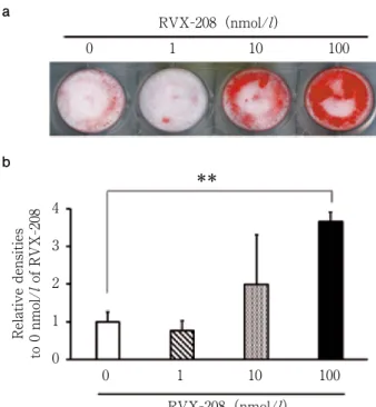
高橋 圭,𠮷𠮷 𠮷𠮷,𠮷𠮷 𠮷人,島田 𠮷史,𠮷𠮷 𠮷𠮷 (519) 生物学的直接覆髄剤開発に向けた生理活性物質RVX‒208の歯髄細胞における機能解析
松本 𠮷大,藤原 千春,島袋 善夫,𠮷本 優,NANTAKEERATIPAT Teerachate
北垣次郎太,八木 寛子,林 聡子,岩𠮷 智明,柏木陽一郎
三木 𠮷史,北村 正博,沢田 啓吾,木下 茉優,村上 伸也 (527) ユニバーサルアドヒーシブへの光線照射の有無が自己接着性レジンセメントの
歯質接着性に及ぼす影響
石井 亮,髙見澤俊樹,野尻 貴絵,辻本 暁正
甘利 佳之,奥脇 岳人,飯島 達也,宮崎 真至 (536) タタキ振動を付与した音波振動歯ブラシのプラーク除去効果および
歯周組織に対する有用性について
鴨井 久博,吉峰 正彌,三浦 悠,濵田真理子 (545)
Influence of Light Irradiation Conditions on the Tensile Bond Strength of Resin Composite Bonding Materials to Dentin
OUCHI Satoko, IWATA Naohiro, LIU Haitao, XIE Zhouqiao, WANG Xiangyu MOROTO Hidetoshi, ISHIDA Shunsuke, KOMASA Reiko, YASUO Kenzo
TANIMOTO Hiroaki, YOSHIKAWA Kazushi and YAMAMOTO Kazuyo (554) 症例報告インプラント周囲炎患者に対しEr:YAGレーザーを併用し外科的治療を行った症例
安田 忠司,𠮷本 敦彦,辰巳 順一 (563)
63巻総目次 (570)
会務報告 (576)
Published
THE JAPANESE SOCIETY OF CONSERVATIVE DENTISTRYby (JSCD)
c/o Oral Health Association of Japan(Kōkūhoken kyōkai)
1‒43‒9, Komagome, Toshima-ku, Tokyo 170‒0003 Japan
THE JAPANESE JOURNAL OF CONSERVATIVE DENTISTRY
Vol. 63, No. 6 DECEMBER 2020
CONTENTS Mini Review
Improving Resin-dentin Bond Durability and a Novel Ultra-high-speed Videography
HOSAKA Keiichi (479)
Original Articles
Study on the Sealing Ability of a New High-penetration Resin Material for Enamel Cracks and Dentinal Tubules
MIURA Tatsuki, YASUO Kenzo, IWATA Naohiro,
YOSHIKAWA Kazushi and YAMAMOTO Kazuyo (483) Influence of Active Application and Etching Mode on Enamel Bond Strength and
Surface Free Energy of a Universal Adhesive
IMAI Arisa, TAKAMIZAWA Toshiki, TAMURA Tomohiko, TAKAMIYA Hiroshi, WAKAMATSU Kengo, TSUJIMOTO Akimasa, SUZUKI Soshi,
SUZUKI Miho and MIYAZAKI Masashi (494) Inhibitory Effects of Sudachitin on the Production of Inflammation-related Molecules in
Human Gingival Fibroblasts Cultured under High Glucose Conditions
NISHIKAWA Yasufumi, NARUISHI Koji, KIDO Jun-ichi and YUMOTO Hiromichi (503) Effect of Taper of Nickel-titanium Rotary Instruments on Cyclic Fatigue Resistance in a Dynamic Model
FUKUMORI Yohei, NAKATSUKASA Taro, MAKI Keiichiro,
KIMURA Shunsuke, EBIHARA Arata, OKIJI Takashi and SUGIZAKI Jumpei (512) Effect of New Decontamination Material on the Dentin Bond Strength of
Saliva-contaminated Hybrid Resin Block for CAD/CAM Crowns
TAKAHASHI Kei, YOSHIYAMA Tomohiro, YOKOYAMA Akihito,
SHIMADA Yasushi and YOSHIYAMA Masahiro (519) The Functional Analysis of RVX-208 in Human Dental Pulp Cells: An Initiative for the
Application of a Bioactive Compound, RV-208, on Direct Pulp Capping
MATSUMOTO Masahiro, FUJIHARA Chiharu, SHIMABUKURO Yoshio, YAMAMOTO Yu, NANTAKEERATIPAT Teerachate, KITAGAKI Jirouta, YAGI Hiroko, HAYASHI Satoko, IWAYAMA Tomoaki, KASHIWAGI Yoichiro, MIKI Koji, KITAMURA Masahiro,
SAWADA Keigo, KINOSHITA Mayu and MURAKAMI Shinya (527) Influence of Light Irradiation of Universal Adhesives on the Bonding Effectiveness of
Self-adhesive Resin Cements
ISHII Ryo, TAKAMIZAWA Toshiki, NOJIRI Kie, TSUJIMOTO Akimasa, AMARI Yoshiyuki,
OKUWAKI Taketo, IIJIMA Tatsuya and MIYAZAKI Masashi (536) The Efficacy of Dental Plaque Removal and Usefulness for Periodontal Tissue of a
Sonic Toothbrush Equipped with Tapping Amplitude
KAMOI Hisahiro, YOSHIMINE Masaya, MIURA Haruka and HAMADA Mariko (545) Influence of Light Irradiation Conditions on the Tensile Bond Strength of
Resin Composite Bonding Materials to Dentin
OUCHI Satoko, IWATA Naohiro, LIU Haitao, XIE Zhouqiao, WANG Xiangyu, MOROTO Hidetoshi, ISHIDA Shunsuke, KOMASA Reiko, YASUO Kenzo,
TANIMOTO Hiroaki, YOSHIKAWA Kazushi and YAMAMOTO Kazuyo (554) Case Report
Surgical Treatment of Peri-implantitis by Using Er: YAG Laser: A Case Report
YASUDA Tadashi, YAMAMOTO Atsuhiko and TATSUMI Junichi (563)
2020年12月 479
はじめに
世界的規模における超高齢化の潮流において,日本で は100歳以上の人口が11万人を突破し,8020達成率も 50%を超えた.そのようななか,現在,根面う蝕・二次 う蝕・トゥースウェアなどへの対応が,歯科保存学・接 着修復学における課題となっている.人生100年時代の 口腔健康を増進させるため,徹底したう蝕予防とう蝕管 理,そして治療介入の際には,低侵襲接着技法を用いた アプローチがますます重要になっている1).近年の目覚 ましい接着歯学の発展に伴い,直接法コンポジットレジ ン修復は,厚生労働省の診療別歯科医療統計でも,この 10年間で治療件数が約20%増え,研究者のみならず臨床 家にとっても注目されている2,3).
接着性レジンの象牙質接着耐久性改良法の探求 接着修復成功の条件はいうまでもなく,良好な歯質接 着性および歯質接着耐久性の獲得である.エナメル質は
リン酸エッチングを行うことにより良好な接着が得られ るが,より有機質・水分を多く含む象牙質への接着はこ れまで難しく,接着耐久性が課題とされてきた.
操作ステップを簡略化した接着システムでは,欧米を 中心に採用されていたウェットボンディングシステムに おいて,リン酸エッチング後の水洗による水分のコント ロールが難しく,界面剝離と残留脱灰象牙質が生じると いう接着劣化につながる問題があった.そこで,著者ら は水洗後にエタノールを塗布し水分と置換するというス テップが,コラーゲン細線維間への疎水性のボンドの浸 透を促進し,良好な樹脂含浸層を作り接着耐久性に寄与 することを見いだした4).現在,世界中で多くの臨床家 に“エタノールウェットボンディングテクニック”とし て応用されている.
一方,マイルドな酸性である機能性モノマーによる脱 灰能をもつセルフエッチングシステムは現在,日本のみ ならず世界のゴールドスタンダードとなっている.しか しながら,切削象牙質上に覆われるスミヤー層が完全に 除 去 さ れ な い こ と に よ っ て 形 成 さ れ るHybridized
smear layerが,接着劣化の一因となる可能性が指摘さ
接着性レジンの象牙質接着耐久性改良法の探求と 接着破壊ダイナミクスの解明
保 坂 啓 一
東京医科歯科大学(TMDU)大学院医歯学総合研究科 医歯学系専攻 口腔機能再構築学講座 う蝕制御学分野
Improving Resin-dentin Bond Durability and a Novel Ultra-high-speed Videography
H
OSAKAKeiichi
Department of Cariology and Operative Dentistry, Division of Oral Health Science, Graduate School of Medical and Dental Sciences, Tokyo Medical and Dental University(TMDU)
キーワード: 象牙質接着耐久性,超高速度イメージング,微小引張接着試験,セルフエッチングシステム,コンポジッ トレジン
日歯保存誌 63(6):479~482,2020
ミニレビュー
本ミニレビューは,2020年度学術賞受賞者へ依頼したものである.
DOI:10.11471/shikahozon.63.479
れている.そこで著者らは,NaOCl水溶液によるスミ ヤー層の改質(有機成分の溶解除去)がスミヤー層中の 有機成分を溶解除去することがHybridized smear layer 形成を防ぎ,接着向上に寄与することを見いだした5). さらに,レジンモノマーの浸透性が向上すること,ミネ ラル密度上昇による化学的接着が向上すること,そして 接着阻害因子の水分量が減少するという3つの利点が考 えられ,直接法でも間接法でも象牙質接着耐久性が向上 する.さらに,脱灰の影響を受け有機質/無機質比率が上 昇したう蝕影響象牙質に対してその効果はより高く6), Smear Layer Deproteinizing Pretreatment(SLDP)と 名付けてその臨床応用を行っている.
SLDPは,次亜塩素酸ナトリウム液と有機質との反応 による酸化物の発生によってボンドの重合阻害を招くこ とから,スルフィン酸塩などを含む還元剤処理が必要で ある.最新の知見では,還元剤処理自体が光重合型ボン ドの重合を促進させる効果も見いだされ,還元剤処理後 にボンドを塗布するワンステップセルフエッチングシス テム(ユニバーサルタイプを含む)では,これらの処理 の効果がより高くなる7).現在,次亜塩素酸ナトリウム 水溶液の代用として,より安全性の高い次亜塩素酸水や 植物由来のタンパク質分解酵素の応用の検討を行ってい る8,9).
超高速度イメージングを応用した 接着破壊ダイナミクスの解明
接着性能・接着耐久性の実験的評価を行うためには,
Sanoらによって開発,1994年に発表された微小引張接 着試験10)が,その汎用性と信頼性の点から今なお多くの 研究で採用されている.接着試験では,測定した接着強 さのみならず,試料の破壊部位に関する情報も重要であ り,接着強さが同じだとしても違う破壊形態を示すこと もある11).破壊面の形態分析は,接着試験後,光学顕微 鏡または走査電子顕微鏡(SEM)を使用して破断した試 験片を観察することによって行われる12).セラミックス のような均質な材料であれば,亀裂の起源を示す兆候が 現れ,破壊の起点と亀裂伝播の方向を決定するための破 面法が利用可能であるが13),複合材料であるコンポジッ トレジンに適用することは困難である.したがって,界 面破壊の性質と複雑さを理解するため,破壊の瞬間のダ イナミクスの検討が必要である.
これまで,接着試験中の試料の破壊観察を行った先行 研究では,標準的なビデオカメラを用いて撮影速度毎秒 25フレーム(fps,frames per second)で,象牙質‒レジ ン接着界面のせん断応力負荷中の破壊の様子14)を観察し ている.同様にわれわれも,標準的なビデオカメラを用
いて微小引張接着試験中の接着性レジンを用いた象牙 質‒レジン接着破壊様相の観察を試みたが,残念ながら 破壊現象を観察することはできなかった.
そこで新しい試みとして,従来のカメラの撮影速度を 超えるハイスピードカメラを使用することとした.
Focal Encyclopedia of Photography15)によると,高速イ メージングは撮影速度によって,4つのカテゴリ,すな わち1)50~500 fpsの「high speed(HS)」,2)500~
10,000 fpsの「very high speed(VHS)」,3)10,000~
10,000,000 fpsの「Ultra high speed(UHS)」,および4)
1000万fpsを超える「Super high speed(SHS)」に区別 される.
われわれの研究では,最高撮影速度1,000,000 fpsの UHSカメラを使用し,試験中に発生した破壊の瞬間を,
撮影速度299,166 fps,256×112ピクセルの解像度で分析 し,亀裂の起点を特定し,その伝播の観察を試みた.撮 影速度に加えて写真の解像度は,イメージングにとって 非常に重要な要素である.一般的に,ビデオカメラは撮 影速度を上げると解像度が下がる.したがって,単純に カメラの最大撮影速度を使用すればよいというわけでは ない.また,適切な照明条件も本イメージングには重要 である.工学分野で一般的に使用される連続光であるメ タルハライドランプは,熱を発生し歯の硬組織標本には 熱損傷を起こす危険性があるため,本研究では,シャッ ターと同期したパルスダイオードレーザーをパルス光源 として使用した(図1).その結果,超高速度接着破壊現 象である亀裂の発生・亀裂進展を,UHSカメラを用いた UHSビデオグラフィーにより可視化することに成功し た(図2)16).亀裂の進展速度は時速1,500 kmを超えて いた.使用した接着システムは,クリアフィルメガボン ド(クラレノリタケデンタル)で,業者指示どおりのセ ルフエッチング(SE)モード,エタノールウェットボン ディングテクニックを用いた(ER)モードの2種類の接 着モードで使用した.微小引張接着試験は,小型卓上試 験機EZ Test(島津製作所)を用いてクロスヘッドスピー ド1 mm/minで行った.
同研究では,UHSビデオグラフィーとSEMによって 決定された破断形態分析による違いについての検証も 行った.SEモードでは,UHSビデオグラフィーとSEM 破壊形態分析との間には有意差が認められなかったが,
ERモードでは,UHSビデオグラフィーとSEM破断面 観察結果は一部異なる結果となった.これは,SEモード における酸性モノマーによるマイルドな脱灰効果と比較 し,ERモードにおけるリン酸による深い脱灰によって 象牙質表面の凝集力が低下し,破壊が象牙質とボンド層 との接着界面・樹脂含浸層直下など複雑な様相で発生し たため,低解像度のUHSビデオグラフィーがSEM破断
2020年12月 接着性レジンの象牙質接着耐久性改良法の探求と接着破壊ダイナミクスの解明 481
面観察ほど正確に破壊位置を特定できなかったこと,お よび観察方向に起因すると考えられる.超高速度イメー ジングの分野は急速に進化しているため,今後,より高 速度・高解像度の撮影技法が開発されると期待される.
観察方向については,複数台のUHSカメラの使用が効 果的かもしれない.
SEモードとERモードとの間には,微小引張接着強さ と同様,UHSビデオグラフィーとSEMによる破壊面形 態において有意差が認められた.これらの結果は,接着 材塗布モードが接着性能と破壊様相に明白な影響を与え ることを示唆しており,接着モードによる接着性能・接 着耐久性への影響を一般化するため,さらなる研究が必 要である.
UHSビデオグラフィーはSEM破壊形態分析と合わせ
ることで破壊ダイナミクスの解明に寄与し,新しい接着 材料開発・接着技法の改良につながることが期待され る.実際,SEモードで作製した試料における接着破壊が ボンド層での破壊が多いということは視覚的に理解しや すく,重合性の改善,ボンド層とコンポジットレジンと の界面における共重合性の改善が行われ,より接着耐久 性の高い接着システムの開発へとつながった17).
超高速度イメージングの応用と今後の展開 直接法コンポジットレジン修復は,臨床的有効性と経 済的効率性の観点から適用を広げ,さまざまな症例に多 く用いられている.特に少数歯欠損における,ファイ バーの補強を行わないシンプルな術式によるダイレクト ブリッジ修復18)は,最新の臨床研究では3年予後成功率 が80%を超えることが報告され19),低侵襲治療として今 後ますます期待されるだろう.
そこで,超高速度イメージングと光弾性分析を組み合 わせることにより,ダイレクトブリッジ構造内部の応力 集中を可視化した(図3).欠損隣接歯が歯槽骨で十分に 支えられている場合,欠損側エナメル質への信頼性の高 い接着システムを使用すると,内部応力がエナメル質の 接着界面ではなくポンティックの中央に集中することが わかり,ポンティック構造を検討することが臨床上非常 に重要であることが示唆された.この知見は,破壊靱性 の高いコンポジットレジンをポンティック内部構造に 使ったダイレクトブリッジへの応用20)につながり,さら に高い臨床予後につながる可能性を秘めている.超高速 図 1 微小引張接着試験機と超高速度(UHS)ビデオグラ
フィーのセッティング
超高速度カメラはPhantom V2512(Research Vision Inc.
US),照明装置はメタルハライドランプだが,歯の硬組織・
高分子材料を使った試料のイメージングでは,熱損傷を避け るため,文献16ではDiode pulse laser(Cavilux HF, Cavitar Ltd, Finland)を同期させて使用した.
図 3 コンポジットレジンダイレクトブリッジ構造の支台 歯の片側に荷重を行った瞬間の光弾性のUHSイメー ジング
ヒト抜去小臼歯(左側)と大臼歯(右側)を石膏に植立後,
エナメルリン酸エッチング,クリアフィルメガボンド2で接 着処理を行いフロアブルコンポジットレジンで連結したダイ レクトブリッジモデル大臼歯支台側に荷重した瞬間を奥側か らの透過光条件でUHSイメージングすると(10,000 fps),接 着界面ではなく,連結部中央に応力ひずみ(白矢印)が集中 していることがわかった.
図 2 クリアフィルメガボンドを使用して作製したコンポ ジットレジン‒象牙質接着界面における破壊の瞬間の イメージング
a :ボンド層内を亀裂が進展している.
b : コンポジットレジンとボンド層の界面中央付近での破壊
が生じ,亀裂が左右両側へ進展したのがわかる16). a
b
度イメージング手法は,歯の破折・セラミックスの破折 など,修復学領域のみならず他領域への応用も期待され るイノベーションである.
おわりに
今後,得られた研究成果を発展させ,健全歯質を最大 限に保存する直接法接着修復の応用可能性を科学的に裏 付け,超高齢社会における患者の歯の延命,ひいては口 腔健康維持と増進にさらに貢献していきたい.
謝 辞
東京医科歯科大学大学院医歯学総合研究科う蝕制御学分野 田上順次教授,中島正俊講師,米国オーガスタ大学名誉教授
Dr. David Pashelyの長年にわたる研究ご指導に対しまして,
深く感謝の意を表します.
文 献
1) Federation FDIWD. FDI policy statement on Minimal Intervention Dentistry(MID)for managing dental car- ies: Adopted by the General Assembly: September 2016, Poznan, Poland. Int Dent J 2017; 67: 6‒7.
2) Chafaie A, Portier R. Anterior fiber-reinforced compos- ite resin bridge: a case report. Pediatr Dent 2004; 26:
530‒534.
3) Wirsching E. Contemporary options for restoration of anterior teeth with composite. Quintessence Int 2015;
46: 457‒463.
4) Hosaka K, Nishitani Y, Tagami J, Yoshiyama M, Brack- ett WW, Agee KA, Tay FR, Pashley DH. Durability of resin-dentin bonds to water- vs. ethanol-saturated den- tin. J Dent Res 2009; 88: 146‒151.
5) Hosaka K, Thanatvarakorn O, Kunawarote S, Takahashi M, Foxton RM, Tagami J, Nakajima M. Smear lay- er-deproteinization: Improving the adhesion of self-etch adhesive systems to caries-affected dentin. Current Oral Health Reports 2018; 5: 168‒177.
6)中島正俊,谷口 玄,Sitthikorn K,保坂啓一,高橋真 広,岩本奈々子,岸川隆蔵,田上順次.う蝕象牙質に対 する2ステップ・セルフエッチ接着システムの接着性能 の改良.日歯保存誌 2008;51:396‒402.
7) Hasegawa M, Tichy A, Hosaka K, Kuno Y, Ikeda M, Nozaki K, Chiba A, Nakajima M, Tagami J. Degree of conversion and dentin bond strengths of light cure multi-mode adhesives pretreated or mixed sulfinate agents. Dent Mater J(in press).
8) Kusumasari C, Abdou A, Tichy A, Hatayama T, Hosaka K, Foxton RM, Wada T, Sumi Y, Nakajima M, Tagami J.
Effect of smear layer deproteinization with chemo-me-
chanical caries removal agents on sealing performances of self-etch adhesives. J Dent 2020; 94: 103300.
9) Paing SY, Tichy A, Hosaka K, Nagano D, Nakajima M, Tagami J. Effect of smear layer deproteinization with HOCl solution on the dentin bonding of conventional and resin-modified glass-ionomer cements. Eur J Oral Sci 2020; 128: 255‒262.
10) Sano H, Ciucchi B, Matthews WG, Pashley DH. Tensile properties of mineralized and demineralized human and bovine dentin. J Dent Res 1994; 73: 1205‒1211.
11) Armstrong SR, Boyer DB, Keller JC. Microtensile bond strength testing and failure analysis of two dentin adhe- sives. Dent Mater 1998; 14: 44‒50.
12) Armstrong S, Geraldeli S, Maia R, Raposo LH, Soares CJ, Yamagawa J. Adhesion to tooth structure: a critical review of“micro”bond strength test methods. Dent Mater 2010; 26: e50‒62.
13) Quinn GD. NIST Recommended Practice Guide Fractog- raphy of Ceramics and Glasses. Report. 2016 May 2016.
Contract No. 960‒16.
14) Griffiths BM, Watson TF, Pagliari DE, Pilecki P, Sherriff M. Video rate confocal microscopic imaging of dentin/
adhesive interfacial failure under load. Am J Dent 2000;
13: 271‒279.
15) Fuller PWW. High-Speed Cinematography. Peres MR.
Focal encyclopedia of photography. 4th ed. Woburn:
Focal Press; 2007. 539.
16) Hosaka K, Tichy A, Ikeda M, Nakagawa K, Sadr A, Ta- gami J, Takahashi M, Sato K, Nishitani Y, Klein-Junior CA, Pashley DH, Nakajima M. Ultra-high-speed videog- raphy of resin-dentin interface failure dynamics under tensile load. Dent Mater 2019; 35: e153‒161.
17) Sato K, Hosaka K, Takahashi M, Ikeda M, Tian F, Ko- mada W, Nakajima M, Foxton R, Nishitani Y, Pashley DH, Tagami J. Dentin bonding durability of two-step self-etch adhesives with improved of degree of conver- sion of adhesive resins. J Adhes Dent 2017; 19: 31‒37.
18)保坂啓一,田代浩史,高橋真広,岸川隆蔵,中島正俊,
大槻昌幸,田上順次.大臼歯部1歯欠損症例にフロアブ ルコンポジットレジンのみを用いた単純化した術式によ りダイレクトブリッジ修復を行った1症例.日歯保存誌 2019;62:47‒53.
19)三木仁志,田代浩史,高垣智博,畑山貴志,保坂啓一,
田上順次.コンポジットレジン直接修復による欠損部回 復の生存解析.第39回接着歯学会学術大会プログラム・
抄録集.2020;38:124,PA‒35.
20) Hosaka K, Tichy A, Hasegawa Y, Motoyama Y, Kanazawa M, Tagami J, Nakajima M. Replacing man- dibular central incisors with a direct resin-bonded fixed dental prosthesis by using a bilayering composite resin injection technique with a digital workflow: A dental technique. J Prosthet Dent 2020.(in press)
2020年12月 483
Study on the Sealing Ability of a New High-penetration Resin Material for Enamel Cracks and Dentinal Tubules
M
IURATatsuki, Y
ASUOKenzo, I
WATANaohiro, Y
OSHIKAWAKazushi and Y
AMAMOTOKazuyo
Department of Operative Dentistry, Osaka Dental University Abstract
Purpose: In line with the increasing number of remaining teeth per person, the incidence of noncariogenic hard tissue diseases other than dental caries and periodontal disease has been increasing. Among noncario- genic diseases, micro cracks of enamel(enamel cracks)may be a cause of hypersensitivity with no substance defect. Furthermore, exposed dentin is a cause of dentin hypersensitivity to transient cold water and abra- sion pain. A new high-penetration resin material for tooth substrates having high flowability, hydrophilicity and wettability also has high penetrability for enamel cracks and dentinal tubules. Considering the sealing of enamel cracks and dentinal tubules with resin-based dentin desensitizers, we measured the permeability inhi- bition rate using a tooth model of hypersensitivity.
Methods: Enamel crack specimens and dentin disc specimens were prepared using healthy human teeth with no dental caries. Using a device prepared following the method reported by Pashley et al., the specimen was connected to the device and the inner pressure was set at 25 mmHg. The materials used in the experi- ment were the new high-penetration resin material for tooth substrates(KE, Kuraray Noritake Dental)and resin-based desensitizers G-Premio BOND(GP; GC)and Scotchbond Universal Adhesive(SU; 3M ESPE). After application of each desensitizer, the enamel crack and dentinal tubule permeability inhibition rate was measured. The surface and longitudinal cross-sectional surface in the enamel crack specimens and dentin disc specimens were observed under SEM, and the penetration depth of resin in enamel cracks was measured.
Results: The permeability inhibition rate of the enamel cracks in the KE, GP and SU groups was 96.0, 96.3 and 94.0%, respectively. The permeability inhibition rate of the dentinal tubules in the KE, GP and SU groups was 90.0, 94.2 and 93.7%, respectively. There was no significant difference in the permeability inhibi- tion rate of the enamel cracks and dentinal tubules between the KE, GP and SU groups, and the permeability inhibition rates of all groups were high. SEM images showed that the superficial layer of the enamel cracks and dentin was covered with a resin coating in all the groups, and that the longitudinal cross-sectional sur- face of the enamel cracks was sealed with resin in all the groups. The penetration depth of resin in the enamel cracks was significantly higher in the KE group than in the GP and SU groups. On the longitudinal cross-sectional surface of the KE group, we observed long resin tags which penetrated into the dentinal tubules. However, we did not observe any resin tags penetrating the dentinal tubules in the GP group, and observed resin tags that penetrated slightly into the dentinal tubules in the SU group.
Conclusion: It is considered that the new high-penetration resin material for tooth substrates delivered a better sealing ability than conventional resin-based desensitizers for enamel cracks and dentinal tubules.
Key words: sealing ability, dentin hypersensitivity model, high-penetration resin material
Corresponding author: Dr. YASUO, Department of Operative Dentistry, Osaka Dental University, 8‒1, Kuzuha-hanazono-cho, Hirakata, Osaka 573‒1121, Japan
TEL: +81‒72‒864‒3077, FAX: +81‒72‒864‒3177, E-mail: [email protected] Received for Publication: July 31, 2020/Accepted for Publication: August 26, 2020 DOI: 10.11471/shikahozon.63.483
日歯保存誌 63(6):483~493,2020
Original Article
Introduction
With the graying of society in Japan, the number of remaining teeth per person is increasing thanks to activities such as the 8020 movement and advances in dental treatment1). As a result, however, the incidence of noncariogenic hard tissue diseases, which are differ- ent from dental caries and periodontal diseases, is increasing. Noncariogenic hard tissue diseases include tooth wear, tooth fracture or cracking, hypoplasia, mal- formation, and abnormal number of teeth2). Among such diseases, two phenomena that can cause hypersen- sitivity are microcracks of enamel(enamel cracks)with no substance defect, and dentin hypersensitivity, which presents with exposure of the dentin. In addition, vari- ous factors such as bruxism, clenching, changes in the environment of the oral cavity due to eating and drink- ing, and decrease in salivary secretion are thought to affect the expansion of enamel cracks and exposure of dentin with substance defect3). It has been reported that the incidence of enamel cracks in healthy teeth increases with age: 60% of those in their teens, 68% of those in their twenties, 90% of those in their thirties4), and more than 95% of those in their forties and older5). Enamel cracks are assumed to cause pigment penetra- tion via the cracks, cosmetic disturbance of the teeth due to secondary caries, expression of dentin hypersen- sitivity-like pain, and disturbance of procedures such as whitening6,7). Enamel cracks may also reach the dentin, allowing stimulation to reach the dental pulp via den- tinal tubules8).
The hydrodynamic theory, which claims that pain occurs as movement of the inner fluid in dentinal tubules stimulates the free nerve endings inside the dental pulp under an external stimulation, which is transmitted through the dentinal tubule openings on the dentin surface, is considered the most likely mechanism of pain expression in dentin hypersensitivity9). Based on the hydrodynamic mechanism similar to dentin hyper- sensitivity, in hypersensitivity which occurs in concur- rence with enamel cracks, it is assumed that pain occurs as stimulation reaches the dentin through the enamel cracks3). Methods of treating dentin hypersensi- tivity include conservative treatments such as applica- tion of medicament, iontophoresis, laser treatment, and coverage with adhesive materials. Similar treatment
methods and external crown repair are also used as methods for treating enamel cracks. The application of medicament is often considered the first choice in the treatment of dentin hypersensitivity due to its simplic- ity and immediate effectiveness. The action mechanism of desensitizers involves sealing of the enamel cracks and dentinal tubules on the exposed dentin surface, pre- venting stimulation from reaching the dental pulp and suppressing the excitation of dental pulp cells. It is also considered to promote calcium deposition to, and encourage active sealing of, enamel cracks and dentinal tubules10). There are many types of desensitization, including solidification of tissue fluid11), crystallization of mineral salt12,13), and sealing with resin-based materi- als14), depending on their action mechanism. However, it has been reported that the cure rate for conservative treatments is only about 60 to 70%15), leaving the possi- bility of symptom recurrence. In comparison, there is no established treatment for enamel cracks. Noninvasive treatment is needed as the concept of minimal inter- vention(MI)has become popular. Furusawa, et al.16)
reported the effectiveness of sealing enamel cracks with dentin desensitizers(desensitizers).
In recent years, coating and sealing of the dentin sur- face with resin-based desensitizers have attracted attention, as indicated by the introduction of insurance coverage for coating treatment using dental sealing/
coating materials to seal the dentinal tubules on the surface of vital abutment teeth17). A prototype new high-penetration resin material for tooth substrates
(new high-penetration resin material), KEC-100, was newly developed by Kuraray Noritake Dental Inc. for the purpose of sealing microcracks in dentin. Its charac- teristics include the incorporation of hydrophilic multi- functional amide-type monomer with high hydrophilic- ity and affinity to dentin, no need for air drying as it contains no solvents such as water and organic sol- vents, low viscosity, and high wettability to dentin. In this study, we examined the ability of this new high-penetration resin material to seal enamel cracks and dentinal tubules by preparing a device that can apply a pressure of 25 mmHg, which is equivalent to the inner pressure of human dental pulp, in accordance with a report by Pashley, et al.18), and used the device and a dentin hypersensitivity model to measure the permeability inhibition rate.
2020年12月 Sealing Ability of High-penetration Resin for Enamel Cracks and Dentinal Tubules 485
Materials and Methods
Table 1 shows the materials used in the experiments and how they were applied. As resin-based desensitiz- ers, we used G-Premio BOND(GP; GC, Tokyo, Japan)
and Scotchbond Universal Adhesive(SU; 3M ESPE, Minnesota, USA). As the prototype new high-penetra- tion resin material for tooth substrates, we used KEC- 100(experimental, KE; Kuraray Noritake Dental, Tokyo, Japan). The groups to which each material was applied are referred to as GP group, SU group and KE group, respectively.
1 .Preparation of enamel crack specimens and dentin disc specimens
As test subject teeth, we used healthy human molars
(human teeth)without caries which had been frozen at
-40℃ in physiological saline for storage after being removed at the Department of Dental Surgery of our University Hospital due to necessity of removal in the course of treatment, and defrosted the teeth immedi- ately before the experiment.
1 )Enamel crack specimens
After cutting the root of the tooth, we cut the crown mesiodistally. We then shaped the surface from the maximum profile part on the labial and lingual sides using a model trimmer to prepare a flat enamel surface.
We polished up to #600 with waterproof abrasive paper, and prepared a cylinder-shaped enamel-dentin disc with the size of 8 mm diameter and 1 mm thickness. After dropping a 200 g weight twice from a height of 2 cm, we identified enamel cracks by phase difference using a confocal laser scanning microscope(VK-X100 series,
×100, KEYENCE, Osaka, Japan). We allowed 10%
phosphoric acid solution(Kishida Chemical, Osaka, Japan)to act on the dentin side of the enamel crack specimen for 30 seconds, then rinsed it under running water for 5 seconds. We obtained the enamel crack specimen by removing the smear layer and rinsing with distilled water in an ultrasonic cleaning machine for 5 minutes to open the dentinal tubules.
2 )Dentin disc specimens
We shaped the surface from the occlusal surface using a model trimmer to prepare a flat dentin surface.
We then cut the root of the tooth near the tooth cervix and removed the dental pulp. We polished it up to #600 with waterproof abrasive paper, and prepared a cylin- der-shaped dentin disc with the size of 8 mm diameter and 1 mm thickness for the exposed dentin surface. We allowed 10% phosphoric acid solution(Kishida Chemi- cal)to act on the occlusal surface of the dentin disc for 30 seconds, and rinsed it under running water for 5 sec- onds. We then allowed 5% sodium hypochlorite solution
(Kishida Chemical)to act on the dental pulp side for 10 seconds, and rinsed it under running water for 5 sec- onds. We obtained the dentin disc specimen by remov- ing the smear layer on both sides and rinsing with dis- tilled water in an ultrasonic cleaning machine for 5 minutes to open the dentinal tubules.
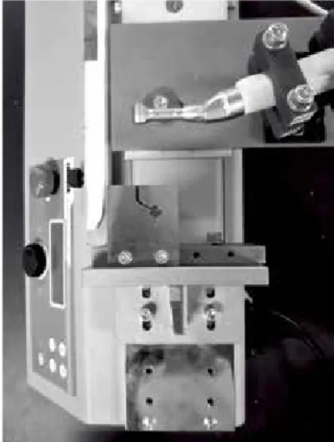
2 .Experiment device
We prepared the dentin hypersensitivity model based on the method by Zennyu, et al19)(Fig. 1).
We set up the specimen stage by placing a rubber ring with 6 mm inner diameter on the dentin side of the enamel crack specimen or the dental pulp side of the dentin disc specimen, and mounting it with a stainless Table 1 Materials used in this study
Materials Composition Application protocol Manufacturer Lot No.
KEC-100(KE)
10-MDP, Bis-GMA, hydrophilic amide monomer, photo initiators
1.Apply bond(waiting time 10 s)
2.No dry
3.Light cure for 10 s
Kuraray Noritake Dental
170804
(experi- mental)
G-Premio BOND
(GP)
10-MDP, 4-META, methacrylate acid ester, MDTP, distilled water, acetone, photo initiators, silica filler
1.Apply adhesive(waiting time 0 s)
2.Dry with max air pressure for 5 s 3.Light cure for 10 s
GC 1710033
Scotchbond Universal Adhesive(SU)
10-MDP, dimethacrylate resins, a-meth- acrylate-modified polyalkenoic acid copolymer, HEMA, filler, ethanol, water, photo initiators, silane
1.Apply adhesive(waiting time 20 s)
2.Dry with gentle air flow for 5 s
3.Light cure for 10 s 3M ESPE 71207A
steel holder to pinch the top and bottom of the speci- men. We ensured fenestration in a square of 5 mm side
(approx. 0.25 cm2 in area)at the center to expose the surface to which to apply the desensitizer. After filling physiological saline assumed as the inner fluid of den- tinal tubules(DF)inside the specimen stage while ensuring that no air was trapped, we connected it to the device for setting the inner pressure, which we pre- pared according to the report by Pashley, et al18). We adjusted the inner pressure of the specimen stage to 25 mmHg, which is equivalent to the inner pressure of human dental pulp, to reproduce the inner pressure of dental pulp in clinical scenes20‒23).
3 .Measurement of permeability inhibition rate First, we cut off the DF from the cylinder direction with three-way cock B and connected it to the glass capillary, which was already connected to the specimen stage and the experiment device. We then blew air on the exposed enamel surface at the central fenestration part of the specimen stage for 60 seconds in this state, left it standing for 30 seconds, and measured the amount of DF movement inside the glass capillary24‒26). We specified the air pressure as 0.3 MPs. We then cut off the DF flowing inside the glass capillary by adjust- ing three-way cock B so that DF could flow only to the specimen stage from the cylinder direction. Next, we injected DF so that it could flow from the cylinder to the pressure gauge and the specimen stage by adjust-
ing three-way cock A, and adjusted the inner pressure to 25 mmHg. We applied each material according to the manufacturer’s instructions, and cured it using Pen- Cure(J. Morita Mfg, Kyoto, Japan). We then blew air for 60 seconds in a similar fashion to that before appli- cation to the specimen, left it standing for 30 seconds, and measured the amount of DF movement again. We measured the permeability inhibition rate for each material based on the amount of DF movement before and after material application. We calculated the rate using Equation(1)below, based on the amount of DF movement before treatment(x)and amount of DF movement after treatment(y):
Permeability inhibition rate(%)={(x-y)/x}×100
………(1)
4 .Laser observation of enamel surface
We used a confocal laser scanning microscope(VK- X100 series, KEYENCE)to observe the enamel cracks on the surface to which desensitizer was applied before and after treatment by phase difference.
5 .SEM observation of sample surface and longi- tudinal section, and measurement of enamel crack sealing depth
We conducted gold deposition on the specimens for surface observation according to the normal method.
We cut each specimen for longitudinal section observa- tion with a low-speed diamond wheel saw(Model 650, South Bay Technology, California, USA)after embed- Fig. 1 Schematic diagram of a dentin hypersensitivity model
A rubber ring with an inner diameter of 6 mm was placed on the dentin side of the enamel crack specimen or the pulp side of the den- tin disc specimen, inserted between the upper and lower stainless steel holders, and used as a specimen stage. The pressure in the spec- imen stage was adjusted to 25 mmHg, equal to human dental pulp, to reproduce the pressure in clinical practice.
specimen stage
glass capillary tube
pressure gauge three-way cock A
glass injection syringe
three-way cock B stainless holder(upper)
enamel crack specimen or dentin disc specimen
rubber ring stainless holder(lower)
DF:physiological saline
2020年12月 Sealing Ability of High-penetration Resin for Enamel Cracks and Dentinal Tubules 487
ding it in epoxy resin. We polished it up to #2000 with waterproof abrasive paper, and soaked it in 1 mol/l hydrochloric acid(Kishida Chemical)prepared with distilled water for 1 minute, then 5% sodium hypochlo- rite solution(Kishida Chemical)for 5 minute to rinse it with water. We then conducted gold deposition accord- ing to the normal method. We observed each specimen with a scanning electron microscope(JSM-5610LV, JEOL, Tokyo, Japan).
We also measured the sealing depth inside the enamel crack by resin based on the longitudinal section SEM images of the enamel crack specimens.
6 .Statistical processing
We set the number of specimens to n=5 in each group, and conducted statistical processing on the per- meability inhibition rate and enamel crack sealing depth which we calculated by using one-way analysis of variance and Tukey’s test(p<0.05).
This study was conducted after obtaining approval from the ethics committee, Osaka Dental University
(Approval No. 110767, May 13, 2020).
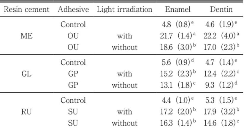
Results
1 .Permeability inhibition rateFigure 2 shows the results of measuring the permea- bility inhibition rate after application of each material on the enamel crack specimens, and Fig. 3 the results after application of each material on the dentin disc specimens.
There was no significant difference in permeability
inhibition rates of the KE, GP and SU groups for the enamel crack specimens and dentin disc specimens, and each group indicated a high permeability inhibition rate.
2 .Laser observation of enamel surface
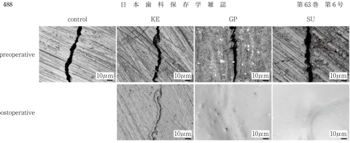
Figure 4 shows the results of laser observation of the enamel surface on the enamel crack specimens.
In all of the KE, GP and SU groups, we observed images in which the resin film coated the enamel crack surface.
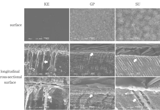
3 .SEM observation of specimen surface and lon- gitudinal section
Figure 5 shows the results of SEM observation of the enamel surface and longitudinal cross-sectional surface of the enamel crack specimens, and Fig. 6 the results of SEM observation of the specimen surface and longitudi- nal cross-sectional surface of the dentin disc specimens.
On the enamel surfaces of the enamel crack speci- mens in all of the KE, GP and SU groups, we observed images in which the resin film coated and sealed the enamel crack surface. On the longitudinal cross-sec- tional surface of all of the KE, GP and SU groups, we observed images in which the insides of enamel cracks were sealed by the penetrating resin.
On the surfaces of the dentin disc specimens in all of the KE, GP and SU groups, we observed images in which the resin film sealed the dentinal tubules and coated the dentin surface. On the longitudinal cross-sec- tional surface of the KE group, we observed long resin tags which penetrated into the dentinal tubules. How- ever, we did not observe any resin tags penetrating the dentinal tubules in the GP group, and observed resin Fig. 2 Measurement of the permeability inhi-
bition rate of each dentin desensitizer in enamel cracks
Same letters indicate no significant differ- ence(p>0.05).
0 20 40 60 80 100 120
KE GP SU
(%)
a a a
permeability inhibition ratio
Fig. 3 Measurement of the permeability inhi- bition rate of each dentin desensitizer in dentin
Same letters indicate no significant differ- ence(p>0.05).
KE GP SU
(%)
a a a
permeability inhibition ratio 0 20 40 60 80 100
tags that penetrated slightly into the dentinal tubules in the SU group.
4 .Enamel crack sealing depth
Figure 7 shows the results of enamel crack sealing depth by resin. The sealing depth of the KE group in enamel cracks was significantly larger than the values of the GP or SU group(p<0.05).
Discussion
KE, which is a prototype new high-penetration resin material for tooth substrates, is intended to seal microc- racks in tooth substrates, and its characteristics include low viscosity and high wettability to tooth substrates.
Since it does not contain solvents such as water and Fig. 4 Laser microscopic observation of enamel surface
In the KE, GP and SU groups, the superficial layer of enamel cracks appeared to be covered with a resin coating.
postoperative preoperative
control KE GP SU
10μm 10μm 10μm 10μm
10μm 10μm 10μm
surface
longitudinal cross-sectional
surface
control KE GP SU
Fig. 5 SEM observation of enamel surface and longitudinal cross-sectional surface of enamel crack
In the KE, GP and SU groups, the superficial layer of enamel cracks appeared to be covered with a resin coating. The specimens were embedded in epoxy resin and SEM observation of the longitudinal cross-sectional surface of the enamel crack specimens was conducted. The inner region of enamel cracks was sealed with resin(white arrows).
2020年12月 Sealing Ability of High-penetration Resin for Enamel Cracks and Dentinal Tubules 489
organic solvents, it does not require air drying after application, and no polymerization failure is expected to occur thanks to no residual solvents even in deeper parts of microcracks. In addition, it quickly adheres to dentin as the hydrophilic multifunctional amide-type monomer that is contained has higher hydrophilicity than 10-MDP or HEMA, features excellent polymeriza-
tion curing property, and is considered resistant to the effects of hydrolysis even after polymerization curing.
The material is also adhesive to tooth substrates27). There are resin-based desensitizer products that apply the one-step self-etching system, which uses dentin adhesive material like the one we used in this experi- ment. Resin-based desensitizers have been reported to deliver higher sealing abilities than other desensitizers as the resin coating adheres well to the dentin and f i n e l y c o v e r s t h e e n a m e l c r a c k s a n d d e n t i n a l tubules14,16,28). Many of the resin-based desensitizers contain hydrophilic monomers, hydrophobic monomers, water, and organic solvents29). Water is necessary for the etching effect of the acidic monomer, and organic solvents are necessary for dissolving the monomer components and water30). However, these solvents may cause resin polymerization to fail, and thus the solvents need to be removed thoroughly by air drying31). On the other hand, air drying during treatment can cause pain for those patients who present hypersensitivity symp- toms. It is also considered difficult to remove solvents that permeate into minute enamel cracks by air drying.
Based on the permeability inhibition rate in enamel surface
longitudinal cross-sectional
surface
KE GP SU
Fig. 6 SEM observation of dentin surface and longitudinal cross-sectional surface of dentin In the KE, GP and SU groups, the superficial layer of dentin appeared to be covered with a resin coating. In the KE group, resin tags penetrated into dentinal tubules(white arrows). In the GP and SU groups, there were no or a few resin tags penetrating into dentinal tubules
(white arrows).
Fig. 7 Penetration depth of resin in enamel crack
Different letters indicate significant differ- ences at p<0.05, as determined by one-way layout analysis of variance and Tukey’s test.
0 100 200 300 400 500 600 700
KE GP SU
(μm)
a
b b
crack specimens, the KE, GP, and SU groups showed favorable enamel crack sealing abilities, and the SEM images of the enamel surface revealed resin films that coated the surface. Based on the SEM images of the longitudinal cross-sectional surface of enamel cracks, the resin permeated more deeply into the cracks in the KE group than in the GP and SU groups, while all three groups showed resin components that permeated inside the enamel cracks. Since KE benefits from the effects of low viscosity and the hydrophilic multifunctional amide- type monomer with high affinity to dentin27), and since it does not contain fillers, it is assumed that it perme- ated more deeply into fine enamel cracks. It is also assumed that sealing with resin was observed even in deeper parts of enamel cracks as sufficient polymeriza- tion occurred thanks to no solvent content, which therefore caused no failure of resin polymerization.
However, due to the low mechanical strength which results from containing no fillers, the results suggest that some resin fell out from inside the enamel cracks during the preparation of specimens for SEM observa- tion. On the other hand, for GP and SU, which are one- step self-etching systems, the viscosity of each material as well as the grain shape and size of the filler con- tained seemed to have affected the penetration depth into the enamel cracks, even though we observed per- meation of resin components into enamel cracks for both systems. Furthermore, since GP contains acetone and water, and SU contains ethanol and water, as sol- vents, it is possible that the air drying did not reach sufficiently into the deep parts of enamel cracks32). Polymerization failure, which occurs due to the pres- ence of unremoved residual solvents33), may also have been a factor that resulted in the difference in penetra- tion depth into enamel cracks.
Based on the permeability inhibition rate in dentin discs, we found that the KE, GP, and SU groups have favorable ability to seal dentinal tubules, with SEM images of the dentin surface showing resin films coat- ing the surface. In the SEM images of the longitudinal section of dentin discs, we observed resin tags that pen- etrated into the dentinal tubules in the KE group. The hydrophilic multifunctional amide-type monomer con- tained in KE has high hydrophilicity, and KE is low in viscosity as it contains no fillers. Therefore, it is assumed that KE permeated even the dentinal tubules to which dental pulp inner pressure was applied, and
that it brought about the formation of resin tags. On the other hand, GP and SU, which are one-step self-etching systems, have been reported to have favorable adhesive properties to dentin34,35). However, we did not observe the penetration of resin tags into the dentinal tubules in the GP group, even though we observed the bonding layer that coated the dentin surface. In addition, the SU group showed only a little penetration of resin tags into the dentinal tubules even though we observed the bonding layer that coated the dentin surface. There is a report which claimed that a coarse resin film with voids was observed due to the effects of the inner fluid of dentinal tubules, which seeps out to the dentin sur- face under the inner pressure of the dental pulp, when a resin-based desensitizer was applied on the dentin surface on which such inner pressure was applied26). The results suggest that resin tags were not observed for GP and SU in this experiment as the bonding com- ponents could not seep into the dentinal tubules due to the effects of the inner fluid of the dentinal tubules under the inner pressure of the dental pulp, even though the adhesiveness between the dentin surface and bonding components was favorable.
Conclusion
In this study, we examined the sealing ability of a prototype new high-penetration resin material for tooth substrates for enamel cracks and dentinal tubules under application of the inner pressure of human dental pulp, and obtained the following results.
1.Regarding the permeability inhibition rate on enamel cracks and dentinal tubules, we observed that the new high-penetration resin material delivered an equally high and favorable rate as conventional res- in-based desensitizers.
2.Compared to conventional resin-based desensitiz- ers, we observed that the new high-penetration resin material permeated more deeply and sealed the enamel cracks and dentinal tubules.
3.The results indicated that the new high-penetra- tion resin material delivered a better sealing ability than conventional resin-based desensitizers.
The authors declare no conflict of interest in this study or report.
2020年12月 Sealing Ability of High-penetration Resin for Enamel Cracks and Dentinal Tubules 491
References
1) Ministry of health, labour and welfare. The survey of dental diseases(2016)Summary of results chart. https://
www.mhlw.go.jp/toukei/list/dl/62-28-02.pdf(cited 2020.6.5)
2) Hirayama S. Hard tissue disease, tooth developmental abnormalities and related diseases. Senda A, Miyazaki M, Hayashi M, Mukai Y, Saito T. Operative dentistry.
7th ed. Ishiyaku publishers: Tokyo; 2019. 27‒35.
3) Han L, Sunada M, Okamoto A, Fukushima M, Okiji T.
The prevalence and related symptoms of enamel cracks: A clinical survey. Jpn J Conserv Dent 2008; 51:
614‒621.(in Japanese)
4) Yamaguchi M, Odaira M, Yamaguchi Y, Tanaka J, Aoki Y, Ikeda T, Meguro D, Kasai K. The evaluation of enamel cracks in adolescence. J Esthet Dent 2008; 21:
1‒5.(in Japanese)
5) Sunada M, Han L, Okamoto A. Crack initiation of tooth with aging and clinical care. Niigata Dent J 2002; 32:
275‒283.(in Japanese)
6) Zachrisson BU, Skogan O, Höymyhr S. Enamel cracks in debonded, debanded, and orthodontically untreated teeth. Am J Orthod 1980; 77: 307‒319.
7) Zachrisson BU, Buyukyimaz T. Bonding in orthodontics.
Graber TM, Vanarsdall RL, Vig KWL. Orthodontics:
Current Principles and Techniques. 4th ed. Elsevier Mosby: St. Louis; 2005. 612‒619.
8) Brännström M, Johnos G, Nordenvall K. Transmission and control of dentinal pain: resin impregnation for the desensitization of dentin. J Am Dent Assoc 1979; 99:
612‒618.
9) Yoshiyama M, Noiri Y, Ozaki K, Uchida A, Ishikawa Y, Ishida H. Transmission electron microscopic characteri- zation of hypersensitive human radicular dentin. J Dent Res 1990; 69: 1293‒1297.
10) Lu L, Yasuo K, Onda K, Yoshikawa K, Yamamoto K.
Influence of various dentin desensitizers on the effect of tooth whitening. J Osaka Dent Univ 2015; 49: 49‒60.
11) Inoue M, Yoshikawa K, Okamoto A, Kota K, Fujii B, Iwaku M. Clinical evaluation of GLUMA3 primer to dentin hypersensitivity. Jpn J Conserv Dent 1996; 39:
768‒776.(in Japanese)
12) Han L, Ishizaki H, Fukushima M, Okiji T. Effect of a prototype fluoride-containing tooth surface coating material on enamel and dentin surfaces. Jpn J Conserv Dent 2012; 55: 53‒59.(in Japanese)
13) Endo H, Kawamoto R, Irokawa A, Takamizawa T, Ando S, Miyazaki M. Evaluation of an experimental desensi- tizing agent by means of ultrasonic device. J J Dent Mater 2012; 31: 116.
14) Shimizu H, Hoshika T, Nishitani Y, Yoshiyama M. Effect of dentin desensitizers on dentin tubules occlusion:
―Influence of dentin and external stimulation―. Jpn J Conserv Dent 2016; 59: 249‒258.(in Japanese)
15) Nagata T, Ishida H, Wakano Y, Ueda M, Imai H, Yama- oka A. Clinical evaluation of potassium nitrate dentifrice for dentinal hypersensitivity. J Jpn Soc Periodontol 1992; 34: 465‒471.(in Japanese)
16) Furusawa K, Yasuo K, Yoshikawa K, Yamamoto K. Seal- ing ability of enamel crack using various dentin desensi- tizers. Jpn J Conserv Dent 2017; 60: 1‒13.
17) Nikaido T, Tagami J, Yatani H, Ohkubo C, Nihei T, Koi- zumi H, Maseki T, Nishiyama Y, Takigawa T, Tsubota Y. Concept and clinical application of the resin-coating technique for indirect restorations. Dent Mater J 2018;
37: 192‒196.
18) Pashley DH, Galloway SE, The effects of oxalate treat- ment on the smear layer of ground surfaces of human dentin. Arch Oral Biol 1983; 30: 731‒737.
19) Zennyu K, Yoshikawa K, Yamamoto K. Effect of laser irradiation on dentin permeability using an in vitro model of hypersensitive dentin. Jpn J Conserv Dent 2008; 51: 48‒62.(in Japanese)
20) Pashley DH, Nelson R, Pashley EL. In-vivo fluid move- ment across dentine in the dog. Arch Oral Biol 1981; 26:
707‒710.
21) Mitchem JC, Terkla LG, Gronas DG. Bonding of resin dentin adhesives under simulated physiological condi- tions. Dent Mater 1988; 4: 351‒353.
22) Nör JE, Feigal RJ, Dennison JB, Edwards CA. Dentin bonding: SEM comparison of the resin-dentin interface in primary and permanent teeth. J Dent Res 1996; 75:
1396‒1403.
23) Sauro S, Pashley DH, Montanari M, Chersoni S, Carvalho RM, Toledano M, Osorio R, Tay FR, Prati C. Effect of simulated pulpal pressure on dentin permeability and adhesion of self-etch adhesives. Dent Mater 2007; 23:
705‒713.
24) Semba T, Tabata S, Wada K, Nakama T. Morphological aspects on the fluid flow in the dentine(II): On blood capillaries in the rat incisor pulp. Ann Kagoshima Dent 1992; 12: 15‒26.(in Japanese)
25) Sasazaki H, Okuda R. Periodic observation of the exuda- tion from the dentinal tubules with time. Jpn J Conserv Dent 1994; 37: 1708‒1718.(in Japanese)
26) Someya Y, Inaba D, Yonemitsu M. The influence of den- tinal fluid flow on remineralization of cavity bottom by glass ionomer cement. J Dent Hlth 2002; 52: 43‒47.(in Japanese)
27) Suyama Y, Yamasa T, Morigami M, Uno S, Sugizaki J.
Interfacial observation with a newly developed univer- sal type adhesive system containing a hydrophilic amide
ester. Adhes Dent 2017; 35: 40‒46.(in Japanese)
28) Nomura Y, Yasuo K, Iwata N, Yoshikawa K, Yamamoto K. Effect of various materials on dentin permeability for the treatment of dentin hypersensitivity. Jpn J Conserv Dent 2013; 56: 516‒525.
29) Van Meerbeek B, Yoshihara K, Yoshida Y, Mine A, De Nlunck J, Van Landuyt KL. State of the art of self-etch adhesives. Dent Mater 2011; 27: 17‒28.
30) Wang Y, Spencer P, Yao X, Brenda B. Effect of solvent content on resin hybridization in wet dentin bonding. J Biomed Mater Res A 2007; 82: 975‒983.
31) Jacobsen T, Soderholm KJ. Some effects of water on dentin bonding. Dent Mater 1995; 11: 132‒136.
32) De Munck J, Arita A, Shirai K, Van Landuyt KL, Coutinho E, Poitevin A, Peumans M, Lambrechts P, Van Meerbeek B. Microrotary fatigue resistance of a
HEMA-free all-in-one adhesive bonded to dentin. J Adhes Dent 2007; 9: 373‒379.
33) Nunes TG, Garcia FC, Osorio R, Carvalho R, Toledano M. Polymerization efficacy of simplified adhesive sys- tems studied by NMR and MRI techniques. Dent Mater 2006; 22: 963‒972.
34) Saito T, Takamizawa T, Ishii R, Tsujimoto A, Hirokane E, Barkmeier WW, Latta MA, Miyazaki M. Influence of application time on dentin bond performance in differ- ent etching modes of universal adhesives. Oper Dent 2020; 45: 183‒195.
35) Kimura S, Hanabusa M, Yamamoto T. Shear bond strengths of a one-step universal dental adhesive con- taining an amide-type monomer. Adhes Dent 2019; 37:
51‒60.