Original Article
Prognostic significance of aromatase and estrogen receptor beta expression in EGFR wild-type lung adenocarcinoma
Kazumi Tanaka1, Kimihiro Shimizu1, Seiichi Kakegawa1, Yoichi Ohtaki1, Toshiteru Nagashima1, Kyoichi Kaira2, Jun Horiguchi1, Tetsunari Oyama3, Izumi Takeyoshi1
Departments of 1Thoracic and Visceral Organ Surgery, 2Medicine and Molecular Science, 3Division of Diagnostic Pathology, Graduate School of Medicine, Gunma University, 3-39-22 Showa-machi, Maebashi, Gunma 371-8511, Japan
Received August 25, 2015; Accepted December 15, 2015; Epub January 15, 2016; Published January 30, 2016 Abstract: Objectives: Based on recent findings of aromatase and estrogen receptor beta (ERβ) expression in non- small-cell lung cancer, we assessed the clinicopathological and prognostic significance of aromatase and ERβ expres- sion and their relationship to epidermal growth factor receptor (EGFR) mutation in lung adenocarcinoma. Materials and methods: We evaluated 150 resected primary lung adenocarcinoma specimens. Expression of aromatase, ERα, ERβ, progesterone receptor (PR), and human epidermal growth factor receptor 2 (HER2) was evaluated by immu- nostaining, and EGFR and KRAS mutations were analyzed. Overall survival (OS) and recurrence-free survival (RFS) were calculated using the Kaplan-Meier method. Results: Expression of aromatase, ERα, ERβ, PR, and HER2 was detected in 88.0%, 1.3%, 79.3%, 2.7%, and 39.3% of specimens, respectively. In patients with EGFR wild-type lung adenocarcinoma, high aromatase expression was an independent predictor of poor OS (hazard ratio [HR]=2.638;
95% confidence interval [CI], 1.173-5.936; P=.019) and RFS (HR=2.505; 95% CI, 1.154-5.434; P=.020). Positive ERβ expression was also an independent predictor of poor RFS (HR=4.013; 95% CI, 1.219-13.207; P=.022). Fur- thermore, high aromatase expression was a significant predictor of poor survival only in females (OS, P=.010; RFS, P=.007), whereas positive ERβ expression was an important predictor of poor survival only in males (OS, P=.073;
RFS, P=.051). No prognostic significance was observed in patients with EGFR mutations. Conclusions: Our findings suggest that EGFR wild-type lung adenocarcinoma is an estrogen-dependent carcinoma, and aromatase expression and ERβ expression are potent prognostic markers for EGFR wild-type lung adenocarcinoma.
Keywords: Aromatase, estrogen receptor beta (ERβ), estrogen signaling pathway, EGFR mutation, lung adenocar- cinoma
Introduction
Lung cancer is one of the most common can- cers globally and is currently the leading cause of death in both females and males [1]. Smoking remains the major cause of lung cancer, but
~53% of all females with lung cancer are non- smokers [2]. Interestingly, a gradual increase in the adenocarcinoma subtype of lung cancer has been reported, despite a decline in the smoking population [3-4]. Therefore, etiologic factors other than tobacco may also play a role in the development of lung adenocarcinoma.
Epidermal growth factor receptor (EGFR) is the most frequently mutated proto-oncogene, par-
ticularly in lung adenocarcinoma of non-smoker females, and its mutations are thought to play an important role in carcinogenesis [5]. How- ever, recent evidence has suggested that es- trogen may also play an important role in the development of non-small cell lung cancer (NSCLC), particularly adenocarcinoma [6]. Se- veral studies have reported that estrogen stim- ulates the proliferation and progression of lung carcinoma cells, functions that were shown to be significantly suppressed by antiestrogenic agents both in vitro and in vivo [7-10].
Estrogen is converted from androgen by aroma- tase, a key enzyme in estrogen biosynthesis. In addition to its expression in the ovary and pla-
centa, aromatase is present in male and fema- le extragonadal tissues, including breast and lung [11]. Aromatase expression is elevated in certain malignancies, such as breast carcino- mas, suggesting that tumor progression caused by stimulation of estrogen signaling pathway could be enhanced by circulating estrogen as well as by localized autocrine or paracrine pro- duction of estrogen by aromatase. Recently, aromatase expression in NSCLC has also been reported [12-14]. Weinberg et al. demonstrated
that aromatase was expressed in NSCLC cell lines, and aromatase inhibitor (AI) suppressed tumor growth in vitro and in vivo [12]. Mah et al.
reported that low aromatase expression was associated with favorable survival in female NSCLC patients, particularly those older than 65 years [13].
In estrogen signaling pathway, estrogens exert their effects mainly via estrogen receptor (ER) [6]. ER is a hormone receptor, as is progester- one receptor (PR). ER has two isoforms, ERα and ERβ, which are encoded by distinct genes and are expressed in various tissues or at vari- ous levels in the same tissue [15]. In the nor- mal lung, ERβ has been reported to be ex- pressed at a higher level than ERα [9]. Althou- gh the expression patterns of ERα and ERβ in NSCLC were highly inconsistent among these reports [6], most of the results showed that there were no or a low (under 10%) rate of ERα- positive cases and a higher rate (over 50%) of ERβ-positive cases [16]. Previously, ERα was considered a tumor promoter, whereas ERβ was believed to inhibit tumorigenesis [17]. Ho- wever, recent studies have demonstrated that ERβ can function as a tumor promoter in the absence of ERα expression [18-22]. The asso- ciation between ERβ expression and the prog- nosis of lung cancer patients remains contro- versial [23-27]. Wu et al. reported that ERβ expression was associated with favorable prog- nosis in NSCLC [23]. However, Stabile et al.
reported that ERβ expression was associated with a poor prognosis in lung cancer [26].
Recent reports have suggested an interaction between EGFR pathway and estrogen signaling pathway in the development of breast and lung cancer; additionally, estrogen signaling path- way is regulated by membrane receptor tyro- sine kinases, including EGFR and human epi- dermal growth factor receptor 2 (HER2) [26, 28-32]. The EGFR pathway becomes activated when estrogen is depleted, and ERβ expression is increased following treatment with EGFR tyrosin kinase inhibitors (EGFR-TKIs) in NSCLC cells [33-35]. Nose et al. reported that strong nuclear expression of ERβ is correlated with EGFR mutations in lung adenocarcinoma [31].
However, the role of aromatase and ERβ in estrogen signaling pathway and the associa- tion between the expression of these proteins and clinicopathological factors, including EGFR Table 1. Clinicopathological factors of the
patients
Characteristics N %
No. of patients 150 100.0
Age (year)
Mean 66
Range 36-84
Sex
Male 63 42.0
Female 87 58.0
Menopause
Premenopausal 4 4.6
Postmenopausal 81 93.1
Unknown 2 2.3
Smoking status
Ever-smoker 68 45.3
Non-smoker 82 54.7
Tumor diameter (mm)
Mean 24
Range 6-70
Pathologic stage
IA 85 56.7
IB 26 17.3
IIA 5 3.3
IIB 6 4.0
IIIA 22 14.7
IIIB 6 4.0
IV 0 0.0
Pleural invasion
Absent 102 68.0
Present 48 32.0
Lymphatic invasion
Absent 101 67.3
Present 49 32.7
Vascular invasion
Absent 104 69.3
Present 46 30.7
mutation, in lung adenocarcinoma, are not we- ll understood.
The purpose of the present study was to exam- ine the correlation between aromatase/ERβ expression and clinicopathological prognostic factors, including EGFR mutations, and to eval- uate the prognostic significance in lung ade- nocarcinoma.
Materials and methods Patients and tissue specimens
One hundred and fifty lung adenocarcinoma specimens were obtained from patients who underwent complete surgical resection consec- utively from 2004 to 2008 at Gunma University Hospital. The clinicopathological factors of the patients are shown in Table 1. The disease stage was determined according to the seventh edition of the TNM classification for lung and pleural tumors [36]. All of the procedures were approved by the Ethics Committee on Human Research of Gunma University Graduate School of Medicine, and written informed consent was obtained from all of the patients before sur- gery.
Immunohistochemistry
Serial tissue sections of 4-µm thickness sliced from paraffin-embedded specimens were used for immunohistochemistry using the labeled streptavidin-biotin method. Immunostaining for ERα, ERβ, PR, aromatase, Ki-67, and HER2 was performed with the antibodies listed in Table 2.
The slides were deparaffinized with xylene and rehydrated with ethanol. For ERα, PR, and Ki-67 analysis, antigen retrieval was performed ac- cording to the manufacturer’s instructions. For ERβ, antigen retrieval was carried out by auto- claving the slides in citrate buffer (0.01 mol/L) at 121°C for 5 min. For HER2, immunohisto-
chemical staining was performed using Ben- chMark XT (Ventana, Tucson, AZ, USA), an au- tomatic immunohistochemical staining system.
Nuclear positive immunoreactivity for ERα, ERβ, and PR was counted among 1000 cells per case and was recorded as “positive” for positive results of more than 10% [14]. Im- munoreactive intensity of ERβ was scored into four phases (0, negative; 1+, weak; 2+, moder- ate; and 3+, strong). For Ki-67, 1000 cells were counted per case, and the proliferative activity was assessed as the percentage of Ki-67-st- ained nuclei (Ki-67 labeling index; LI) in each sample. Cytoplasmic staining for aromatase in over 10% of the cancer lesion was recorded as
“positive”, and immunoreactive intensity was scored into four phases (0, negative; 1+, weak;
2+, moderate; and 3+, strong) [6, 13]. HER2 immunoreactivity was evaluated using the DA- KO HercepTest scoring system (DakoCytoma- tion), and over 2+ was considered “positive”.
Two observers (K.T. and T.O.) who were unaware of the clinical data independently reviewed all pathological slides.
Gene mutation analysis
We examined EGFR and KRAS mutations in the present study. Genomic DNA was extracted from a 3- to 5-mm cube of tumor tissue using a DNA Mini Kit (Qiagen, Hilden, Germany) and subsequently diluted to 20 ng/µL. KRAS and EGFR mutations were analyzed by sequencing as described previously [37, 38].
Statistical analysis
Statistical analyses were performed using IBM SPSS Statistics 21.0 (IBM Co., Armonk, NY, USA). Student’s t-test and chi-squared test we- re used to compare percentages and mean val- ues, respectively. Survival was calculated using the Kaplan-Meier method and confirmed using the log-rank test. Overall survival (OS) was Table 2. Primary antibodies used in the present study
Antigen Clone Dilution Source
ERα 1D5 1:50 DakoCytomation, Glostrup, Denmark
ERβ 14C8 1:200 GeneTex, CA, USA
PR PGR636 1:800 DakoCytomation, Glostrup, Denmark
Aromatase #677/H7 1:1000 Contributed by Dr. Evans DB, Novartis, Basel, Switzerland
Ki-67 MIB1 1:150 DakoCytomation, Glostrup, Denmark
HER2 4B5 1:1 Ventana, Tucson, AZ, USA
ER, estrogen receptor; PR, progesterone receptor; HER2, human epidermal growth factor receptor 2.
determined as the time from tumor resection to death from any cause. Recurrence-free survival (RFS) was defined as the time between tumor resection and first disease recurrence or death.
The median follow up for survivors was 65.5 months (average, 63.7 months; range, 1-117 months). Variables with P value less than .05 after univariate analysis were entered into mul- tivariate analysis using the Cox proportional hazards model. P < .05 was deemed to indicate statistical significance. The midpoint and medi- an intensity (1.5) was used to define low and high aromatase expression as previously de- scribed [13]. For univariate and multivariate analyses, each continuous variable (age and Ki-67 LI) was dichotomized at the median va- lue.
Results
Immunohistochemical analysis
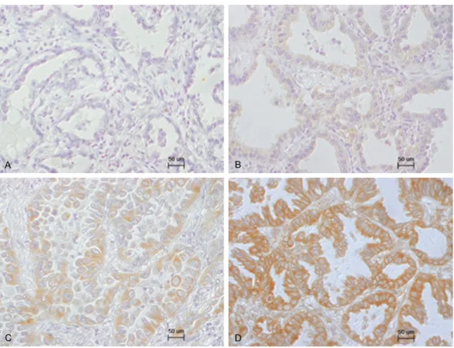
Among the 150 cases, ERα, ERβ, PR, aroma- tase, and HER2 were detected in 2 (1.3%), 119 (79.3%), 4 (2.7%), 132 (88.0%), and 7 (4.7%) cases, respectively. Figure 1 shows represen- tative staining for ERα, ERβ, PR, and aroma- tase. Regarding the immunoreactive intensity of aromatase, 18 (12.0%) cases were scored as 0, 60 (40.0%) cases were scored as 1+, 51 (34.0%) cases were scored as 2+, and 21 (14.0%) cases were scored as 3+ (Figure 2).
Therefore, low expression group comprised 78 (52.0%) cases, and high expression group com- prised 72 (48.0%) cases. ERβ staining was Figure 1. Representative immunohistochemical staining of ERα, ERβ, PR, and aromatase in lung adenocarcinoma.
A. Negative staining of ERα; B. Positive staining of ERα; C. Negative staining of ERβ; D. Positive staining of ERβ; E.
Negative staining of PR; F. Positive staining of PR; G. Negative staining of aromatase; H. Positive staining of aroma- tase.
Figure 2. Representative immunohistochemical staining pattern of aromatase in lung adenocarcinoma. Specimens were assigned one of four scores (0, negative; 1+, weak; 2+, moderate; and 3+, strong) according to the intensity of immunoreactivity. A, 0; B, 1+; C, 2+; D, 3+.
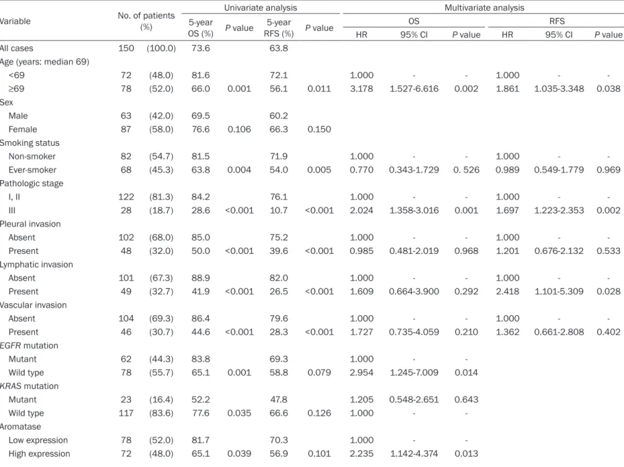
Table 3. Univariate and multivariate analyses of prognostic factors in all patients
Variable No. of patients
(%)
Univariate analysis Multivariate analysis
5-year
OS (%) P value 5-year
RFS (%) P value OS RFS
HR 95% CI P value HR 95% CI P value
All cases 150 (100.0) 73.6 63.8
Age (years: median 69)
<69 72 (48.0) 81.6 72.1 1.000 - - 1.000 - -
≥69 78 (52.0) 66.0 0.001 56.1 0.011 3.178 1.527-6.616 0.002 1.861 1.035-3.348 0.038
Sex
Male 63 (42.0) 69.5 60.2
Female 87 (58.0) 76.6 0.106 66.3 0.150
Smoking status
Non-smoker 82 (54.7) 81.5 71.9 1.000 - - 1.000 - -
Ever-smoker 68 (45.3) 63.8 0.004 54.0 0.005 0.770 0.343-1.729 0. 526 0.989 0.549-1.779 0.969
Pathologic stage
I, II 122 (81.3) 84.2 76.1 1.000 - - 1.000 - -
III 28 (18.7) 28.6 <0.001 10.7 <0.001 2.024 1.358-3.016 0.001 1.697 1.223-2.353 0.002
Pleural invasion
Absent 102 (68.0) 85.0 75.2 1.000 - - 1.000 - -
Present 48 (32.0) 50.0 <0.001 39.6 <0.001 0.985 0.481-2.019 0.968 1.201 0.676-2.132 0.533
Lymphatic invasion
Absent 101 (67.3) 88.9 82.0 1.000 - - 1.000 - -
Present 49 (32.7) 41.9 <0.001 26.5 <0.001 1.609 0.664-3.900 0.292 2.418 1.101-5.309 0.028
Vascular invasion
Absent 104 (69.3) 86.4 79.6 1.000 - - 1.000 - -
Present 46 (30.7) 44.6 <0.001 28.3 <0.001 1.727 0.735-4.059 0.210 1.362 0.661-2.808 0.402
EGFR mutation
Mutant 62 (44.3) 83.8 69.3 1.000 - -
Wild type 78 (55.7) 65.1 0.001 58.8 0.079 2.954 1.245-7.009 0.014
KRAS mutation
Mutant 23 (16.4) 52.2 47.8 1.205 0.548-2.651 0.643
Wild type 117 (83.6) 77.6 0.035 66.6 0.126 1.000 - -
Aromatase
Low expression 78 (52.0) 81.7 70.3 1.000 - -
High expression 72 (48.0) 65.1 0.039 56.9 0.101 2.235 1.142-4.374 0.013
ERα
Negative 148 (98.7) 73.9 64.0
Positive 2 (1.3) 50.0 0.405 50.0 0.568
ERβ
Negative 31 (20.7) 76.9 74.2
Positive 119 (79.3) 72.7 0.447 61.0 0.145
PR
Negative 146 (97.3) 73.6 63.5
Positive 4 (2.7) 75.0 0.724 75.0 0.573
HER2
Negative 143 (95.3) 74.5 64.9 1.000 - -
Positive 7 (4.7) 57.1 0.091 42.9 0.015 3.093 1.151-8.311 0.025
Ki-67 LI (%: median 11.5)
Low score 75 (50.0) 87.7 84.0 1.000 - - 1.000 - -
High score 75 (50.0) 59.3 <0.001 43.3 <0.001 2.591 1.115-6.021 0.027 2.620 1.299-5.284 0.007 OS, overall survival; RFS, recurrence-free survival; HR, hazard ratio; CI, confidence interval; EGFR, epidermal growth factor receptor; ER, estrogen receptor; PR, progesterone receptor;
HER2, human epidermal growth factor receptor 2; LI, labeling index.
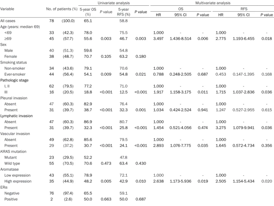
Table 4. Univariate and multivariate analyses of prognostic factors in patients with EGFR wild-type lung adenocarcinoma Variable No. of patients (%)
Univariate analysis Multivariate analysis
5-year OS
(%) P value 5-year
RFS (%) P value OS RFS
HR 95% CI P value HR 95% CI P value
All cases 78 (100.0) 65.1 58.8
Age (years: median 69)
<69 33 (42.3) 78.0 75.5 1.000 - - 1.000 - -
≥69 45 (57.7) 55.6 0.003 46.7 0.003 3.497 1.436-8.514 0.006 2.775 1.193-6.455 0.018
Sex
Male 40 (51.3) 59.6 54.8
Female 38 (48.7) 70.7 0.105 63.2 0.180
Smoking status
Non-smoker 34 (43.6) 79.1 70.6 1.000 - - 1.000 - -
Ever-smoker 44 (56.4) 54.1 0.009 54.8 0.021 0.788 0.248-2.505 0.687 0.453 0.147-1.395 0.168
Pathologic stage
I, II 62 (79.5) 77.2 71.0 1.000 - - 1.000 - -
III 16 (20.5) 18.8 <0.001 12.5 <0.001 1.917 1.158-3.175 0.011 1.715 1.037-2.836 0.036
Pleural invasion
Absent 47 (60.3) 82.9 76.4 1.000 - - 1.000 - -
Present 31 (39.7) 38.7 <0.001 32.3 0.001 1.034 0.424-2.524 0.941 1.247 0.527-2.955 0.615
Lymphatic invasion
Absent 47 (60.3) 86.9 80.7 1.000 - - 1.000 - -
Present 31 (39.7) 32.3 <0.001 25.8 <0.001 1.454 0.521-4.056 0.474 3.275 1.079-9.941 0.036
Vascular invasion
Absent 49 (62.8) 85.6 79.5 1.000 - - 1.000 - -
Present 29 (37.2) 30.7 <0.001 24.1 <0.001 2.893 1.076-7.775 0.035 1.645 0.572-4.734 0.356
KRAS mutation
Mutant 23 (29.5) 52.2 47.8
Wild type 55 (70.5) 70.6 0.473 63.4 0.430
Aromatase
Low expression 43 (55.1) 78.9 72.1 1.000 - - 1.000 - -
High expression 35 (44.9) 48.2 0.005 42.9 0.010 2.638 1.173-5.936 0.019 2.505 1.154-5.434 0.020
ERα
Negative 76 (97.4) 65.5 59.1
Positive 2 (2.6) 50.0 0.663 50.0 0.687
ERβ
Negative 19 (24.4) 78.9 78.9 1.000 - -
Positive 59 (75.6) 60.5 0.079 52.3 0.031 4.013 1.219-13.207 0.022
PR
Negative 75 (96.2) 65.0 58.5
Positive 3 (3.8) 66.7 0.638 66.7 0.637
HER2
Negative 74 (94.9) 66.1 59.5
Positive 4 (5.1) 50.0 0.060 50.0 0.133
Ki-67 LI (%: median 11.5)
Low score 36 (46.2) 83.0 80.6 1.000 - - 1.000 - -
High score 42 (53.8) 49.5 <0.001 40.1 <0.001 1.634 0.501-5.334 0.416 2.239 0.752-6.666 0.148 OS, overall survival; RFS, recurrence-free survival; HR, hazard ratio; CI, confidence interval; ER, estrogen receptor; PR, progesterone receptor; HER2, human epidermal growth factor receptor 2; LI, labeling index.
seen in both the cytoplasm and nucleus. Re- garding the immunoreactive intensity of ERβ, 31 cases were scored as 0 (20.7%), 98 as 1+
(65.3%), 18 as 2+ (12.0%), and 3 as 3+ (2.0%).
Additionally, among the tumors that stained positive for ERβ, almost all of the tumor tissue Figure 3. Kaplan-Meier survival curves of 78 patients with EGFR wild-type lung adenocarcinoma according to the immunoreactivity results for aromatase and ERβ. A. Overall survival (OS) stratified by high versus low expression of aromatase. B. Recurrence-free survival (RFS) stratified by high versus low expression of aromatase. C. OS stratified by positive versus negative expression of ERβ. D. RFS stratified by positive versus negative expression of ERβ. E. OS stratified by combined high expression of aromatase/positive expression of ERβ versus combined low expression of aromatase/negative expression of ERβ versus others. F. RFS stratified by combined high expression of aromatase/
positive expression of ERβ versus combined low expression of aromatase/negative expression of ERβ versus others.
Figure 4. Kaplan-Meier survival curves of male and female patients with EGFR wild-type lung adenocarcinoma ac- cording to the immunoreactivity results for aromatase and ERβ. A. Recurrence-free survival (RFS) of males stratified by high versus low expression of aromatase. B. RFS of females stratified by high versus low expression of aroma- tase. C. RFS of males stratified by positive versus negative expression of ERβ. D. RFS of females stratified by positive versus negative expression of ERβ. E. RFS of males stratified by combined high expression of aromatase/positive expression of ERβ versus combined low expression of aromatase/negative expression of ERβ versus others. F. RFS of females stratified by combined high expression of aromatase/positive expression of ERβ versus combined low expression of aromatase/negative expression of ERβ versus others.
stained positive. Therefore, ERβ immunoreac- tivity was not scored using scoring system such as the Allred score [39].
Association between aromatase/ERβ expres- sion and clinicopathological factors, including EGFR and KRAS mutation
In 10 of the 150 cases, DNA could not be extracted because the specimen was too small.
Among the remaining 140 cases, EGFR and KRAS mutations were found in 62 (44.3%) and 23 (16.4%) cases, respectively. The occurrenc- es of these mutations were mutually exclusive.
Aromatase expression status was significantly associated with pleural invasion (P=.037), and ERβ expression was not significantly associat- ed with clinicopathological factors (Table S1).
Survival analysis
On univariate analysis, 10 variables were found to be significantly associated with poor OS. For RFS, eight variables were identified as statisti- cally significant factors (Table 3). Multivaria- te analysis demonstrated that older age, ad- vanced pathological stage, EGFR wild-type sta- tus, high aromatase expression, and high Ki-67 LI score were significant independent predic- tors of poor OS; additionally, older age, ad- vanced pathological stage, lymphatic invasion, HER2 expression, and high Ki-67 LI score were significant independent predictors of poor RFS (Table 3).
Survival analysis according to EGFR/KRAS mu- tation status
To clarify the prognostic significance of aroma- tase/ERβ expression according to EGFR/KRAS mutation status, we examined survival analysis stratified by EGFR/KRAS mutation status. No relationship was found between survival and KRAS mutation status (data not shown); how- ever, a significant relationship was found be- tween survival and EGFR mutation status. On univariate analysis of the EGFR wild-type popu- lation, eight variables were significantly associ- ated with poor OS. Regarding RFS, nine vari- ables were identified as statistically significant factors (Table 4). Multivariate analysis showed that older age, advanced pathological stage, vascular invasion, and high aromatase expres- sion were significant independent predictors of poor OS. Concerning RFS, older age, advan- ced pathological stage, lymphatic invasion, hi-
gh aromatase expression, and ERβ positive sta- tus were significant independent predictors (Table 4).
Figure 3 shows the survival curves of 78 patients with EGFR wild-type lung adenocarci- noma according to aromatase and ERβ ex- pression, respectively. Interestingly, differences in survival became clearer when patients we- re stratified by aromatase and ERβ expre- ssion. Patients with high aromatase expres- sion had poor prognosis in both OS (Figure 3A;
P=.005) and RFS (Figure 3B; P=.010). Patients with ERβ-positive also had poor prognosis in terms of RFS (Figure 3D; P=.031); an identical tendency was observed for OS (Figure 3C;
P=.079). Furthermore, patients with high ex- pression of aromatase and ERβ-positive had a poorer prognosis than patients with low expres- sion of aromatase and ERβ-negative in terms of both OS (Figure 3E; P =.004) and RFS (Figure 3F; P=.002).
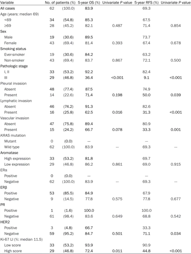
Conversely, no significant difference was noted in the survival of patients with EGFR mutant lung adenocarcinoma according to aromatase and ERβ expression (Table S2 and Figure S1).
Survival analysis according to sex in patients with EGFR wild-type lung adenocarcinoma Next, we performed a survival analysis strati- fied by sex to clarify the prognostic impact of hormonal effect on sex in patients with EGFR wild-type lung adenocarcinoma. Figure 4 sho- ws the RFS curves of males and females wi- th EGFR wild-type lung adenocarcinoma ac- cording to aromatase and ERβ expression, respectively. High aromatase expression was significantly associated with poor prognosis only in females (Figure 4B; P=.007), whereas ERβ-positive had a tendency for poor prognosis only in males (Figure 4C; P=.051). Furthermore, patients with high expression of aromatase and ERβ-positive had a poorer prognosis than patients with low expression of aromatase and ERβ-negative, but only among females (Figure 4F; P=.008).
Discussion
Understanding the role of estrogen and EGFR pathways in lung adenocarcinoma is necessary to develop new preventative and treatment strategies. We report here for the first time that aromatase and ERβ expression are indepen-
dent, unfavorable prognostic factors in EGFR wild-type lung adenocarcinoma. Furthermore, regarding EGFR wild-type lung adenocarcino- ma, we showed that high aromatase expres- sion was a significant predictor of poor survival only in females, whereas ERβ expression was an important predictor of poor survival only in males. These observations indicate that these pathways are important to a different extent in males and females with lung adenocarcinoma.
In our study, PR was detected only in 2.7%. The rate of PR expression in lung tumors varies among reports, ranging from no expression to marginal (22%-35%) or even high expression (39%-63%) [40]. This difference may be related to the antibody used in each study. The anti- body we used is a representative PR antibody widely used for breast cancer research and to guide clinicians’ choice of therapy. However, its use remains limited for lung tumors, and the most appropriate antibody for PR staining in lung tumors is yet to be identified.
Recent studies linking ER expression status with EGFR mutation have suggested that con- sidering these signaling pathways together may provide important insight into lung cancer biol- ogy [31, 41-42]. Thus, we analyzed the associa- tion of aromatase/ERβ expression with EGFR mutation status. Interestingly, our subgroup analysis showed a significant difference in OS and RFS according to aromatase expression, and a significant difference in RFS according to ERβ expression, only in EGFR wild-type lung adenocarcinoma.
Mah et al. reported that low aromatase expres- sion was found to be associated with favorable survival in female NSCLC patients in the United States [13]. However, in reports based on Asian populations, aromatase expression had no as- sociation with prognosis, although aromatase was expressed in more than 60% of lung can- cer patients [14, 27]. The result of Mah’s report [13] is consistent with that of ours, although those of later reports are not. However, those previous studies grouped adenocarcinoma and other histological types of NSCLC together.
Furthermore, EGFR mutation status was not analyzed in those studies. The precise mecha- nism underlying the worse survival regarding the association of aromatase expression with EGFR mutation status in our study is unknown.
However, the frequency of EGFR mutations in
NSCLC varies among races: from 27 to 60% in Asians and from 8 to 16% in Europeans, Af- ricans, and Caucasian Americans [43-44]. Ba- sed on these observations and our findings, we speculate that the prognostic discrepancy in relation to the aromatase expression level am- ong previous reports might be attributable to the difference in EGFR mutation status in each study.
To further examine aromatase and ERβ as a predictor of survival, and to assess the impor- tance of sex, we analyzed the association be- tween aromatase/ERβ expression and sex in EGFR wild-type patients according to survival.
As expected, our result showed that high aro- matase expression was a significant predictor of poor survival only in females, and we found no predictive value for aromatase expression levels in males. By contrast, our result showed that ERβ-positive was a significant predictor of poor survival only in males, and we found no predictive value for ERβ expression levels in females.
The differences in these sex-related results seem to depend on the difference in the status of, and sensitivity to, reproductive hormones according to sex. In postmenopausal females, circulating estrogen levels are decreased due to the decline in estrogen production by the ovaries [45]. Under these conditions, local es- trogen production through aromatase might be an important determinant of estrogen levels. In fact, Niikawa et al. demonstrated that the intra- tumoral estradiol concentration was signifi- cantly higher in NSCLC than in nonneoplastic lung tissue and was positively correlated with intratumoral aromatase expression [46]. In our study, we did not distinguish females according to menopause status because only one of the females with EGFR wild-type lung adenocarci- noma was premenopausal. In other words, our result indicated that aromatase expression level is prognostic factor in postmenopausal females with EGFR wild-type lung adenocarci- noma. Therefore, intratumoral aromatase ex- pression is associated with tumor progression via estrogen signaling pathway in postmeno- pausal females, particularly in EGFR wild-type lung adenocarcinoma.
In contrast to females, circulating estrogen lev- els in males are almost the same as those in postmenopausal females, and this condition
remains relatively constant with age [47]. Ma- rtin et al. demonstrated that ER-positive breast cancer cells are hypersensitive to low doses of estrogen with long-term estrogen deprivation [48]. Furthermore, in males, a higher level of androgen, which is a substrate of estrogen syn- thesis, is present than in females [47]. Based on these observations, at low expression lev- els, aromatase may produce an amount of es- trogen sufficient for proliferation of ER-positive cells. In the present study, 92.1% of lung ade- nocarcinoma expressed aromatase at lower levels in males. Therefore, these findings sup- port our hypothesis that lung adenocarcinoma in males supplies sufficient levels of estrogen to activate ER for tumor cell maintenance, and that the level of tumor proliferation activity- mediated via estrogen signaling pathway-is more dependent on ERβ than aromatase ex- pression in males.
The role of AIs in lung adenocarcinoma is unclear. However, many studies have reported that AIs demonstrated significant anti-tumor effects in NSCLC expressing aromatase both in vitro and in vivo [12, 16, 33, 46, 49]. These observations and our findings revealed that EGFR wild-type lung adenocarcinoma patients with high aromatase expression are a suitable subset for AI treatment, particularly in post- menopausal females. Currently, a phase I clini- cal trial of the irreversible steroidal AI exemes- tane in combination with chemotherapy for la- te-stage lung cancer in postmenopausal fe- males is underway (NCT01664754). We are awaiting the results of this phase I study, which may reveal that AI treatment is effective for lung cancer in postmenopausal females.
Stabile et al. reported that increased EGFR sig- naling might be caused by depletion of estro- gen signals induced by endocrine therapy, and targeting both pathways could be beneficial for therapy [34]. A phase II trial of erlotinib (EGFR- TKI) or erlotinib + fulvestrant in previously treat- ed male and female advanced NSCLC has been completed [Garon EB, Siegfried JM, Dubinett SM, Elashoff RM, Park DJ, Parikh RJ, Patel R, Hu EH, Reckamp KL, Adams B, Martinez D, Wang HJ, Kabbinavar F, Dacic S, Brennan M, Laux I, Márquez-Garban DC, Stabile LP, Slamon DJ, Pietras RJ. Results of TORI-L-03, a random- ized, multicenter phase II clinical trial of erlo- tinib (E) or E + fulvestrant (F) in previously treat- ed advanced non-small cell lung cancer (NS-
CLC). Presented at the 104th Annual Meeting of the American Association for Cancer Research;
Washington DC. PA, April 6-10, 2013. p. Abs- tract 4664; Unpublished results]. Interestingly, the clinical benefit rate was significantly higher among patients treated with the combination compared with erlotinib alone among patients with EGFR wild-type tumors, although the sur- vival and response rates were similar between the two treatment arms in unselected patients.
The latter finding supports our suggestion that estrogen signaling pathway plays an important role in the development of EGFR wild-type lung adenocarcinoma. Thus, endocrine therapy co- uld also be beneficial for EGFR wild-type lung adenocarcinoma as a combination therapy with EGFR-TKI.
The limitations of present study include se- lection of antibodies, the retrospective design, and relatively small number of patients, all of whom were Japanese. Thus, large, population- based prospective studies with ethnically di- verse populations are warranted to elucidate the role of growth factor pathways, including EGFR and/or estrogen signaling pathways, in lung adenocarcinoma.
In conclusion, we demonstrated that aroma- tase and ERβ expressions are independent negative prognostic factors in EGFR wild-type lung adenocarcinoma and that high aromatase expression is a significant predictor of poor sur- vival only in females, whereas ERβ-positive is an important predictor of poor survival only in males. We suggest that EGFR wild-type lung adenocarcinoma is a hormone-related carci- noma.
Acknowledgements
We thank Toshiaki Hikino (Division of Diagnostic Pathology, Graduate School of Medicine, Gun- ma University) for skillful technical assistance.
The first author was supported by Grant-in-Aid for Young Scientists (B) (24791454) from Japan Society for the Promotion of Science (JSPS).
Disclosure of conflict of interest None.
Address correspondence to: Kimihiro Shimizu, De- partment of Thoracic and Visceral Organ Surgery, Gunma University Graduate School of Medicine, 3-39-22 Showa-machi, Maebashi, Gunma 371-85- 11, Japan. Tel: 81-27-220-8245; Fax: 81-27-220- 8255; E-mail: [email protected]
References
[1] Jemal A, Siegel R, Ward E, Hao Y, Xu J, Thun MJ. Cancer statistics, 2009. CA Cancer J Clin 2009; 59: 225-249.
[2] Subramanian J, Govindan R. Lung cancer in never smokers: a review. J Clin Oncol 2007;
25: 561-570.
[3] Charloux A, Quoix E, Wolkove N, Small D, Pauli G, Kreisman H. The increasing incidence of lung adenocarcinoma: reality or artifact? A re- view of the epidemiology of lung adenocarci- noma. Int J Epidemiol 1997; 26: 14-23.
[4] Lam B, Lam WK, Lam CL, Ooi GC, Ho JCM, Wong MP, Tsang KW. Adenocarcinoma of the lung in Chinese patients: a revisit and some perspectives from the literature. Postgrad Med J 2001; 77: 708-712.
[5] Kakegawa S, Shimizu K, Sugano M, Miyamae Y, Kaira K, Araki T, Nakano T, Kamiyoshiha- ra M, Kawashima O, Takeyoshi I. Clinicopatho- logical features of lung adenocarcinoma with KRAS mutations. Cancer 2011; 117: 4257- 4266.
[6] Verma MK, Miki Y, Sasano H. Aromatase in hu- man lung carcinoma. Steroids 2011; 76: 759- [7] Stabile LP, Davis AL, Gubish CT, Hopkins TM, 764.
Luketich JD, Christie N, Finkelstein S, Siegfried JM. Human non-small cell lung tumors and cells derived from normal lung express both estrogen receptor α and β and show biological responses to estrogen. Cancer Res 2002; 62:
2141-2150.
[8] Hershberger PA, Vasquez AC, Kanterewicz B, Land S, Siegfried JM, Nichols M. Regulation of endogenous gene expression in human non- small cell lung cancer cells by estrogen recep- tor ligands. Cancer Res 2005; 65: 1598-1605.
[9] Omoto Y, Kobayashi Y, Nishida K, Tsuchiya E, Eguchi H, Nakagawa K, Ishikawa Y, Yamori T, Iwase H, Fujii Y, Warner M, Gustafsson JÅ, Hayashi S. Expression, function, and clinical implications of the estrogen receptor β in hu- man lung cancers. Biochem Biophysl Res Com- mun 2001; 285: 340-347.
[10] Mollerup S, Jorgensen K, Berge G, Haugen A.
Expression of estrogen receptors α and β in human lung tissues and cell lines. Lung Can- cer 2002; 37: 153-159.
[11] Simpson ER, Mahendroo MS, Means GD, Kilgore MW, Hinshelwood MM, Graham-Lor- ence S, Amarneh B, Ito Y, Fisher CR, Michael MD, Mendelson CR, Bulun SE. Aromatase cyto- chrome P450, the enzyme responsible for es- trogen biosynthesis. Endocr Rev 1994; 15:
342-344.
[12] Weinberg OK, Márquez-Garban DC, Fishbein MC, Goodglick L, Hermes J. Garban, Dubinett
SM, Pietras RJ. Aromatase inhibitors in human lung cancer therapy. Cancer Res 2005; 65:
11287-11291.
[13] Mah V, Seligson DB, Li A, Márquez DC, Wistuba II, Elshimali Y, Fishbein MC, Chia D, Pietras RJ, Goodglick L. Aromatase expression predicts survival in women with early-stage non-small cell lung cancer. Cancer Res 2007; 67: 10484- 10490.
[14] Abe K, Miki Y, Ono K, Mori M, Kakinuma H, Kou Y, Kudo N, Koguchi M, Niikawa H, Suzuki S, Ev- ans DB, Sugawara S, Suzuki T, Sasano H. High- ly concordant coexpression of aromatase and estrogen receptor β in non-small cell lung can- cer. Hum Patol 2009; 41: 190-198.
[15] Gustafsson JÅ. Estrogen receptor beta - a new dimension in estrogen mechanism of action. J Endocrinol 1999; 163: 379-383.
[16] Miki Y, Suzuki T, Abe K, Suzuki S, Niikawa H, Iida S, Hata S, Akahira J, Mori K, Evans DB, Kondo T, Yamada-Okabe H, Sasano H. Intratu- moral localization of aromatase and interac- tion between stromal and parenchymal cells in the non-small cell lung carcinoma microenvi- ronment. Cancer Res 2010; 70: 6659-6669.
[17] Bardin A, Boulle N, Lazennec G, Vignon F, Pujol P. Loss of ERβ expression as a common step in estrogen-dependent tumor progression. En- docr Retal Cancer 2004; 11: 537-551.
[18] Skliris GP, Leygue E, Watson PH, Murphy LC.
Estrogen receptor α negative breast cancer pa- tients: estrogen receptor β as a therapeutic target. J Steroid Biochem Mol Biol 2008; 109:
1-10.
[19] Gruvberger-Saal SK, Bendahl PO, Saal LH, Laakso M, Hegardt C, Edén P, Peterson C, Malmström P, Isola J, Borg Å, Fernö M. Estro- gen receptorβ expression is associated with tamoxifen response in ERα-negative breast carcinoma. Clin Cancer Res 2007; 13: 1987- 1994.
[20] Cheng G, Weihua Z, Warner M, Gustafsson JÅ.
Estrogen receptors ERα and ERβ in prolifera- tion in the rodent mammary gland. Proc Natl Acad Sci U S A 2004; 101: 3739-3746.
[21] Leav I, Lau KM, Adams JY, McNeal JE, Taplin ME, Wang J, Singh H, Ho SM. Comparative studies of the estrogen receptor β and α and the androgen receptor in normal human pros- tate glands, dysplasia, and in primary and me- tastatic carcinoma. Am J Pathol 2001; 159:
79-92.
[22] Matsuyama S, Ohkura Y, Eguchi H, Kobayashi Y, Akagi K, Uchida K, Nakachi K, Gustafsson JÅ, Hayashi S. Estrogen receptor β is expressed in human stomach adenocarcinoma. J Cancer Res Clin Oncol 2002; 128: 319-324.
[23] Wu CT, Chang YL, Shih JY, Lee YC. The signifi- cance of estrogen receptor β in 301 surgically
treated non-small cell lung cancers. J Thoracic Cardiovasc Surg 2005; 130: 979-986.
[24] Schwartz AG, Prysak GM, Murphy V, Lonardo F, Pass H, Schwartz J, Brooks S. Nuclear estro- gen receptor beta in lung cancer: expression and survival differences by sex. Clin Cancer Res 2005; 11: 7280-7287.
[25] Skov BG, Fisher BM, Pappot H. Oestrogen re- ceptor beta over expression in males with non- small cell lung cancer is associated with better survival. Lung Cancer 2008; 59: 88-94.
[26] Stabile LP, Dacic S, Land SR, Lenzner DE, Dhir R, Acquafondata M, Landreneau RJ, Grandis JR, Siegfried JM. Combined analysis of estro- gen receptor β-1 and progesterone receptor expression identifies lung cancer patients with poor outcome. Clin Cancer Res 2011; 17: 154- [27] Verma MK, Miki Y, Abe K, Nagasaki S, Niikawa 164.
H, Suzuki S, Kondo T, Sasano H. Co-expression of estrogen receptor beta and aromatase in Japanese lung cancer patients: Gender-depen- dent clinical outcome. Life Sci 2012; 91: 800- [28] Osborne CK, Schiff R. Mechanisms of endo-808.
crine resistance in breast cancer. Annu Rev Med 2011; 62: 233-247.
[29] Musgrove EA, Sutherland RL. Biological deter- minants of endocrine resistance in breast can- cer. Nat Rev Cancer 2009; 9: 631-643.
[30] Miki Y, Abe K, Suzuki S, Suzuki T, Sasano H.
Suppression of estrogen actions in human lung cancer. Mol Cell Endocrinol 2011; 340:
168-174.
[31] Nose N, Sugio K, Oyama T, Nozoe T, Uramoto H, Iwata T, Onitsuka T, Yasumoto K. Associa- tion between estrogen receptor-β expression and epidermal growth factor receptor mutation in the postoperative prognosis of adenocarci- noma of the lung. J Clin Oncol 2009; 27: 411- 417.
[32] Pietras RJ, Márquez DC. Membrane-associ- ated estrogen receptor signaling pathways in human cancers. Clin Cancer Res 2007; 13:
4672-4676.
[33] Koutras A, Giannopoulou E, Kritikou I, Antona- copoulou A, Evans TR, Papavassiliou AG, Kalo- fonos H. Antiproliferative effect of exemestane in lung cancer cells. Mol Cancer 2009; 8: 109.
[34] Stabile LP, Lyker JS, Gubish CT, Zhang W, Gran- dis JR, Siegfried JM. Combined targeting of the estrogen receptor and the epidermal growth factor receptor in non-small cell lung cancer shows enhanced antiproliferative effects. Can- cer Res 2005; 65: 1459-1470.
[35] Shen H, Yuan Y, Sun J, Gao W, Shu YQ. Com- bined tamoxifen and gefitinib in non-small cell lung cancer shows antiproliferative effects.
Biomed Pharmacother 2010; 64: 88-92
[36] Leslie Sobin MG CW (ed). TNM Classification of Malignant Tumors, 7th Ed. Geneva, UICC Inter- national Union Against Cancer; 2009. pp. 136- [37] Araki T, Shimizu K, Nakamura T, Baba M, 146.
Kawai Y, Nakamura K, Mitani Y, Obayashi K, Aomori T, Fujita Y, Miyamae Y, Kakegawa S, Kaira K, Lezhava A, Hayashizaki Y, Takeyoshi I, Yamamoto K. Clinical screening assay for EGFR exon 19 mutations using PNA-clamp smart amplification process version 2 in lung adenocarcinoma. Oncol Rep 2011; 26: 1213- 1219.
[38] Miyamae Y, Shimizu K, Mitani Y, Araki T, Kawai Y, Baba M, Kakegawa S, Sugano M, Kaira K, Lezhava A, Hayashizaki Y, Yamamoto K, Takey- oshi I. Mutation detection of epidermal growth factor receptor and KRAS genes using the smart amplification process version 2 from formalin-fixed, paraffin-embedded lung cancer tissue. J Mol Diagn 2010; 12: 257-264.
[39] Allred DC, Harvey JM, Berardo M, Clark GM.
Prognostic and predictive factors in breast cancer by immunohistochemical analysis. Mod Pathol 1998; 11: 155-168.
[40] Kishi S, Yokohira M, Yamakawa K, Saoo K, Imaida K. Significance of the progesterone re- ceptor and epidermal growth factor receptor, but not the estrogen receptor, in chemically induced lung carcinogenesis in female A/J mice. Oncol Letters 2014; 8: 2379-2386 [41] Raso MG, Behrens C, Herynk MH, Liu S, Prud-
kin L, Ozburn NC, Woods DM, Tang X, Mehran RJ, Moran C, Lee JJ, Wistuba II. Immunohisto- chemical expression of estrogen and proges- terone receptors identifies a subset of NSCLCs and correlates with EGFR mutation. Clin Can- cer Res 2009; 15: 5359-5368.
[42] Toh CK, Ahmad B, Soong R, Chuah KL, Tan SH, Hee SW, Leong SS, Tan EH, Lim WT. Correlation between epidermal growth factor receptor mu- tation and expression of female hormone re- ceptors in East-Asian lung adenocarcinomas. J Thorac Oncol 2010; 5: 17-22.
[43] Tam IYS, Chung LP, Suen WS, Wang E, Wong MCM, Ho KK, Lam WK, Chiu SW, Girard L, Min- na JD, Gazdar AF, Wong MP. Distinct epidermal growth factor receptor and KRAS mutation pat- terns in nonsmall cell lung cancer patients with different tobacco exposure and clinico- pathologic features. Clin Cancer Res 2006;
12: 1647-1653.
[44] Ahrendt SA, Decker PA, Alawi EA, Zhu Yr YR, Sanchez-Cespedes M, Yang SC, Haasler GB, Kajdacsy-Balla A, Demeure MJ, Sidransky D.
Cigarette smoking is strongly associated with mutation of the K-ras gene in patients with pri- mary adenocarcinoma of the lung. Cancer 2001; 92: 1525-1530.
[45] Folkerd EJ, Martin LA, Kendall A, Dowsett M.
The relationship between factors affecting en- dogenous oestradiol levels in postmenopausal women and breast cancer. J Steroid Biochem Mol Biol 2006; 102: 250-255.
[46] Niikawa H, Suzuki T, Miki Y, Suzuki S, Nagasaki S, Akahira J, Honma S, Evans DB, Hayashi S, Kondo T, Sasano H. Intratumoral estrogens and estrogen receptors in human non-small cell lung carcinoma. Clin Cancer Res 2008; 14:
4417-4426.
[47] Vermeulen A, Kaufman JM, Goemaere S, van Pottelberg I. Estradiol in elderly men. Aging Male 2002; 5: 98-102.
[48] Martin LA, Farmer I, Johnston SRD, Ali S, Dow- sett M. Elevated ERK1/ERK2/estrogen recep- tor cross-talk enhances estrogen-mediated signaling during long-term estrogen depriva- tion. Endocrine-Related Cancer 2005; 12:
S75-S84.
[49] Márquez-Garban DC, Chen HW, Goodglick L, Fishbein MC, Pietras RJ. Targeting aromatase and estrogen signaling in human non-small cell lung cancer. Ann NY Acad Sci 2009; 1155:
194-205.
Table S1. Association between clinicopathologicalfactors and immunoreactivity for aromatase in 150 lung adenocarcinomas
Aromatase ERβ
High expression Low expression
P value Positive Negative
P value N = 72 % (range) N = 78 % (range) N = 119 % (range) N = 31 % (range)
Age (years: median 69) 68 (40–83) 65 (36–84) 0.077 67 (40–83) 63 (36–84) 0.066
Sex
Male 33 22.0 30 20.0 48 32.0 15 10.0
Female 39 26.0 48 32.0 0.361 71 47.3 16 10.7 0.272
Smoking status
Ever-smoker 35 23.3 33 22.0 54 36.0 14 9.3
Non-smoker 37 24.7 45 30.0 0.438 65 43.4 17 11.3 0.573
Tumor diameter (mm) 25 (7–53) 23 (6–70) 0.377 25 (7–70) 20 (6–60) 0.050 Pathologic stage
IA 38 25.3 47 31.4 62 41.3 23 15.4
IB 14 9.3 12 8.0 23 15.4 3 2.0
IIA 2 1.3 3 2.0 5 3.3 0 0.0
IIB 4 2.7 2 1.3 4 2.7 2 1.3
IIIA 12 8.0 10 6.7 20 13.3 2 1.3
IIIB 2 1.3 4 2.7 0.763 5 3.3 1 0.7 0.207
Pleural invasion
Absent 43 28.7 59 39.3 79 52.6 23 15.4
Present 29 19.3 19 12.7 0.037 40 26.7 8 5.3 0.589
Lymphatic invasion
Absent 48 32.0 53 35.3 79 52.6 22 14.7
Present 24 16.0 25 16.7 0.867 40 26.7 9 6.0 0.399
Vascular invasion
Absent 47 31.3 57 38.0 80 53.3 24 16.0
Present 25 16.7 21 14.0 0.301 39 26.0 7 4.7 0.191
EGFR mutation
Mutant 33 23.6 29 20.7 53 37.9 9 6.4
Wild type 35 25.0 43 30.7 0.326 59 42.1 19 13.6 0.108
KRAS mutation
Mutant 10 7.1 13 9.3 19 13.6 4 2.9
Wild type 58 41.4 59 42.2 0.593 93 66.4 24 17.1 0.493
Aromatase
High expression 60 40.0 12 8.0
Low expression 59 39.3 19 12.7 0.168
ERα
Positive 1 0.7 1 0.7 2 1.3 0 0.0
Negative 71 47.3 77 51.3 0.731 117 78.0 31 20.7 0.628
ERβ
Positive 60 40.0 59 39.3
Negative 12 8.0 19 12.7 0.168
PR
Positive 2 1.3 2 1.3 4 2.7 0 0.0
Negative 70 46.7 76 50.7 0.659 115 76.7 31 20.6 0.392
HER2
Positive 4 2.7 3 2.0 7 4.7 0 0.0
Negative 68 45.3 75 50.0 0.455 112 74.7 31 20.6 0.190
Ki-67 LI (%) 18.9 (1.0–63.1) 17.3 (0.2–93.6) 0.582 18.2 (1.0–66.4) 17.5 (0.2–93.6) 0.865 EGFR, epidermal growth factor receptor; ER, estrogen receptor; PR, progesterone receptor; HER2, human epidermal growth factor receptor 2; LI, labeling index.