ORIGINAL ARTICLE
RECONSTITUTION OF THE IMMUNE SYSTEM AFTER MURINE ALLOGENEIC UMBILICAL CORD BLOOD TRANSPLANTATION
Hideaki Sato
1),Masashi Sato
2),Makie Chiba
2),Kyoko Ito
1)and Koichi Ito
1, 3)Abstract The ability of murine allogeneic umbilical cord blood cells (UCBCs) to reconstitute the immune system was investigated. UCBCs obtained from fetuses of C57BL/6 (B6; H-2b) mice, which were transgenic for green fluorescent protein (GFP), were transplanted into RAG2 (-/-) BALB/c mice (H-2d). After transplantation, flow cytometric analysis revealed successful reconstitution of phenotypically mature GFP-positive immune cells of donor origin, including T cells, B cells, monocytes, and granulocytes in the peripheral blood of the recipient mice. Analysis of functional maturation of lymphocytes revealed that 2,4,6-trinitrophenyl-keyhole limpet hemocyanin (TNP-KLH)
-immunized UCBC-transplanted recipients produced both TNP-specific IgM and IgG antibodies. These results indicated that the recipient mice were capable of mounting antibody responses to T-dependent antigens; further, Ig class switching from IgM to IgG confi rmed that both B cells and CD4+ helper T cells derived from allogeneic UCBCs were immunologically competent. Furthermore, mice transplanted with allogeneic UCBCs accepted skin grafts from both B6 and BALB/c mice. However, these chimeric mice completely rejected skin grafts from third party C3H/
HeJ (H-2k) mice, indicating the presence of functional CD8+ killer T cells as well as CD4+ helper T cells. In terms of potential clinical application, our results indicate that allogeneic UCBC transplantation can enable recovery of the normal immune system in recipients.
Hirosaki Med.J. 61:35―45,2010
Key words: umbilical cord blood; reconstitution of immune system; allogeneic UCBC transplantation.
原 著
マウス同種異系臍帯血移植による免疫系の再構築
佐 藤 英 明
1)佐 藤 雅
2)千 葉 真希枝
2)伊 藤 京 子
1)伊 藤 巧 一
1, 3)抄録 本研究では,組織適合性抗原(MHC)不適合の臍帯血移植により構築される免疫系細胞の成熟とその機能を検証し た.致死線量照射 RAG2 欠損 BALB/c マウスに,GFP 遺伝子導入 C57BL/6 マウス胎児の臍帯血を移植した.移植後,
レシピエントマウスの免疫系は,そのすべてがドナー由来の緑色蛍光を示す T 細胞,B 細胞,単球および顆粒球で構築 されていた.さらにこのキメラマウスは,TNP-KLH 免疫に対し,特異的 IgM および IgG で応答した.この結果は,ドナー 由来 B 細胞の正常な抗体産生能だけでなく,Ig クラススイッチに必要な T-B 相互作用の存在も裏付けている.またキメ ラマウスは,第三者の C3H/HeJ の移植皮膚片を拒絶したことから,ドナー由来キラー T 細胞とヘルパー T 細胞の相互 作用を伴う正常な機能が示された.以上の結果は,MHC 不適合臍帯血移植で構築される免疫系の正常な機能的成熟性 を示唆する.
弘前医学 61:35―45,2010
キーワード:臍帯血;免疫系再構築;不適合臍帯血移植.
1)Department of Biomedical Sciences, Hirosaki University Graduate School of Health Sciences
2)Department of Medical Technology, Hirosaki University School of Health Sciences
3)Research Center for Biomedical Sciences, Hirosaki University Graduate School of Health Sciences Correspondence: K. Ito
Received for publication, November 10, 2009 Accepted for publication, January 4, 2010
1)弘前大学大学院保健学研究科生体機能科学分野
2)弘前大学医学部保健学科検査技術科学専攻
3)弘前大学大学院保健学研究科生体応答科学研究セン
ター
別刷請求先: 伊藤巧一 平成21年11月10日受付 平成22年 1 月 4 日受理
INTRODUCTION
Umbilical cord blood cell (UCBC) transplan- tation has been applied as a strategy for the treatment of various hematological diseases
1-3). UCBC transplantation has several advantages over bone marrow cell (BMC) transplantation, including the much larger size of the available donor pool, the rich proportion of hematopoietic progenitor cells
4), the low content of mature T cells that might cause a graft-versus-host reaction
5,6), and the low risk of cytomegalovirus transmission
7). In clinical situations, a perfect major histocompat ibility complex (M HC)
matching cannot be expected in UCBC trans- pla ntat ion, which is based essent ia lly on unrelated donor-recipient combinations. Although the low content of mature T cells allows the use of even MHC-mismatched UCBCs
6,8), the extent to which lymphocytes derived from MHC- mismatched UCBC transplantation recover their immune function remains unclear because of the lack of convenient animal models. In humans, the analysis of the differentiation capacity of allogeneic UCBCs has been limited to clinical observation after transplantation in patients
1-3). To resolve this issue, we evaluated the functional maturation of lymphocytes derived from UCBC-hematopoietic stem cell (HSC) in a fully MHC-mismatched combination by using a murine model for UCBC transplantation developed in a previous study
9). For comparison, a llogeneic BMC t ra nspla ntat ion was a lso performed. In the present study, immunological competence of allogeneic UCBC-derived T and B lymphocytes was investigated with
in vivoantigenic stimulation. The aim of the clinical application of allogenic UCBC transplantation is to not only expand the donor pool but also provide rapid treatment without the need for MHC-matching in patients.
MATERIALS and METHODS
Animals
Female C57BL/6 (B6; H-2
b), BALB/c (H-2
d) and C3H/HeJ (C3H; H-2
k) mice were purchased from CLEA Japan Inc., C57BL/6 -TgN (act- EGFP) OsbY01 (green fl uorescent protein [GFP]
transgenic mice on a B6 (H-2
b) background
10)that were denoted as GFP.B6 in this study) and RAG2-knockout BALB/c mice (T cell- and B cell- defi cient mice on a BALB/c (H-2
d) background that were denoted as RAG2 (-/-) BALB/c in this study) were provided by Dr. Masaru Okabe
(Osaka University) and Dr. Michio Shimamura
(Mitsubishi Kagaku Institute of Life Sciences), respectively. Mice were maintained in a specifi c pathogen-free facility at Hirosaki University.
The experiment was approved by the Animal Research Committee of Hirosaki University and performed in accordance with the Guidelines for Animal Experimentation, Hirosaki University.
Staining reagents
Biotinylated lineage-specific monoclonal antibodies against CD3e (T cells) , CD45R/
B220 (B cells) , CD11b (monocytes) , Ly-6G and Ly-6C (granulocytes) , and TER119 (erythroid cells) were purchased as Mouse Lineage Panel
(BD Biosciences, CA, USA) . Phycoerythrin
(PE) -labeled anti-CD4, PE-labeled anti-CD45R/
B220, PerCP-Cy5.5 -labeled anti-CD8, PerCP- Cy5.5-labeled anti-IgM antibodies and PE-labeled streptavidin were also purchased from BD Biosciences. Unlabeled anti-CD4 (GK1.5) , anti-CD8
(83-12-5) , and anti-Fc
γRII/III (2.4G2) antibodies were purifi ed from hybridoma culture supernatant in our laboratory. To prepare anti-asialo GM1 antiserum, rabbits were intracutaneously injected with asialoganglioside GM1 isolated from bovine brain
11)(Sigma-Aldrich Co., MO, USA) .
Preparation of UCBCs and BMCs
UCBCs were collected from (GFP.B6 × B6)
F1 fetuses at 18.5 days of gestation as described previously
12). BMCs were collected from the femur and pelvic bones of (GFP.B6 × B6) F1 adults. Both UCBCs and BMCs were obtained from (GFP.B6 × B6) F1 fetuses and adults, because the strong fl uorescence emitted by GFP homozygous cells was not detected with GFP negative cells in the same plotting fi eld on fl ow cytometry. Thereafter, mature T cells in UCBC and BMC populations were depleted by the induction of complement-dependent cytotoxicity with anti-CD4 (GK1.5) and anti-CD8 (83-12-5)
monoclonal antibodies
13,14).
Transplantation
To deplete natural killer (NK) cells, which interfere with the engraftment of HSCs
15), RAG2
(-/-) BALB/c recipients were intraperitoneally administered 50
μl of rabbit anti-asialo GM1antiserum prepared in our laboratory 1 day before transplantation. On the next day, the recipients were lethally irradiated with 8 Gy using an X-ray irradiator (MBR-1505R2; Hitachi Medico Co., Tokyo, Japan) with a filter (Cu: 0.5 mm, Al: 2 mm) . Thereafter, UCBCs or BMCs were transplanted from a tail vein of the irradiated mice at high dose (1 × 10
6cells) , medium dose
(0.5 × 10
6cells) , or low dose (0.1 × 10
6cells) .
Flow cytometric analysis
Cells were stained in and washed with ice- cold phosphate-buffered saline (PBS) solution containing 0.5 % bovine serum albumin (BSA;
Sigma-Aldrich Co., MO, USA) and 0.1 % sodium azide. Before staining, all the cells were treated with anti-FcγRII/III (2.4G2) antibody. To confi rm the profiles of UCBCs and BMCs used for transplantation, cells with or without treatment for T-cell depletion were stained with biotinylated antibodies against lineage markers, including CD3e (T cells) , CD45R/B220 (B cells) , CD11b
(monocytes) , Ly-6G and Ly-6C (granulocytes) , and TER119 (erythroid cells) , followed by PE-
labeled streptavidin. After transplantation, the engrafted cells in the peripheral blood of the RAG2 (-/-) BALB/c recipients were also analyzed using the above staining reagents except for biotinylated anti-TER119 antibody. In addition, the developmental process of lymphocytes in the recipients was examined by staining thymocytes with PE-labeled anti-CD4 and PerCP-Cy5.5-labeled anti-CD8 antibodies and BMCs with PE-labeled anti-CD45R/B220 and PerCP-Cy5.5-labeled anti- IgM antibodies. Subsequently, the stained cells were washed and analyzed on an EPICS XL fl ow cytometer using EXPO32 software (Beckman Coulter Inc., CA, USA) .
Immunization and enzyme-linked immunosorbent assay
More than 16 weeks after transplantation, the chimeric RAG2 (-/-) BALB/c mice were immunized with 2 biweekly intraperitoneal injections of 100
μg 2,4,6-trinitrophenyl-keyhole limpet hemocya nin (TN P-K LH ; Biosea rch Technologies Inc., CA, USA) , initially with complete Freundʼs adjuvant and the second time without the adjuvant. These immunized mice were bled from the tail vein 2 weeks after the second immunization. Anti-TNP antibody production was examined by performing an enzyme-linked immunosorbent assay (ELISA) with TNP-BSA- coated 96-well plates. TNP-BSA conjugates were also purchased from Biosearch Technologies Inc. Serial 2-fold dilutions of sera were added to the TNP-BSA-coated plates, and bound TNP- specif ic IgM and IgG were detected using horseradish peroxidase (HRP) -conjugated rabbit anti-mouse IgM and anti-mouse IgG antibodies
(Zymed Laboratories, CA, USA) , respectively, followed by the addition of O-phenylenediamine dihydrochloride. Optical density (OD) was measured at 490 nm.
Skin grafting
For skin grafting, dermis was harvested from
the tail of BALB/c, B6 and C3H mice and placed on the shaved back of the chimeric RAG2 (-/-)
BALB/c mice that had survived for more than 16 weeks after transplantation. These grafts were protected by immediately wrapping them with a bandage. After 7 days, the bandage was removed to allow observation. Rejection time was calculated as the number of days until complete detachment of the grafted skin.
Statistical analysis
Statistical diff erences were determined using the log rank test or Tukeyʼs test. Diff erences at p
< 0.05 were considered signifi cant.
RESULTS
Profi le of UCBCs and BMCs used for transplantation To deplete mature T cells, UCBCs and BMCs were subjected to complement-dependent cytotoxi- city by using anti-CD4 and anti-CD8 antibodies.
Subsequently, the treated and untreated cells were separately stained with biotinylated antibodies against lineage markers, including CD3e (T cells), CD45R/B220 (B cells), CD11b
(monocytes), Ly-6G and Ly-6C (granulocytes), and TER119 (erythroid cells), followed by PE-labeled streptavidin. The percentages of cells of each lineage were determined by flow cytometric analysis (Table 1). Lineage-negative cells represented those cells that could not be detected with the panel of lineage antibodies.
Values are expressed as the % mean ± standard deviation (SD) of 3 independent experiments.
UCBC population did not contain mature T cells
(before T-cell depletion, 0.9 ± 0.4; after T-cell depletion, 0.8 ± 0.4). The percentage of T cells among BMCs decreased to a level similar to that among UCBCs after T-cell depletion (before T-cell depletion, 3.8 ± 1.7; after T-cell depletion, 0.8 ± 0.2). However, even after T cell depletion, the UCBC population contained approximately 4 times as many lineage-negative cells as in the BMC population (among UCBCs, 26.9 ± 1.3;
among BMCs, 7.3 ± 3.0), indicating that UCBC population was rich in hematopoietic progenitor cells. T cell-depleted UCBCs and BMCs with the above phenotypic profi les were transplanted into allogeneic recipients.
Survival rate after transplantation
RAG2 (-/-) BALB/c recipients were lethally irradiated with 8 Gy and then given a transplant containing a high dose (1 × 10
6cells), medium dose (0.5 × 10
6cells) or low dose (0.1 × 10
6cells) of UCBCs or BMCs obtained from (GFP.
B6 × B6) F1 fetuses or adults, respectively.
There were 5 recipients in each dose group.
All the recipients transplanted with a low dose of UCBCs died within 2 weeks after trans- plantation, as did 5 control mice that had been irradiated but not given a transplant (Figure 1A). In contrast, the recipients transplanted with a low dose of BMCs achieved a high survival rate of 40 % with significant differences until 16 weeks after transplantation (Figure 1B).
However, no signifi cant diff erence was observed
Table 1 Profi le of donor cells used for transplantation
BMC UCBC
T-cell depletion
% Before After Before After
Lineage- cells 5.8 ± 2.4 7.3 ± 3.0 23.2 ± 1.3 26.9 ± 1.3
CD3e+ 3.8 ± 1.7 0.8 ± 0.2 0.9 ± 0.4 0.8 ± 0.4
CD45R/B220+ 25.4 ± 6.3 25.0 ± 10.6 10.2 ± 2.9 10.4 ± 2.7
CD11b+ 65.5 ± 3.8 67.6 ± 7.6 64.1 ± 4.8 61.4 ± 4.1
Ly-6G & Ly-6C+ 50.1 ± 3.8 47.2 ± 9.5 31.8 ± 2.1 31.9 ± 1.5
TER119+ 2.5 ± 0.5 0.4 ± 0 11.6 ± 2.3 9.7 ± 4.7
in the survival rate between UCBC- and BMC- transplanted recipients in the medium-dose or high-dose group. Essentially, the survival rate was proportional to the number of transplanted cells in both UCBC- and BMC-transplanted recipients. The relatively slow engraftment of UCBCs compared with that of BMCs has been suggested as a drawback of UCBC transplan- tation. This tendency was also observed in this study because lower survival rates were achieved in recipients that had received a low dose of UCBCs than in those that had received a low dose of BMCs, as described above. However, the lower survival rate in UCBC-transplanted recipients was recovered when larger number of UCBCs were transplanted; this suggests that the difference in the recovery of the hematopoietic system between UCBC- and BMC-transplanted recipients is attributable to the number of HSCs in the transplanted cell population
16)and not to the nature of HSCs. Collectively, these results suggest that UCBC-HSCs and BMC-HSCs are essentially similar with respect to their capacity for self-renewal and their ability to diff erentiate into cells of all hematopoietic lineages, even in an allogeneic environment.
Reconstitution of the hematopoietic system
RAG2 (-/-) BALB/c mice that had been irradiated with 8 Gy and received a medium dose (0.5 × 10
6) of UCBCs or BMCs derived from (GFP.B6 × B6) F1 mice were analyzed 16 weeks after transplantation (Figure 2) . Engrafted cells were detected in the peripheral blood of the recipient mice on flow cytometric analysis that was performed using the biotinylated lineage- specific antibodies, followed by PE -labeled streptavidin. The analysis revealed that the transplanted UCBCs and BMCs had developed into T cells, B cells, monocytes, and granulocytes in allogeneic recipients. Each cell lineage consisted of more than 95 % GFP-positive cells, indicating that these phenotypically mature cells developed from donor-derived HSCs. Similar results were obtained for recipients that received other doses of UCBCs or BMCs (data not shown) .
Development of T cells in the thymus and B cells in the bone marrow
The development of T and B cells in lymphoid organs was also examined. The thymus and bone marrow were harvested from the chimeric
Figure 1 Eff ect of cell dose on survival after transplantation. Survival curves for the recipients receiving UCBCs (A) or BMCs (B) of high dose (1.0 × 10⁶ cells, n=5), medium dose (0.5 × 10⁶ cells, n=5), and low dose (0.1 × 10⁶ cells, n=5) up to 16 weeks after transplantation. The negative control group was comprised of 9 X-ray-irradiated RAG2
(-/-)BALB/c mice without any transplantation. Statistical diff erences were determined using the log rank test.
Survival rate of the low dose UCBC group signifi cantly diff ered from that of the low dose BMC group (p < 0.05).
RAG2 (-/-) BALB/c mice that survived for more than 16 weeks after transplantation of allogeneic UCBCs or BMCs (Figure 3). Double staining with anti-CD4 and anti-CD8 antibodies revealed
that GFP-positive thymocytes in the recipient mice consisted of all the 4 populations, namely double negative, double positive, CD4 and CD8 single-positive populations, observed in normal
Figure 2 Reconstitution of the hematopoietic system in allogenic recipients by transplantation of donor cells. At 16 weeks after transplantation, peripheral blood cells of the RAG2(-/-)BALB/c recipients were analyzed using fl ow cytometry with biotinylated antibodies against CD3e (T cells), CD45R/B220 (B cells), CD11b (monocytes), and Ly-6G and Ly-6C (granulocytes), followed by the addition of streptavidin-PE. Representative data of UCBC- transplanted (upper 4 panels) or BMC-transplanted recipients (lower 4 panels) shows that reconstituted-T cells, B cells, monocytes, and granulocytes were GFP positive cells of donor origin.
Figure 3 Development of T cells and B cells in lymphoid organs. Both the thymus and bone marrow were harvested from the recipients of transplanted UCBCs and BMCs at 16 weeks after transplantation and stained with PE-labeled anti- CD4 and PerCP-Cy5.5-labeled anti-CD8 antibodies against T lineage cells, and with PE-labeled anti-CD45R/B220 and PerCP-Cy5.5-labeled anti-IgM antibodies against B lineage cells. The development of T cells in the thymus (upper 3 panels) and B cells in the bone marrow (lower 3 panels) in a representative recipient of transplanted UCBCs and BMCs was similar to that in a normal B6 mouse.
B6 mice. This confi rmed the normal intrathymic development of T lineage cells derived from UCBC-HSCs and BMC-HSCs of the donors
(Figure 3, upper 3 panels). In addition, 2 distinct populations of B lineage cells
17), pro/pre B cells
(CD45R/B220
+IgM
-) and newly matured B cells (CD45R/B220
+IgM
+), were detected using flow cytometry. This finding indicated that the normal development of UCBC- and BMC-derived B lineage cells occurred in the bone marrow of the recipients. (Figure 3, lower 3 panels).
Similar results were observed in other recipients
(data not shown). These results suggest that lymphocytes derived from UCBC-HSCs as well as those derived from BMC-HSCs can undergo normal maturation in the lymphoid organs, even after transplantation between fully MHC- mismatched individuals. Next, functional analysis of the lymphocytes that had developed in the chimeric mice was performed.
Function of UCBC-derived B cells
The most important aspect of this study was to determine whether allogeneic UCBC-derived lymphocytes attain functional maturity. First, the functional maturity of allogeneic UCBC- or BMC-derived B cells in RAG2 (-/-) BALB/
c mice was examined by the intraperitoneal injections of TNP-KLH, a T-dependent antigen.
The production of antibodies against TNP was determined using ELISA of serum samples obtained 2 weeks after the last immunization.
In the absence of contaminating host-derived B cells (Figure 2), both IgM and IgG antibody responses to TNP were successfully induced in both UCBC and BMC chimeric mice (Figure 4).
Needless to say, the production of TNP-specifi c antibody was not induced in the nonchimeric RAG2 (-/-) BALB/c mice. These results indicate that in recipient mice, the reconstituted B cells derived from allogeneic donor HSCs are capable of inducing antibody responses to T-dependent antigens such as TNP-KLH. Further, Ig class
Figure 4 B cell function in UCBC-transplanted recipients. Over 16 weeks after allogeneic UCBC or BMC transplantation, the chimeric RAG2(-/-)BALB/c mice were immunized with TNP-KLH twice. Two weeks after the last immunization, TNP-specifi c IgM (A) and IgG (B) antibody titers in serial 2-fold dilutions of serum samples were determined using ELISA. The mean OD values at 490 nm obtained for 7 mice in each group were plotted. IgM and IgG antibody responses to TNP were successfully induced in both UCBC and BMC chimeric mice. These results that indicate the ability of recipients to induce antibody responses to T-dependent antigen such as TNP-KLH and undergo Ig class switching confi rmed that both B cells and helper T cells derived from allogeneic UCBCs were immunologically competent. Both of the chimeric mice showed higher titers of TNP-specifi c IgM and inversely lower titers of TNP- specifi c IgG than those observed in normal BALB/c mice, indicating that Ig class switching from IgM to IgG was delayed in both of the chimeric mice.
†p < 0.05, ‡p < 0.01 by Tukeyʼs test; normal BALB/c mice versus UCBC chimeric mice.
*p < 0.05, **p < 0.01 by Tukeyʼs test; normal BALB/c mice versus BMC chimeric mice.
switching from IgM to IgG confi rmed that both helper T cells and B cells derived from allogeneic UCBCs were immunologically competent. For antibody production, several events should be accomplished during the T-B interaction
18), including antigen-presentation by activated B cells and recognition of the presented antigen by T cells, and the consequent induction of CD40/
CD40L interaction and cytokine production.
However, we found that TNP-specifi c IgM titers were signifi cantly higher and TNP-specifi c IgG titers were signifi cantly lower in the UCBC and BMC chiemeric mice than in normal BALB/c mice in serial 2-fold dilutions of serum samples, indicating that Ig class switching from IgM to IgG in response to TNP was delayed in these chimeric mice when compared to that in normal BALB/c mice.
Function of UCBC-derived T cells
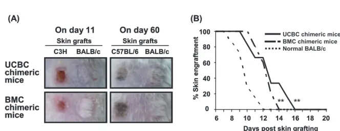
To investigate the functional competence of allogeneic UCBC- or BMC-derived T cells, the chimeric RAG2 (-/-) BALB/c mice were grafted
the skin from BALB/c, B6, and C3H mice. Both of the chimeric mice accepted the skin graft from BALB/c and B6 as autoantigens, but rejected to that from C3H mice as a third-party alloantigen in the absence of contaminating host-derived T cells. This fi nding indicates the presence of donor- derived functional CD8
+killer T cells and CD4
+helper T cells that activated killer T cells via interleukin-2 secretion and the ability of both T cells to differentiate between self and nonself antigens (Figure 5A) . In both of the chimeric RAG2 (-/-) BALB/c mice, the T cell function was restricted by MHCs from both BALB/c and B6 mice. In addition, the rejection time for skin grafts from C3H mice was compared among 6 UCBC, 5 BMC chimeric mice and 7 normal BALB/c mice (Figure 5B) . Although both of the chimeric mice completely rejected the allogeneic C3H skin graft, their rejection time was signifi cantly longer than that of normal BALB/c mice. No signifi cant differences were observed between UCBC and BMC chimeric mice with regard to the rejection time for C3H skin grafts.
Figure 5 T cell function in UCBC-transplanted recipients. Over 16 weeks after allogenic UCBC or BMC transplantation, a piece of skin harvested from the tail of BALB/c and B6 or C3H mice was simultaneously grafted on the shaved back of the chimeric RAG2(-/-)BALB/c mice. (A) Representative photographs showed that the skin graft from C3H mice was rejected as a third-party alloantigen on day 11; however, the skin graft from BALB/c and B6 mice was accepted as an autoantigen on day 60 by UCBC and BMC chimeric mice. This indicates that T cells reconstituted from allogeneic UCBCs were functionally competent. (B) The time taken for the rejection of the C3H skin graft was compared among 6 UCBC chimeric, 5 BMC chimeric mice, and 7 normal BALB/c mice.
Although both of the chimeric mice completely rejected the skin graft from C3H mice, the time taken for the rejection of the skin graft was signifi cantly longer than that taken by normal BALB/c mice. The time taken for the rejection of the C3H skin graft did not signifi cantly diff er between UCBC and BMC chimeric mice.
*p < 0.05, **p < 0.01 by log rank test; normal BALB/c mice versus UCBC or BMC chimeric mice.
DISCUSSION
In our experiment, cell lineages derived from HSCs were clearly identified as GFP-positive cells in the recipients. In addition, RAG2 (-/-)
BALB/c mice were used as allogeneic recipients because the examination of whole body immune response of UCBC-derived T and B cells was not informative in the co-existence of the BALB/c
(host type) bone marrow-derived T and B cells.
HSCs among GFP-positive allogeneic UCBCs differentiated into phenotypically mature T cells, B cells, monocytes, and granulocytes in RAG2 (-/-) BALB/c mice. In addition, normal development of the GFP-positive T and B lineages was observed in the thymus and bone marrow, respectively. Furthermore, in RAG2 (-/-)
BALB/c mice, the reconstituted B cells derived from UCBCs of B6 mice mounted specific antibody responses to T-dependent antigen and exhibited Ig class switching in the absence of host-derived T and B cells. This finding confirmed that both B cells and helper T cells derived from UCBCs were as immunologically competent as those derived from BMCs. T and B cell interaction is essential for these events.
Further, rejection of the allogeneic skin graft by both of the chimeric mice indicates the functional maturity of both CD8
+killer and CD4
+helper T cells derived from UCBC-HSCs and BMC-HSCs.
In an allogeneic environment, low-dose UCBC transplantation could not rescue the X-ray- irradiated recipients. However, transplantation with medium or high dose of UCBCs achieved a high survival rate comparable to that with BMCs. This observation possibly corresponds to that of previous studies suggesting the different numbers of potent HSCs in UCBCs
(1 cell per 4 × 10
4UCBCs)
9)and in BMCs (1 cell per 2.5 × 10
4BMCs)
12)that contribute to long-term engraftment. In addition, allogeneic UCBC transplantation showed survival rates
comparable to syngeneic UCBC transplantation
[(GFP.B6 × B6) F1 > RAG2 (-/-) B6] at 3 different doses (data not shown). Collectively, our results indicate that UCBC-HSCs have potential to reconstitute the hematopoietic system equal to BMC-HSCs even in an allogeneic environment.
Although our results indicate that T and B cells derived from allogeneic UCBCs are immunologically competent, the responsiveness of reconstituted lymphocytes in both of the chimeric mice was signifi cantly lower than that in normal mice, as indicated by a delayed Ig class switching in B cells (Figure 4) and T cell- induced skin graft rejection (Figure 5B). Chen et al.
19)have reported delayed hematopoietic engraftment and impaired immune reconstitution in normal BALB/c mice that received allogeneic UCBCs harvested from B6 fetuses. This fi nding is possibly attributable to decreased numbers of reconstituted peripheral T, B, and splenic dendritic cells. However, we detected no decrease in the number of reconstituted immune cells in UCBC-transplanted recipients, which indicates the presence of other defects in the immune system (data not shown). Talvensaari et al.
20)suggested that UCBC transplantation leads to a broad T-cell repertoire diversity and a favorable long term immune reconstitution in patients. At present, we are examining the detailed functions of reconstituted T and B cells in UCBC- and BMC-transplanted recipients by using an in vitro assay.
Because the quantity of individual cord blood samples is limited, the use of pooled cord blood might be required in clinical practice.
In mixed UCBC transplantation
21, 2 2), MHC restriction can be more complex. As shown in Figure 5A, the chimeric RAG2 (-/-) BALB/
c mice reconstituted with UCBCs harvested
from B6 mice accepted skin grafts from B6 and
BALB/c mice. In these recipients, the repertoire
of T and B cell receptors were composed by
antigen-presenting cells from both BALB/
c and B6 mice. Thus, in such cases, a clear understanding of the mechanisms underlying MHC restriction is necessary. Problems related to MHC incompatibility can be solved only by in
vivo experiments. We expect that murine UCBCtransplantation will facilitate the exploration of these issues in future studies.
ACKNOWLEDGEMENTS
The authors thank Dr. Nobukata Shinohara
(Kitasato University) for providing helpful suggestions on the experimental design. This work was supported by a Grant for Co-medical Education Program in Radiation Emergency Medicine by the Ministry of Education, Culture, Sport, Science and Technology, Japan, and by a Grant for Hirosaki University Institutional Research.
REFERENCES
1)Gluckman E, Broxmeyer HE, Auerbach AD, Friedman HS, Douglas GW, Devergie A, Esperou H, et al. Hematopoietic reconstitution in a patient with Fanconiʼs anemia by means of umbilical-cord blood from an HLA-identical sibling. N Engl J Med 1989;321:1174-1178.
2)Gluckman E, Rocha V, Boyer-Chammand A, Locatelli F, Arcese W, Pasquini R, Ortega J, et al.
Outcome of cord-blood transplantation from related and unrelated donors. N Engl J Med 1997;337:373- 381.
3)Broxmeyer HE, Douglas GW, Hangoc G, Cooper S, Bard J, English D, Arny M, et al. Human umbilical cord blood as a potential source of transplantable hematopoietic stem/progenitor cells. Proc Natl Acad Sci USA 1989;86:3828-3832.
4)Wang JCY, Doedens M, Dick JE. Primitive human hematopoietic cells are enriched in cord blood compared with adult bone marrow or mobilized peripheral blood as measured by the quantitative in vivo SCID-repopulating cell assay. Blood 1997;
89:3919-3924.
5)Bofill M, Akber AN, Salmon M, Robinson M,
Burford G, Janossay G. Immature CD45RAlowROlow T cells in the human cord blood. J Immunol 1994;
152:5613-5623.
6)Madrigal JA, Cohen SBA, Gluckman E, Charron DJ. Does cord blood transplantation result in lower graft-versus host disease? Hum Immunol 1997;56:1- 5.
7)Rubinstein P, Rosenfi eld RE, Adamson JW, Stevens CE. Stored placental blood for unrelated bone marrow reconstitution. Blood 1993;81:1679-1690.
8)Wagner JE, Rosenthal J, Sweetman R, Shu XO, Davies SM, Ramsay NK, McGlave PB, et al.
Successful transplantation of HLA-matched and HLA-mismatched umbilical cord blood from unrelated donors: analysis of engraftment and acute graft-versus-host disease. Blood 1996;88:795- 802.
9)Oikawa A, Ito K, Seguchi H, Okabe M, migishima F, Eshima K, Azuma S, et al. Development of im- munocompetent lymphocytes in vivo from murine umbilical cord blood cells. Transplantation 2007;84:
23-30.
10)Okabe M, Ikawa M, Kominami K, Nakanishi T, Nishimune Y. ʻGreen Miceʼ as a source of ubiqui- tous green cells. FEBS Lett 1997;407:313-319.
11)Kasai M, Iwamori M, Nagai Y, Okumura K, Tada T. A glycolipid on the surface of mouse natural killer cells. Eur J Immunol 1980;10:175-180.
12)Migishima F, Oikawa A, Kondo S, Ema H, Morita Y, Nakauchi H, Yokoyama M, et al. Full reconsti- tution of hematopoietic system by murine cord blood. Transplantation 2003;75:1820-1826.
13)Park SY, Kojima M, Suzuki H, Shinohara N. Ef- fective blocking of natural cytotoxicity of young rabbit serum on murine thymocytes by high con- centration of glucose in complement-dependent c y t o t o x i c i t y m e t h o d . J I m m u n o l M e t h o d 1992;154:109-119.
14)Ho VT, Soiff er RJ. The history and future of T-cell depletion as graft-versus-host disease prophylaxis for allogeneic hematopoietic stem cell transplanta- tion. Blood 2001;98:3192-3204.
15)Tiberghien P, Longo DL, Wine JW, Alvord WG, Reynolds CW. Anti-asialo GM1 antiserum treat-
ment of lethally irradiated recipients before bone marrow transplantation: evidence that recipient natural killer depletion enhance survival, engraftment, and hematopoietic recovery. Blood 1990;76:1419-1430.
16)Uchida N, Tsukamoto A, Dongping He, Friera AM, Roland S, Weissman IL. High doses of purified stem cells cause early hematopoietic recovery in syngeneic and allogeneic hosts. J Clin Invest 1998;101:961-966.
17)Hardy RR, Carmack CE, Shinton SA, Kemp JD, Hayakawa K. Resolution and characterization of pro-B and pre-pro-B cell stages in normal mouse bone marrow. J Exp Med 1991;173:1213-25.
18)Kawabe T, Naka T, Yoshida K, Tanaka T, Fujiwara H, Suematsu S, Yoshida N, et al. The immune responses in CD40-deficient mice: impaired im- munoglobulin class switching and germinal center formation. Immunity 1994;1:167-78.
19)Chen BJ, Cui X, Sempowski GD, Gooding ME, Liu C, Haynes BF, Chao NJ. A comparison of murine
T-cell-depleted adult bone marrow and full-term fetal blood cells in hematopoietic engraftment and immune reconstitution. Blood 2002;99:364-371.
20)Talvensaari K, Clave E, Douay C, Rabian C, Garderet L, Busson M, Garnier F, et al. A broad T-cell repertoire and an effi cient thymic function indicate a favorable long-term immune reconsti- tution after cord blood stem cell transplantation.
Blood 2002;99:1458-1464.
21)Barker JN, Weisdolf DJ, DeFor TE, Blazar BR, McGlave PB, Miller JS, Verfaillie CM, et al.
Transplantation of 2 partially HLA-matched umbilical cord blood units to enhance engraftment in adults with hematologic malignancy. Blood 2005;105:1343-1347.
22)Nauta AJ, kruisselbrink AB, Lurvink E, Mulder A, Claas FH, Noort WA, Willemze R, et al. Enhanced engraftment of umbilical cord blood-derived stem cells in NOD/SCID mice by cotransplantation of a second unrelated cord blood unit. Exp Hematol 2005;33:1249-1256.