NOVEL RAT MIDDLE CEREBRAL ARTERY OCCLUSION MODEL: TRANS- FEMORAL ARTERY APPROACH COMBINED WITH PRESERVATION OF
THE EXTERNAL CAROTID ARTERY
Norihito Shimamura M.D., Ph.D. and Hiroki Ohkuma M.D., Ph.D.
Abstract We developed a novel trans-femoral artery approach to the rat middle cerebral artery occlusion model
(TF-MCAO) without sacrifi cing the external carotid artery (ECA) with/without the pterygopalatine artery, which is important for chewing food. To make the TF-MCAO we fi rst dissect the left common carotid artery (CCA), ECA, and internal carotid artery (ICA). Transient occlusion clips are applied to the proximal ECA and the pterygopalatine artery; we never sacrifi ce the ECA branch. A 24-gage catheter is inserted into the left femoral artery. We insert a slightly bent 0.014 inch hydrophilically-coated guide wire via a haemostasis valve. Anatomically, the left common carotid artery is located rostral to the descending aorta, permitting a straight-forward, blind approach to the CCA. The guide wire is gently advanced about 17 mm from the bifurcation of the CCA until slight resistance is encountered. The guide wire and temporary occlusion clips are withdrawn after 90 minutes. Rats were sacrifi ced 24 h after reperfusion. Eleven rats were examined. One rat died before occlusion due to deep anesthesia. The success rate for producing infarction was 80%. The mean infarction volume of the basal ganglia was 94.4 ㎣ ± 9.4 se and mean infarction volume of the cerebral cortex was 124.2 ㎣ ± 21.6 se. No rat died due to cerebral infarction and no rat suff ered subarachnoid hemorrhage. We conclude that TF-MCAO was useful for producing a cerebral infarction.
Hirosaki Med.J. 61, Supplement:S142―S146,2010
Key words: Cerebral ischemia; micro guide wire; neurological evaluation; rat; trans-femoral artery
approach.
Introduction
The rat intraluminal suture method of middle cerebral artery occlusion (MCAO)
was first reported by Koizumi et al. in 1986
1). Following that, a large number of reports on the rat intraluminal suture MCAO model has been published
2-5). All of those methods must sacrifice the external carotid artery (ECA)
with or without sacrificing the pterygopalatine artery. Sacrificing the ECA induces ischemic tissue damage of the arterial territory, causing impaired mastication and swallowing
6).
We previously reported that commercially available silicon coated 4-0 nylon suture was suitable to produce rat MCAO
7). But we had also
Department of Neurosurgery, Hirosaki University School of MedicineCorresponding author: Norihito Shimamura M.D., Ph.D.
Department of Neurosurgery
Hirosaki University School of Medicine
5-Zaihuchou, Hirosaki Aomori pref. JAPAN. 036-8562 Tel: +81-172-39-5115 Fax : +81-172-39-5116
e-mail: [email protected]
sacrificed the ECA and pterygopalatine artery to make the rat MCAO model. If we can instead preserve all branches of the ECA, nutrition of MCAO rats improves and long term survival study projects that require a therapeutic diet can be carried out.
We have now created a novel rat MCAO
model via a trans-femoral artery approach (TF-
MCAO). This model preserves the ECA and
pterygopalatine artery. In this paper we evaluate
the steady result of the infarction volume with a
0.014 inch hydrophilic occlusion guide wire and
correlate the neurological evaluation and the
infarction volume.
Material and Methods
All experiments were approved by the Hirosaki University Animal Care Committee and conformed to the guidelines provided by the American Academy of Accreditation of Laboratory Animal Care.
Eleven male Sprague-Dawley rats weighing 29 0 to 310 g were housed under identical, controlled 12 h light-cycling conditions. The rats had free access to water and food and were anesthetized with 0.4 mg/100 g body weight
(BW) intra-peritoneal (ip) atropine sulfate hydrate (Mitsubishi Tanabe Pharma Co., Osaka, Japan), 1 mg/100 g BW ip xylazine (Bayer Schering Pharma, Osaka, Japan) and 8 mg/100 g BW ip ketamine hydrochloride (Daiichi Sankyo Co., Tokyo, Japan).
MCAO model
After general anesthesia rats were placed in the supine position on the operating table
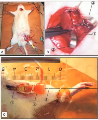
(Fig. 1A). Rectal temperature of rats was kept at 37 ± 1 ℃ from the start of anesthesia to awakening. Initially, we exposed the left common carotid artery (CCA), ECA, and the internal carotid artery (ICA) through a midline incision.
Temporary occlusion clips were applied to the proximal ECA and the pterygopalatine artery;
we never sacrifi ced the ECA branch (Fig. 1B).
Next, a left femoral 1 cm skin incision was performed and a 24-gage catheter (Angiocath
®, Becton Dickinson Co., Fukushima Japan, 0.7 x 1.9 mm) was inserted into the left femoral artery under a microscope (Fig. 1C). We inserted a slightly bent 0.014 inch hydrophilically-coated guide wire (Transend EX Platinum
®, Boston Scientifi c, Fairmont, CA, USA) via a haemostasis valve (Radifocus
®Haemostasis ValveⅡ, Terumo, Tokyo, Japan) (Fig. 2). Anatomically, the left common carotid artery is located rostral to the descending aorta, permitting a straight- forward, blind approach to the CCA without
high resolution fluoroscopy. The guide wire was gently advanced about 17 mm from the bifurcation of the CCA until slight resistance was encountered (Fig. 1C). Transient ligation of the CCA was performed to interrupt the CCA blood flow and fixation of the occlusion guide wire (Fig. 1B). After 90 minutes occlusion time, we removed the transient ligation and withdrew the occlusion wire. The temporary clips were withdrawn, and all of the ICA and ECA blood flow recovered. The skin was sutured and the rats were allowed to wake up. Twenty four hours later we evaluated neurological defi cit and sacrifi ced the rats. We did not use heparin at all during the procedure to make the MCAO.
Figure 1 Intraoperative view
A: Overview of trans-femoral middle cerebral artery occlusion. B: Operative filed of left neck. ①Transient ligation of common carotid artery. ② Temporary occlusion clip on external carotid artery. ③Temporary occlusion clip on pterygopalatine artery. ④ Occlusion micro guide wire. C: Cannulation of catheter. ① Twenty-four- gage canula inserted into the left femoral artery.
② Haemostatic valve. ③ 0.014 inch occlusion wire (Transend EX platinum tip). ④ Seventeen millimeter marker that prevents over-insertion of the wire. Numeric measure shows centimeters.
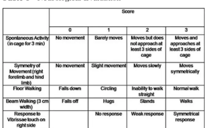
Neurological Evaluation
We carried out a neurological evaluation 24 h after the cerebral ischemia. Scoring was done blindly by MN on individual animals with a neurological score that was reported previously
(Table 1)
7). The range of our neurological score was 1 to 15 and a value of 15 indicated no neurological defi cit.
Measurement of Infarct Volume
The rats were decapitated under deep anesthesia . The bra ins were immediately removed and placed in ice-cold phosphate- buffered saline (PBS, Mitsubishi Chemical Medience Co. Tokyo, Japan) for 15 min. Brain tissue was cut coronally to a 2 mm thickness with a brain slicer (Harvard Apparatus; South Natick, MA). Brain slices were immersed in 2%
2,3,5-triphenyl - 2H - tetrazolium chloride (TTC, Kanto Chemical Co., Tokyo, Japan) solution at 37 °C for 5 min, followed by 10% formaldehyde solution. The infarct area was traced and quantifi ed by an image analysis system (Image J 1.33u).
Statistical analysis
Infarction volumes are expressed as mean
± S.E.M. and neurological scores are expressed as median with I.Q.R. The correlation between infarction volume and neurological score was analyzed by JMP 8.01(SAS institute Inc. Cary, NC), and a p-value below 0.05 was accepted as statistically signifi cant.
Results
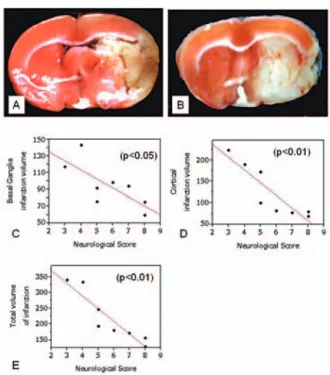
One rat died before occlusion due to deep anesthesia. Two rats showed no neurological deficit or cerebral infarction. The success rate for producing infarction was 80% (Fig. 3A, B).
No rat suff ered subarachnoid hemorrhage and no rat died due to cerebral infarction.
The total infarction volume for this model was 218.6 ㎣ ± 28.5 se, infarction volume of basal ganglia was 94.4 ㎣ ± 9.4 se, infarction volume of the cortex was 124.2 ㎣ ± 21.6 se.
Median neurological score was 5.5 (4.25 ‒ 7.75)
(n = 8).
The correlation between infarction volume and neurological score was statistically signifi cant
(p<0.05, Fig. 3 C, D, E) .
Discussion
We have developed a new rat MCAO model without sacrifi cing the ECA. This novel method is reproducible and reliable. Previously, all rat transient intraluminal suture MCAO models sacrificed the ECA with or without sacrificing the pterygopalatine artery. This sacrifi ce of the
Figure 2 Occlusion micro guide wire.
A: Overview of the wire. The tip of the wire is slightly bent.
B: Magnification of wire. Tip of wire is round.
Parallel marker shows 1 millimeter.
Table 1 Neurological Evaluation
ECA induces ischemic tissue damage of the arterial territory, causing impaired mastication and swallowing
6). We can avoid sacrificing the ECA via a trans-femoral approach to the ICA that is commonly used in neuro-interventional examination and treatment. Surgical approach to make the MCAO
8,9)also preserves the ECA, but such models require dissection of the temporal muscle, exposure of the cerebral cortex, and a change in intracranial pressure. Our new model does not require craniofacial surgical manipulation; this point is a major advantage.
The diameter of the 0.014 inch wire is 0.36 mm and the diameter of the end of the ICA is about 0.3 mm, and the diameter of the entrance to the MCA and ACA is about 0.2 mm. We can thus occlude the orifi ce of the MCA with 0.014 inch diameter guide wire. In a pilot study we
used 0.016 inch diameter wire, but this wire could not reach the end of ICA safely due to the large caliber. Also, we tried a soft tip 0.014 inch diameter wire, but this very smooth guide wire easily perforated the end of the ICA or the orifi ce of the ACA. In this study we used a 0.014 inch diameter platinum tip guide wire with a 3 cm length grooved platinum coil tip. This grooved tip helps us maintain precise insertion of the occlusion wire during ligation of the CCA during occlusion. We required approximately 35 minutes from the incision of the neck to the beginning of MCA occlusion.
Rarely, the guide wire advanced to the left cardiac ventricle, causing arrhythmia, and we quickly withdrew the guide wire. If we use high resolution fluoroscopy, we will be able to approach the left ICA safely and quickly.
In the pilot experiment we used laser Doppler CBF monitoring, but in this study we did not use a CBF monitor to avoid complications and to maintain of the TF-MCAO advantage. Measuring CBF of t he rat requires a cra niotomy or translucent cranium. Either of these maneuvers can cause cortical injury. Intracranial pressure decreases due to a craniotomy, so CBF increases even with steady perfusion pressure. The invasiveness of the operation is increased if we add the skin incision for the CBF monitor.
Additionally, dissection of the temporal muscle causes masticatory dysfunction that leads to an inadequate rat diet.
The success rate, volume of infarction, and neurological defi cit were similar to our previous report
4,5,7). Infarction volume and neurological score also correlated statistical significantly, and this new TF-MCAO method is useful as a routine MCAO model. Moreover, we can guide the micro catheter to the ICA via the trans- femoral artery by this method. Super selective drug delivery and infusion of neural stem cells
10,11)
for a small animal can be done with this method. Our new method leads to a more useful
Figure 3 2,3,5-triphenyltetrazolium chloride hydrate staining and Correlation between infarction volume and neurological score.
A: Cerebral infarction with hemorrhagic change.
B: Diffuse basal ganglia infarction occurred. C:
Infarction volume of basal ganglia. D: Infarction volume of cerebral cortex. E: Total volume of infarction. Infarction volume correlated statistically signifi cantly with the neurological score.
model to study the basic neuroscience of rat ischemia.
Acknowledgements
This study was supported by a Hirosaki University Grant for Exploratory Research by Young Scientists. The authors have no fi nancial interest in any of the drugs or materials described in this report. Micro guide wire was donated by Boston Scientifi c, Japan. This paper is already accepted by Journal of Neuroscience Methods.
References
1)Koizumi J YYNTOG. Experimental studies of ischemic brain edema 1. A new experimental model of cerebral embolism in ratsin which recirculation can be introduced in the ischemia area. Jpn J Stroke 1986;8:1-8.
2)Garcia JH, Wagner S, Liu KF, Hu XJ. Neurological defi cit and extent of neuronal necrosis attributable to middle cerebral artery occlusion in rats.
Statistical validation. Stroke 1995;26:627-34.
3)Longa EZ, Weinstein PR, Carlson S, Cummins R. Reversible middle cerebral artery occlusion without craniectomy in rats. Stroke 1989;20:84-91.
4)Shimamura N, Matchett G, Yatsushige H, Calvert JW, Ohkuma H, Zhang J. Inhibition of integrin alphavbeta3 ameliorates focal cerebral ischemic damage in the rat middle cerebral artery occlusion model. Stroke 2006;37:1902-9.
5)Shimamura N, Matchett G, Solaroglu I, Tsubokawa T, Ohkuma H, Zhang J. Inhibition of integrin alphavbeta3 reduces blood-brain barrier break- down in focal ischemia in rats. J Neurosci Res 2006;
84:1837-47.
6)Dittmar M, Spruss T, Schuierer G, Horn M.
External carotid artery territory ischemia impairs outcome in the endovascular filament model of middle cerebral artery occlusion in rats. Stroke 2003;34:2252-7.
7)Shimamura N, Matchett G, Tsubokawa T, Ohkuma H, Zhang J. Comparison of silicon- coated nylon suture to plain nylon suture in the rat middle cerebral artery occlusion model. J Neurosci Methods 2006;156:161-5.
8)Buchan AM, Xue D, Slivka A. A new model of temporary focal neocortical ischemia in the rat.
Stroke 1992;23:273-9.
9)Coyle P. Middle cerebral artery occlusion in the young rat. Stroke 1982;13:855-9.
10)Honma T, Honmou O, Iihoshi S, Harada K, Houkin K, Hamada H, Kocsis JD. Intravenous infusion of immortalized human mesenchymal stem cells protects against injury in a cerebral ischemia model in adult rat. Exp Neurol 2006;199:56-66.
11)Kamiya N, Ueda M, Igarashi H, Nishiyama Y, Suda S, Inaba T, Katayama Y. Intra-arterial trans- plantation of bone marrow mononuclear cells immediately after reperfusion decreases brain injury after focal ischemia in rats. Life Sci 2008;83:
433-7.