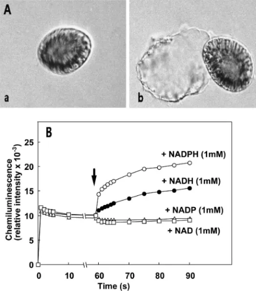
• 検索結果がありません。

Possible Factors Responsible for the Fish-Killing Mechanisms ofthe Red Tide Phytoplankton, Chattonella marina andCochlodinium polykrikoides

N/A
N/A
Protected

Academic year: 2021

シェア "Possible Factors Responsible for the Fish-Killing Mechanisms ofthe Red Tide Phytoplankton, Chattonella marina andCochlodinium polykrikoides"

Copied!
24
0
0

読み込み中.... (全文を見る)

全文

(1)

245

© by TERRAPUB and Nagasaki University, 2010.

Possible Factors Responsible for the Fish-Killing Mechanisms of the Red Tide Phytoplankton, Chattonella marina and

Cochlodinium polykrikoides

Daekyung K IM

1

and Tatsuya O DA

2

1

Jeju Center, Korea Basic Science Institute (KBSI), 66 Jejudaehakno, Jeju-Si,

Jeju Special Self-Governing Province 690-756, Korea

2

Division of Biochemistry, Faculty of Fisheries, Nagasaki University, Nagasaki 852-8521, Japan

Abstract—Generation of reactive oxygen species (ROS) such as superoxide (O

2

) and hydroxyl radical (·OH) by Chattonella marina could be confirmed by ESR spin trapping method. Several lines of evidence suggested that cell surface structure of C. marina, glycocalyx, has NADPH-dependent O

2

generation system. Immunohistochemical analysis of gill lamellae from yellowtail exposed to C. marina using antiserum against crude glycocalyx demonstrated that there were the antigns recognaised by the antiserum on the surface of gill lamellae. The results suggest that continuous accumulation of discharged glycocalyx on the gill surface occurs during C. marina exposure, which may be responsible for the ROS- mediated severe gill tissue damage leading to fish death. As compare to C. marina, the levels of O

2

and H

2

O

2

detected in Cochlodinium polykrikoides were trace levels. Furthermore, no significant increase in O

2

generation by C. polykrikoides was observed in the presence of lectins or fish mucus prepared from skin and gill of yellowtail, whereas C. marina generated increased level of O

2

responding to these stimuli. The cell-free aqueous extract prepared from C. polykrikoides showed toxic effect on HeLa cells, but the extract of C. marina had almost no effect.

Furthermore, gradual accumulation of polysaccharides in the medium was observed during the culture of C. polykrikoides, and the medium gradually became viscous, but no such change was observed in the medium of C. marina. These results suggest that ichthyotoxic mechanisms of C. marina and C. polykrikoides are different. ROS may play a significant role in the fish-killing activity of C. marina, whereas in the case of C. polykrikoides, certain toxic substances or polysaccharides are mainly responsible for the toxicity rather than ROS.

Keywords: red tide, Chattonella marina, Cochlodinium polykrikoides, glycocalyx, reactive oxygen species (ROS), cytotoxicity, mucus, toxin

1. INTRODUCTION

There has been a global increase in the frequency, magnitude, and geographical

extent of harmful algal blooms (HABs). The increase appears to be correlated with

degree of coastal pollution, utilization of coastal water for aquaculture, or global

(2)

warming. In general, the mass mortality of fish caused by HABs not only results in economic loss but also contributes to pollution of coastal waters (Hallegraeff et al., 1998). Chattonella marina is one of the major toxic phytoplankton species, which is highly toxic to fish especially to yellowtail Seriola quinqueradiata and causes serious damage to fish farming in Japan (Okaichi, 1989). Previous studies demonstrated that a decrease in oxygen partial pressure of arterial blood is the earliest physiological disturbance observed in fish after exposure to C. marina (Ishimatsu et al., 1990, 1991;

Tsuchiyama et al., 1992). In addition, physiological and histological studies of fish exposed to C. marina suggested that the blockage of respiratory water flow through the gill lamellae caused by excessive mucus interferes with O

2

transfer, resulting in asphyxia (Ishimatsu et al., 1996a; Hishida et al., 1997). Although the precise mechanism of the toxic action of C. marina is still controversial, suffocation due to gill tissue damage is generally supposed to be the direct cause of the fish death by this flagellate (Sakai et al., 1986; Endo et al., 1988).

We and other groups have found that Chattonella spp. generate reactive oxygen species (ROS) such as O

2

, H

2

O

2

, and hydroxyl radical (·OH) (Shimada et al., 1989, 1991, 1993; Tanaka et al., 1992, 1994; Oda et al., 1992a, 1994, 1995a, 1998; Kawano et al., 1996). Since ROS are generally considered to induce various detrimental effects on living organisms (Halliwell and Gutteridge, 1984; Oda et al., 1989), the ROS generated by C. marina may be involved in gill tissue injury leading to eventual fish death. This hypothesis may be supported by our previous finding that one strain of C. marina, which produces very low levels of O

2

, was less toxic against yellowtail than an other strain with higher O

2

producing ability (Ishimatsu et al., 1996b).

Furthermore, we have found that C. marina exhibited ROS-mediated toxic effect on a marine bacterium, Vibrio alginolyticus (Oda et al., 1992b).

Regarding the cell surface structure of Raphidophycean flagellate, it has been demonstrated the presence of a glycocalyx, a polysaccharide-containing common structure on the cell surface of Chattonella antiqua and Heterosigma akashiwo (Yokote and Honjo, 1985; Yokote et al., 1985). We have found that the glycocalyx is easily discharged from C. marina cells by the addition of fish mucus substances or concanavalin A (Con A) (Okamoto et al., 2000). In addition to these chemical stimuli, physical stimulation such as agitation also causes the dissociation of the glycocalyx from the flagellate cells. Laser scanning microscopic study using a specific fluorescence probe for detecting O

2

suggested that O

2

is generated at the “verruciform protrusions”

located in the glycocalyx of C. antiqua (Shimada et al., 1991). Taken together, these findings suggest that the glycocalyx somehow involves in ichthyotoxicity of C. marina.

Similar to C. marina, Cochlodinium polykrikoides is also known to be highly

toxic to fish (Onoue and Nozawa, 1989; Yuki and Yoshimatsu, 1989). Red tides due

to this dinoflagellate have been reported in Japan, Korea, and other countries, and

frequently cause severe damage to fish farming (Onoue and Nozawa, 1989; Yuki and

Yoshimatsu, 1989; Whyte et al., 2001). It has been speculated that C. polykrikoides

shows harmful effects on fish through certain toxic compounds (Onoue and Nozawa,

1989; Onoue et al., 1985). In addition to these hypotheses, Kim et al. (1999) reported

(3)

that C. polykrikoides generates O

2

and H

2

O

2

. Based on the observation, they proposed that ROS generated from C. polykrikoides are causative factors responsible for the oxidative damage of gill tissue leading to fish kills.

Although the precise toxic mechanisms of C. marina and C. polykrikoides are still unclear, there seems to be a similarity between these red tide plankton species such as the potent fish-killing activity. To gain insight into the possible toxic factors of these plankton species, in this study, comparative studies on C. marina and C. polykrikoides were conducted especially in terms of the involvement of ROS in the fish-killing activity.

2. MATERIALS AND METHODS 2.1. Materials

5,5-Dimethyl-1-pyrroline-N-oxide (DMPO) and nitric oxide -reactive fluorescent probe diaminofluorescein-FM dictate (DAF-FM DA) were purchased from Dojin Chemical Laboratories Co., Ltd. (Kumamoto, Japan). Fluorescein isothiocyanate (FITC)-conjugated goat anti-rat IgG antibody was purchased from Wako Pure Chemical Industry, Co., Ltd. (Osaka, Japan). Superoxide dismutase (Cu,Zn-SOD) (3800 units/mg of protein, from bovine erythrocytes) and horseradish peroxidase (100 units/mg of protein) were purchased from Wako Pure Chemical Industry, Co., Ltd., (Osaka, Japan). 2-Methyl-6-(p-methoxyphenyl)-3,7-dihydroimidazo[1,2-]

pyrazin-3-one (HCl salt) (MCLA) was obtained from Tokyo Kasei Kogyo, Co., Ltd.

(Tokyo, Japan). p-Hydroxyphenyl acetic acid (PHPA) was from Sigma Chemical Co.

(St. Louis, MO, USA). 5-(and-6)-Carboxy-2 ′ ,7 ′ -dichlorodihydrodihydrofluorescein dictate, acetyl ester (CM-H

2

DCFDA) was from Molecular Probes, Inc. (Eugene, The Netherlands). Other chemicals were of the highest grade commercially available.

2.2. Plankton culture

Chattonella marina was generously provided by Kagoshima Prefectural Fisheries Experimental Station, Japan. Cochlodinium polykrikoides was isolated in Yatsusiro Sea (32 ° 20 ′ N; 130 ° 19 ′ E) in Japan. Axenic culture of each clonal strain of these algae was maintained at 26 ° C in sterilized Erd-Schreiber modified (ESM) medium (pH 8.2) under illumination from a fluorescent lamp (30 µ E/m

2

/S) with a cycle of 12 h light and 12 h dark. ESM medium was prepared as described previously (Oda et al., 1992b).

In brief, 120 mg of NaNO

3

, 5 mg of K

2

HPO

4

, 0.1 mg of vitamin B

1

, 0.01 mg of vitamin B

12

, 0.001 mg of biotin, 0.26 mg of EDTA-Fe

3+

, 0.33 mg of EDTA-Mn

2+

, and 1 g of tris(hydroxymethyl)aminomethane were dissolved in 1 liter of seawater and the pH was adjusted to 8.2, followed by autoclaving (121 ° C, 15 min). All cultivations were done using sterilized instruments. Cells were counted with a hemocytometer.

2.3. Electron spin resonance (ESR) spin trapping

To detect oxygen radicals such as superoxide and hydroxyl radical in C. marina

cell suspension, ESR spin trapping method was employed as descried previously

(Oda et al., 1992a). In brief, C. marina cell suspension (1.5–2.5 × 10

4

cells/mL in ESM

(4)

medium) was mixed with 5,5-dimethyl-1-pyrroline-N-oxide (DMPO) as a spin trap, and then the mixture was immediately transferred to the ESR quartz flat cell and the cell was placed in a JEOL JES-RE1X ESR spectrometer (JEOL Co., Ltd., Akishima, Japan). ESR spectra were then recorded at room temperature.

2.4. Preparation of crude glycocalyx fraction

The cell-free supernatant was prepared by gentle agitation of flagellate cell suspension (2.0–2.5 × 10

4

cells/mL) for 30 s at room temperature and subsequent centrifugation (3000 × g) for 1 min. Preliminary experiments indicated that the glycocalyx was discharged from the plankton cells by this treatment without disruption of cells. Thus, the supernatant is supposed to contain glycocalyx as a main constituent derived from C. marina. The cell-free supernatant was concentrated by ultrafiltration using a membrane (Amicon Model 8400) which passes molecules with molecular weight of less than 10,000. The protein content was then quantified by the method of Lowry et al. (1951) with bovine serum albumin as a standard.

2.5. Preparation of antiserum to crude glycocalyx fraction

Crude glycocalyx fraction prepared from C. marina cell suspension was dialyzed against distilled water for 3 days, and then lyophilized. The lyophilized powder (20 mg) was resuspended in 1 mL of phosphate buffered saline (PBS) and emulsified with 1 mL Freund’s complete adjuvant. Two healthy rats (4–5-week- male) were injected subcutaneously three times at ten day intervals with 10 mg of lyophilized supernatant in Freund’s complete adjuvant. Ten days after the last injection, the serum was obtained by aorta puncture of the abdominal region. The antiserum was stored at –30 ° C until use.

2.6. Preparation of gill tissue of yellowtail after C. marina exposure.

Yellowtails weighing from 570 to 620 g (N = 2) were used in this experiment.

Maintenance of fish and experimental protocols were similar to those reported previously (Ishimatsu et al., 1990). In brief, the fish were subjected to the following two treatments. To the first tank, 10 liters of ESM medium was added at the onset of an experiment (control). To the second tank, 10 liters of C. marina cell suspension in ESM medium was added to attain a final cell concentration of 5,700 cells/mL.

Dissolved oxygen levels of these two tanks were maintained at near saturation levels by bubbling the water with pure oxygen. Fish were killed by a sharp blow on the head when moribund (C. marina-exposed) or at the end of an experiment (3 h, control). The first gill arches on the left side were immediately cut, rinsed in diluted isotonic seawater, and were transferred into vials of fixative (4 % paraformaldehyde, pH 7.4).

The time from killing to fixation was less than 20 s. Water temperature was kept at

25 ± 1 ° C. The vials, containing gill tissue, were left in a rotating mixer at room

temperature for 72 h before immunohistochemical analysis.

(5)

2.7. Indirect immunofluorescence

Fixed gill tissues were incubated with anti-glycocalyx antiserum as a primary antibody (1:100 dilution) in PBS for 1 h at 25 ° C. After washing twice with PBS, the gill tissues were incubated with FITC-conjugated goat anti-rat IgG as a secondary antibody (1:10,000 dilution) for 30 min at 25 ° C. After a final washing with PBS, the gill tissues were observed with fluorescence microscopy (Olympus BX60). Control experiments were done by using normal rat serum or the secondary antibody alone.

2.8. Chemiluminescence assay for measurement of O

2

Generation of superoxide anion (O

2

) by flagellates was measured by chemiluminescence method using 2-methyl-6(p-methoxyphenyl)-3,7- dihydroimidazo[1,2-]pyrazin-3-one (MCLA) as a superoxide-specific chemiluminescent probe as previously described (Lee et al., 1995; Oda et al., 1998).

MCLA was dissolved in distilled water and stored at –80 ° C until use. After the addition of MCLA to a flagellate cell suspension, the chemiluminescence response was recorded immediately with a 1254 luminometer. The reaction mixtures typically consisted of 145 µ L flagellate cell suspension (0.1–2.5 × 10

4

cells/mL), 50 µ L MCLA solution (final 5 µ g/mL), and 5 µ L SOD (final 100 units/mL) or ESM medium. The mixture with ESM medium but without flagellate cells was used as control. All chemiluminescence analysis was done at 26 ° C. The detection of O

2

in the cell-free supernatant was also conducted by chemiluminescence method using MCLA.

2.9. Fluorescence assay for measurement of H

2

O

2

Detection of H

2

O

2

in the flagellate cell suspension was done by PHPA assay method at 26 ° C (Hyslop and Sklar, 1984). After the addition of PHPA (final 1 mM) and horseradish peroxidase (final 100 µ g/mL) to the flagellate cell suspension in ESM medium, an increase in fluorescence intensity during the first min of incubation was measured with a fluorescence spectrophotometer (Hitachi Model 650-60) at an excitation wavelength of 317 nm and an emission wavelength of 400 nm in the presence or absence of 500 units/mL catalase. The catalase-inhibitable increase of fluorescence intensity was considered to reflect actual H

2

O

2

. The concentration of H

2

O

2

was estimated by using a standard curve of H

2

O

2

in cell-free ESM medium. The standard solution of H

2

O

2

in ESM medium was prepared from reagent H

2

O

2

. Under these assay conditions, the increase of fluorescence intensity was proportional to the concentration of H

2

O

2

.

2.10. Fluorescence microscopic observation

Flagellate cells were incubated with MCLA (final concentration, 100 µ M) for

15 min at 27 ° C. After incubation, flagellate cells were observed with a fluorescence

microscope (Carl Zeiss Axiovert 200). The location of H

2

O

2

production in flagellates

was detected by 5-(and-6)-carboxy-2 ′ ,7 ′ -dichlorodihydrodihydrofluorescein dictate,

acetyl ester (CM-H

2

DCFDA) (final concentration, 10 µ M) which is H

2

O

2

-specific

cell-permeable agent that is trapped intracellularly after cleavage by cellular esterases.

(6)

Fluorescence produced by the oxidation of 2 ′ ,7 ′ -dichlorodihydrodihydrofluorescein (DCF) with H

2

O

2

was observed with a fluorescence microscope (Carl Zeiss Axiovert 200) at 27 ° C (Mahadev et al., 2001). For the microscopy observation of nitric oxide (NO) production, flagellate cells were incubated with DAF-FM DA (final concentration, 10 µ M) under normal growth conditions and were observed in live condition with a fluorescence microscope (Carl Zeiss Axiovert 200). DAF-FM DA is a membrane-permeable ester derivative of DAF-FM, which has been used to load living cells, in which DAF-FM DA is hydrolyzed by intracellular esterases and converted to membrane-impermeable DAF-FM, which is further converted to the fluorescent triazole derivative DAF-FM T after specifically reacting with NO.

2.11. Preparation of aqueous extracts of C. marina and C. polykrikoides

Harvested cells from 200 mL of the culture of C. marina (1 × 10

4

cells/mL) or Cochlodinium polykrikoides (5 × 10

3

cells/mL) by centrifugation (1,000 × g for 10 min at 4 ° C) was resuspended in 100 µ L of distilled water and vigorously agitated by sonication for 2 min at room temperature. Each lysate was centrifuged at 13,000 × g for 10 min at 4 ° C and the supernatant was withdrawn and used immediately as aqueous cell-free extract.

2.12. Cytotoxicity against HeLa cells

HeLa (human epithelial carcinoma) cells obtained from Riken Cell Bank, Tsukuba, Japan, were cultured in α -minimal essential medium ( α -MEM) supplemented with 10% fetal bovine serum (FBS), 10 µ g each of adenosine, guanosine, cytidine, and thymidine per mL of medium, penicillin (100 µ g/mL), and streptomycin (100 µ g/mL) as described (Oda and Wu, 1993). The cytotoxicities of the cell-free extracts prepared from algal cells against HeLa cells were measured by the inhibition of colony formation as described previously with some modifications (Oda et al., 1995b). In brief, adherent cells (200 cells/well) in 24-well plates were preincubated with varying concentrations of the extract in phosphate buffered saline (PBS) at 37 ° C for 3 h, and then PBS containing the extract in each well was replaced with α -MEM containing 10% FBS and cultured for 5 days under the usual growth conditions. The numbers of colonies formed were counted after staining with 1%

methylene blue in 50% methanol. Clusters of 30 or more cells were considered colonies.

2.13. Detection of polysaccharides

Total polysaccharide levels in the culture supernatants of C. polykrikoides and C. marina were determined by the phenol-sulfuric acid method (Dubois et al., 1956).

3. RESULTS

3.1. Detection of oxygen radicals by ESR spin trapping in C. marina

The generation of oxygen radicals in C. marina suspension was measured by

ESR spin trapping. The superoxide and hydroxyl radicals react with a spin trap

(7)

DMPO to yield a DMPO-OOH signal and a DMPO-OH signal, respectively. As shown in Fig. 1, both DMPO-OOH adducts and DMPO-OH adducts were produced in the suspension of intact C. marina. Analysis of the time course of the spin trap signal showed that the DMPO-OOH signal gradually changed to the DMPO-OH signal. The addition of 500 U/mL of SOD resulted in the disappearance of the DMPO- OOH signal and also a decrease in the DMPO-OH signal to a negligible level.

Furthermore, the generation of oxygen radicals from C. marina depended on the viability of the plankton cells; no spin adduct signal was observed when the algal cells were ruptured by sonication.

3.2. Possible presence of NADPH-dependent oxygen radical generation system in glycocalyx of C. marina

Regarding the cell surface structure of C. marina, the presence of a glycocalyx as an carbohydrate- protein complex on the surface of algal cells has been demonstrated (Yokote and Honjo, 1985). Recently, we have found that the glycocalyx of C. marina is easily discharged from the cells by simple agitation (Fig. 2A). Furthermore, several lines of evidence suggest that an enzymatic system of superoxide generation may be located on the glycocalyx (Shimada et al., 1991; Oda et al., 1998). Thus, it is of interesting to examine if the glycocalyx fraction has the superoxide generating activity. As shown in Fig. 2B, the addition of NADPH to the crude glycocalyx fraction resulted in a potent chemiluminescence response. NADH was also effective but NAD

+

and NADP

+

had no effect. The NADPH-dependent chemiluminescence response of the crude glycocalyx fraction was inhibited by SOD (100 U/mL) (Kim et

Fig. 1. ESR spectra of DMPO spin adducts obtained with suspensions of intact (A and B) or ultrasonic-

treated (C) C. marina. (A) The spectra were recorded at 1, 3.5, and 6 min after addition of DMPO

to the algal suspension. (B) The spectrum at 1 min after addition of DMPO in the presence of SOD

(500 U/mL). (C) The spectrum of the ultrasonic-treated algal suspension at 1 min. In this last case,

plankton cells were completely ruptured during 60-s sonication. Reprinted from Marine Biotechnology

Kenkyu Kaiho, 8, Oda and Ishimatsu, 15–28, 1995, Fig. 3.

(8)

al., 2000). These results suggest that C. marina has an NADPH-dependent superoxide generation system in the glycocalyx.

3.3. Immunohistochemical analysis of gill tissues of yellowtails exposed to C. marina For the immunohistochemical analysis, the fixed gill tissues from control and Chattonella-exposed fish were incubated with the anti-crude glycocalyx antiserum for 1 h, and then washed with PBS. After incubation with FITC-labeled secondary antibody and subsequent washing with PBS, the specimens were observed with phase-contrast or fluorescence microscopy. In phase-contrast microscopy, slight

Fig. 2. Morphological changes of C. marina after mild agitation (A) and the effects of NADPH, NADH,

NADP

+

or NAD

+

on MCLA-mediated chemiliminescence responses in the concentrated cell-free

supernatant of C. marina. (A) Photomicrographs of normal C. marina (a) and morphologically

changed C. marina (b) after gentle agitation for 30 s at 27

°

C. (B) After the addition of MCLA

(5

µ

g/mL), each reagent (final 1 mM) was added to the cell-free supernatant at the point indicated

by arrow and chemiluminescence responses were measured at 27

°

C. Redrawn after Biochimica et

Biophysica Acta 1524, Kim et al., Mechanism of superoxide anion generation in the toxic red tide

phytoplankton Chattonella marina: possible involvement of NAD(P)H oxidase, 220–227, 2000,

Figs. 2 and 4, with permission from Elsevier.

(9)

morphological changes of gill lamellae were observed in the Chattonella-exposed fish as compared to the gill from the control fish (Figs. 3A, C). The fluorescence microscopic observation of the same specimens showed intense fluorescence staining on the surface of interlamellae region of the gill tissue of the Chattonella-exposed fish (Fig. 3D), whereas no such staining was observed in the control fish (Fig. 3C). Neither normal rat serum nor the secondary antibody alone produced fluorescence even in the Chattonella-exposed fish (data not shown).

3.4. Comparison of the generation of reactive oxygen species (ROS) by C. marina and C. polykrikoides

Cell suspensions of C. marina and C. polykrikoides kept in ESM medium under normal growth conditions were assayed for chemiluminescence. When MCLA was added to C. marina cell suspension, a rapid chemiluminescence response was observed without a lag time, and the chemiluminescence response was inhibited by SOD (100 units/mL) to a background level of ESM medium alone (Fig. 4A). The intensity of integrated emission during the first 10 s was dependent on C. marina cell concentrations (Fig. 4B). However, no significant chemiluminescence responses

Fig. 3. Indirect immunofluorescence staining of the fixed gill tissues of control (A, C) or C. marina-

exposed yellowtail (B, D). After 1 h incubation without (A, C) or with (B, D) anti-glycocalyx

antiserum, the gill lamellae were washed twice with PBS, and then reacted with FITC-labeled

secondary antibody. After washing twice with PBS, the specimens were observed with phase-

contrast (A, B) or fluorescence microscopy (C, D). Reprinted with kind permission from Springer

Science + Business Media: Marine Biology, Possible involvement of the glycocalyx in the ichthyotoxity

of Chattonella marina (Raphidophyceae): immunological approach using antiserum against cell

surface structures of the flagellate, 139, 2001, 625–632, Kim et al., Fig. 4. © Springer-Verlag.

(10)

were observed in C. polykrikoides even at a maximum level of cell concentration (Figs. 4A, B). To further examine whether or not C. polykrikoides generates ROS, we employed PHPA and phenol red assay methods for the detection of H

2

O

2

. In PHPA assay, the level of H

2

O

2

detected in the C. polykrikoides even at nearly maximum cell density was found to be a trace level as compared to the level in C. marina (Table 1).

In addition, red color of phenol red in C. marina cell suspension was almost completely discolored in the presence of horseradish peroxidase during 24 h incubation under usual culture conditions, while no such change in color of phenol red in C. polykrikoides cell suspension was observed even after additional 3 days culture (data not shown). The change in color of phenol red in C. marina cell

Fig. 4. MCLA-dependent chemiluminescence responses of C. polykrikoides and C. marina.

(A) Immediately after the addition of MCLA (final 5

µ

g/mL) to each cell suspension, the chemiluminescence response was recorded during first 30 s at 26

°

C. The concentrations of cells used were 8.0

×

10

3

cells/mL for C. polykrikoides (

) and 2.0

×

10

4

cells/mL for C. marina (

). (

);

Luminescence of MCLA in ESM medium alone. (

); Luminescence response of C. marina (2.0

×

10

4

) in the presence of SOD (100 units/mL). (B) After addition of MCLA to indicated concentrations of C. polykrikoides (

) or C. marina (

) cell suspension, chemiluminescence responses which were expressed in terms of relative intensity of integrated emission during the first 10 s were measured.

Each point represents the average of triplicate measurements. Reprinted from Comparative

Biochemistry and Physiology Part C, 132, Kim et al., Possible factors responsible for the toxicity of

Cochlodinium polykrikoides, a red tide phytoplankton, 415–423, 2002, Fig. 1, with permission from

Elsevier.

(11)

suspension was prevented by the addition of catalase (data not shown). It has been known that phenol red is oxidized with H

2

O

2

concomitant with discoloration under the catalytic activity of horseradish peroxidase (Pick and Keisari, 1980). Thus, these results suggest that C. marina continuously releases H

2

O

2

into the medium during culture, whereas C. polykrikoides may not release H

2

O

2

at least under normal physiological culture conditions.

3.5. Effects of lectins and fish mucus substances on the chemiluminescence responses in C. marina and C. polykrikoides

Our previous studies have demonstrated that fish mucus substances or lectins such as concanavalin A (Con A) and wheat germ agglutinin (WGA) stimulate C. marina to generate markedly increased levels of O

2

(Nakamura et al., 1998; Oda et al., 1998). Thus, we examined the effects of fish mucus prepared from skin and gill of yellowtail (Seriola quinqueradiata) and lectins such as Con A and WGA on the production of O

2

by C. polykrikoides and C. marina. As shown in Fig. 5, increased chemiluminescence responses were detected in C. marina in the presence of these stimuli as previously reported, whereas no significant chemiluminescences were observed in C. polykrikoides even in the presence of these stimuli.

3.6. Fluorescence microscopic analysis of ROS generation in C. marina and C. polykrikoides

As shown in Fig. 6B, the strong fluorescence was observed on the cell surface of C. marina after incubation with MCLA. The result suggests that O

2

generation system of C. marina was located on the cell surface. On the other hand, only a slight fluorescence emission was observed in C. polykrikoides (Fig. 6D). We also investigated H

2

O

2

generation in C. marina and C. polykrikoides using 5-(and-6)-carboxy-2 ′ ,7 ′ -

Table 1. Detection of H

2

O

2

in the cell suspensions of C. marina and C. polykrikoides. C. marina (1

×

10

4

cells/mL) or C. polykrikoides (8

×

10

3

cells/mL) cell suspensions at exponential growth phase were subjected to PHPA assay in the presence or absence of catalase (500 units/mL).

Flagellate species H

2

O

2

detected (nmol/min/10

4

cells) C. marina

None 1.07

±

0.05

+ Catalase 0.11

±

0.01

C. polykrikoides

None 0.05

±

0.01

+ Catalase ND*

*ND; not detectable

Reproduced from Comparative Biochemistry and Physiology Part C, 132, Kim et al., Possible factors

responsible for the toxicity of Cochlodinium polykrikoides, a red tide phytoplankton, 415–423, 2002,

Table 1, with permission from Elsevier.

(12)

Fig. 6. Fluorescence microscopic observation of C. marina (A, B) and C. polykrikoides (C, D) after incubation with MCLA as a specific fluorescenct probe for O

2

. Phase-contrast (A, C) and fluorescence micrographs (B, D) of plankton cells. Note the strong fluorescence was observed on the cell surface of C. marina, whereas such strong fluorescence was not observed in C. polykrikoides.

Redrawn after Journal of Plankton Research, Kim et al., 29: 241–247, 2007, Fig. 1. © Oxford University Press.

Fig. 5. Effects of Con A, WGA, gill mucus, and skin mucus on chemiluminescence responses in

C. polykrikoides and C. marina. After simultaneous addition of MCLA (final 5

µ

g/mL) and each

sample (final 100

µ

g/mL) to C. polykrikoides (

) or C. marina (

) cell suspensions, intensities of

integrated emission during the first 10 s were measured. Each data represents the average of triplicate

measurements. The luminescence of MCLA in ESM medium alone is also shown (

). Reprinted

from Com. Biochem. Shys. Part C, 132, Kim et al., 415–423, Fig. 2, 2002. © Elsevier.

(13)

dichlorodihydrodihydrofluorescein dictate, acetyl ester (CM-H

2

DCFDA), which is specific fluorescent probe for detecting H

2

O

2

. Interestingly, the both plankton cells showed intense fluorescence in the entire intracellur area after the incubation with CM-H

2

DCFDA (Fig. 7), suggesting that both C. marina and C. polykrikoides are producing H

2

O

2

intracellularly.

3.7. Cytotoxic effects of aqueous extracts prepared from C. polykrikoides and C. marina on HeLa cells

Previous studies have reported that three different solvent fractions such as neurotoxic, hemolytic and hemagglutinating were obtained from Cochlodinium spp.

(Onoue et al., 1985; Onoue and Nozawa, 1989). Thus, we prepared cell-free aqueous extract from C. polykrikoides cells, and its cytotoxicity on HeLa cells was examined.

As shown in Fig. 8, 24 h treatment with the extract (final 10%) in α -MEM containing 10% FBS resulted in the cytotoxic morphological changes, and many cells were detached. Colony formation assay also revealed that the extract exhibits cell killing activity against HeLa cells in a concentration-dependent manner (Fig. 9). In contrast to C. polykrikoides, no significant cytotoxicity was found in the extract of C. marina prepared by the same way (Figs. 8, 9).

Fig. 7. Fluorescence microscopic observation of C. marina (A, B) and C. polykrikoides (C, D) after

incubation with CM-H

2

DCFDA as a specific fluorescenct probe for H

2

O

2

. Phase-contrast (A, C) and

fluorescence micrographs (B, D) of plankton cells. Note the strong fluorescence was observed in the

entire cytoplasm area in both flagellate cells. Redrawn afterJournal of Plankton Research, Kim

et al., 29: 241–247, 2007, Fig. 2. © Oxford University Press.

(14)

3.8. Extracellular production of polysaccharides by C. polykrikoides during the growth

An increase in viscosity of the water due to mucus substances produced by flagellates has been suggested as a possible cause of death of fish (Jenkinson, 1989).

In addition, suffocation from the mucus secreted by C. polykrikoides has also been reported as a potential cause of fish kill (Lee, 1996). Since both C. polykrikoides and C. marina are known to produce extracellular polysaccharides (Matsusato and Kobayashi, 1974; Yokote and Honjo, 1985; Hasui et al., 1995), we compared the polysaccharide levels in the medium of these flagellates during the growth. As shown in Fig. 10, continuous accumulation of extracellular polysaccharide was observed in the medium of C. polykrikoides during the growth, and ethanol fixation suggested that C. polykrikoides cells at exponential growth phase were surrounded by polysaccharide substances (Fig. 11B). In the culture of C. marina, only a small

Fig. 8. Effects of aqueous extract prepared from C. polykrikoides and C. marina on cellular morphology in HeLa cells. Adherent HeLa cells in 96-well plate (2

×

10

4

cells/well) were incubated with final 10%

of each cell-free extract in α-MEM containing 10% FBS for 24 h at 37

°

C. Phase contrast micrographs

of the cells treated with C. polykrikoides extract (A) or with C. marina extract (B). Reproduced from

Comparative Biochemistry and Physiology Part C, 132, Kim et al., Possible factors responsible for

the toxicity of Cochlodinium polykrikoides, a red tide phytoplankton, 415–423, 2002, Fig. 3, with

permission from Elsevier.

(15)

amount of extracellular polysaccharides was detected even at later exponential growth phase (Fig. 10).

3.9. Detection of nitric oxide (NO) in C. marina and C. polykrikoides

To examine NO generation in plankton cells, we used NO-reactive fluorescent probe diaminofluorescein-FM diacetate (DAF-FM DA). The plankton cell suspension was incubated with DAF-FM DA (final 10 µ M) for 3 h at 27 ° C. The fluorescence from diaminofluorescein-FM T (DAF-FM T), the reaction product of DAF-FM with NO, was observed with a fluorescence microscope. Clear intense fluorescence emission was observed in the cytoplasm of C. marina (Fig. 12B). The emission of bright fluorescence from the plankton cells disappeared in the presence of specific NO scavenger, carboxy-PTIO (data not shown). On the other hand, no significant fluorescence emission from C. polykrikoides was observed (Fig. 12D). These results suggest that C. marina has a specific NO generating system, but C. polykrikoides does not.

4. DISCUSSION

Despite the fact that Chattonella shows the lethal effect on various fish species especially yellowtail (Seriola quinqueradiata) and causes massive mortalities of cultured fish, the exact toxic mechanism is still unclear. In previous several studies, histological analysis in the fish exposed to Chattonella have been done. Shimada et al. (1983) have reported that most of the mucus goblet cells and mucus coat

Fig. 9. Cytotoxicity of the aqueous extracts prepared from C. polykrikoides and C. marina on HeLa cells.

Adherent HeLa cells in 24-well plate (200 cells/well) were preincubated with varying concentration of C. polykrikoides (

) or C. marina (

) extract in PBS for 3 h at 37

°

C, then the PBS containing the extracts were replaced with _-MEM containing 10% FBS and continued to culture for 5 days at 37

°

C.

The number of colonies formed in each well were counted as described under Materials and Methods.

Each point represents an average of triplicate measurements. Each bar represents standard deviation.

Reproduced from Comparative Biochemistry and Physiology Part C, 132, Kim et al., Possible factors

responsible for the toxicity of Cochlodinium polykrikoides, a red tide phytoplankton, 415–423, 2002,

Fig. 4, with permission from Elsevier.

(16)

Fig. 10. Accumulation of polysaccharides in the medium during the growth of C. polykrikoides and C. marina. Each flagellate cell suspension at exponential growth phase was inoculated into 100 mL of ESM medium with a final cell density of 1000–1200 cells/mL and cultured under the conditions described under Materials and Methods. Viable cell count (A) and measurement of polysaccharides in each culture supernatant (B) were done at 2-day intervals until the growth reached the stationary phases. (A) Growth curves of C. polykrikoides (

) and C. marina (

). Each point represents an average of triplicate measurements. Each bar represents standard deviation. (B) The concentrations of polysaccharides in the culture supernatant of C. polykrikoides (

) or C. marina (

) prepared by centrifugation (10,000

×

g, 5 min) were estimated by phenol sulfuric acid method with glucose as a standard. Each point represents an average of duplicate measurements. Reproduced from Comparative Biochemistry and Physiology Part C, 132, Kim et al., Possible factors responsible for the toxicity of Cochlodinium polykrikoides, a red tide phytoplankton, 415–423, 2002, Fig. 5, with permission from Elsevier.

disappeared from the gill lamellae of yellowtail exposed to C. antiqua. Since there was concomitant shrinkage of the gill epithelium, they proposed that loss of mucus layers impaired osmoregulatory function. The resultant osmotic imbalance would lead to edema formation which in turn reduces the gas exchange capacity of the gills.

Toyoshima et al. (1985) observed histological alterations of chloride cells in the gill

of fish exposed to C. antiqua, when the gill samples were taken immediately after

death due to Chattonella exposure. They also considered that these changes might be

related to edema formation. Endo et al. (1985) also reported that the gills taken from

moribund fish that had been exposed to Chattonella showed edema and hypertrophy

of the epithelia in many secondary lamellae. Accordingly, it seems likely that the gill

is a primary target tissue of Chattonella attack, and edema formation in gill lamellae

(17)

is a typical lesion observed in dying fish after Chattonella exposure. Since it has been reported that the gills of fish dying from environmental hypoxia showed very few lesions and were histologically hardly discernible from the control gills, it seems evident that epithelial separation (edema) and other morphological changes observed in fish dying from Chattonella exposure were truly attributable to direct effects of Chattonella and not by severe hypoxemia. Consistent with these reports, morphological changes of gill lamellae of fish exposed to C. marina were observed in this (Fig. 3) and our previous study (Ishimatsu et al., 1996a).

Ishimatsu et al. (1990, 1991) demonstrated that the earliest physiological change observed in the yellowtail after Chattonella exposure was a rapid drop of arterial oxygen partial pressure (PaO

2

). Furthermore, they found that the epithelial separation

Fig. 11. Photomicrographs of C. polykrikoides before and after 50% ethanol fixation. (A) Normal motile

cells of C. polykrikoides. (B) After fixation with 50% ethanol, fiber-like precipitates were emerged

around C. polykrikoides cells. Reproduced from Comparative Biochemistry and Physiology Part C,

132, Kim et al., Possible factors responsible for the toxicity of Cochlodinium polykrikoides, a red tide

phytoplankton, 415–423, 2002, Fig. 6, with permission from Elsevier.

(18)

had not developed when the tissues were sampled immediately after the onset of hypoxemia, and the only histological alteration detectable at the time of PaO

2

drop was a considerable accumulation of mucus substances between the filaments and lamellae. Based on those findings, they proposed that the initial PaO

2

drop during Chattonella exposure is due to blockade by mucus of respiratory water flow through the gills and not from edema formation as previously proposed. Therefore, the accumulation of mucus substances on the gill surface appears to be the earliest event leading to gill tissue injury caused by Chattonella exposure. However, the origin of the mucus substances accumulated between gill lamellae is still controversial.

Matsusato and Kobayashi (1974) claimed that fish kill by Chattonella is due to the interception of respiratory water flow by mucus-like substance from the alga itself.

They reported that Chattonella readily produced a mucus-like substance by filtration

Fig. 12. Microscopic observation of nitric oxide (NO) production in C. marina (A, B) and C.

polykrikoides (C, D). Phase-contrast (A, C) and fluorescence micrographs (B, D) of the plankton cells. Each plankton cell suspension was incubated with NO-specific fluorescence probe diaminofluorescein-FM dictate (DAF-FM DA) for 3 h at 27

°

C, and then observed microscopically.

Redrawn after Journal of Plankton Research, Kim et al., 28: 613–620, 2006, Fig. 3. © Oxford

University Press.

(19)

through a silk net whose mesh size (95 × 95 µ m) was a little larger than the fish gill sieve. This phenomenon may be closely related to our finding that glycocalyx is easily discharged from C. marina cells responding to physical stimulation as well as stimulation with gill tissue mucus or lectin (Shimada et al., 1993; Okamoto et al., 2000). Thus, one possibility is that the mucus substances on the gill surface are derived from Chattonella cells. This notion is partly supported by the fact that the anti-glycocalyx antiserum stained the gill surface of Chattonella-exposed fish (Fig. 3). At least our present result clearly indicates that certain substances originated from Chattonella cells attach to gill tissue. Another possibility which we prefer is that Chattonella might stimulate gill mucus cells and induce excessive mucus secretion from these cells, which in turn leads to blockade of respiratory water flow. Since ROS are known to induce mucin secretion from mucus cells in guinea pig gallbladder (LaMont, 1989) and rat gastric mucosa (Hiraishi et al., 1991), it is conceivable that ROS produced by Chattonella induce mucus secretion from gill mucus cells. Based on the laser scanning microscopic observation at high magnification, Shimada et al.

(1993) and Tanaka et al. (1994) reported the evidence that O

2

was generated in small particles or “verruciform protrusions”, located on the surface of Chattonella cells.

Our microscopic observation using ROS-specific fluorescent prove also suggested that O

2

was generated on the cell surface of C. marina (Fig. 6). More interestingly, they observed that these structures were released from the Chattonella cells when mucus substances prepared from yellowtail gill were added to cell suspension.

Although the relationship between the verruciform protrusions and glycocalyx is unclear, we also observed that extracellular addition of yellowtail gill mucus enhanced O

2

generation by C. marina concomitant with the discharge of the glycocalyx (Nakamura et al., 1998; Okamoto et al., 2000). Furthermore, the discharge of the glycocalyx can occur even by gentle agitation (Fig. 2). Thus, it may be possible that the glycocalyx with O

2

generation system is discharged when C. marina cells are inhaled into fish mouth and contact with gill surface, and then the glycocalyx sticks to the gill surface where continuous O

2

generation takes place. ROS generated by the glycocalyx on the gill surface may induce excess amount of mucus secretion from gill mucus cells that may contribute to blockade of respiratory water flow.

Chattonella cells trapped in gill lamellae may also participate in further aggravation of gill situation (Ishimatsu et al., 1996a).

Similar to Chattonella, Cochlodinium polykrikoides is also known to be a noxious red tide dinoflagellate with potent fish-killing activity, and the blooms due to this dinoflagellate associated with massive mortality of fish have been recorded in a worldwide scale (Yuki and Yoshimatsu, 1989; Brusle, 1995; Chang and Kim, 1997;

Kim, 1997; Whyte et al., 2001). For instance, an extensive bloom of C. polykrikoides

in the summer of 1995 in Korean coastal waters caused losses of aquacultured fish

amounting to $ 95.5 million (Kim, 1997). More recently, blooms of Cochlodinium

sp., monitored for the first time on the west of Vancouver Island in 1999, caused

substantial mortality to farmed salmon (Whyte et al., 2001). Although it has been

considered that C. polykrikoides produces certain ichthyotoxic agents (Brusle, 1995),

the detailed toxic mechanism is still totally unknown.

(20)

Recently, it has been reported that C. polykrikoides isolated in Korea produced O

2

and H

2

O

2

, and ROS-mediated ichthyotoxic mechanism of this dinoflagellate has been proposed (Kim et al., 1999). However, in the present study, we found that the levels of O

2

and H

2

O

2

detected in C. polykrikoides isolated in Japan were almost trace levels as compared to those of C. marina, and no comparable levels of ROS reported in Korean strain of C. polykrikoides were detected (Fig. 4, Table 1). In the fluorescence microscopic observation using ROS-specific probe, no significant level of O

2

was detected in C. polykrikoides, while potent fluorescence emission derived from O

2

was observed in C. marina (Fig. 6). Furthermore, no increase in O

2

generation was observed in our strain of C. polykrikoides in the presence of Con A, WGA, or fish mucus substances, although the generation of O

2

by C. marina was markedly increased by these stimuli (Fig. 5). The reason for this discrepancy is unclear now, but one possible explanation may be differences between the strains used. Regarding this point, our previous study has demonstrated that even the levels of ROS produced by C. marina were different between the strains (Ishimatsu et al., 1996b; Oda et al., 1997).

Regarding H

2

O

2

, the fluorescence microscopic observation using H

2

O

2

-specific probe suggested that C. polykrikoides produces H

2

O

2

intracellularly similar to C.

marina (Fig. 7). Since H

2

O

2

could not be detected in C. polykrikoides cell suspension by PHAP assay as described above (Table I), the intracellular H

2

O

2

may not be released into the medium, while C. marina can release a part of intracellularly produced H

2

O

2

into the medium. To clarify the biological roles of H

2

O

2

in C. marina and C. polykrikoides, further studies are required.

Previous studies have suggested that Cochlodinium spp. may secrete some ichthyotoxic substances consisting of three different solvent fractions such as neurotoxic, hemolytic and hemagglutinating (Onoue et al., 1985; Onoue and Nozawa, 1989). In agreement with this, we found that aqueous extract prepared from C. polykrikoides showed cytotoxic effect on HeLa cells, whereas no significant toxicity was observed in the extract prepared from C. marina by the same way (Figs. 8, 9). Although the detailed characteristics of such toxic substances in the extract are still unclear, these results support the notion that C. polykrikoides may cause harmful effect on fish through certain toxic substances.

Histological examination by light microscopy of fish exposed to Cochlodinium sp. suggested that no pathogenic abnormalities to internal organs, but gill edema and separation of the lamellar epithelium were evident (Whyte et al., 2001). Interestingly, it has been reported that similar gill damage also occurs with exposure of fish to solvent extracts of Cochlodinium. Namely, the red seabream (Chrysophrys major), when exposed to the neurotoxin obtained from Cochlodinium by organic solvent extraction, displayed edema of the secondary lamellae (Onoue and Nozawa, 1989).

Furthermore, Onoue et al. have reported that the supernatant prepared from Cochlodinium red tide seawater by centrifugation showed intense fish toxicity (Onoue et al., 1985).

In addition to the direct toxic effects via certain ichthyotoxic agents described

above, suffocation from the mucus secreted by C. polykrikoides has also been

(21)

reported as a potential cause of fish kills (Lee, 1996). In Costa Rica and Panama, blooms of Cochlodinium catenatum were implicated in coral mortality, which was partly attributed to smothering by the mucus produced by the dinoflagellate (Guzmán et al., 1990). Consistent with these reports, continuous accumulation of polysaccharides was observed in C. polykrikoides during the culture (Fig. 10), and ethanol fixation revealed that C. polykrikoides cells were surrounded by large amount of polysaccharides (Fig. 11B). Although the polysaccharide production by C. marina has also been reported (Matsusato and Kobayashi, 1974; Yokote and Honjo, 1985), the amounts of polysaccharides detected in the medium of C. marina was almost negligible level as compared to that of C. polykrikoides at least during the culture periods tested. Thus, the extracellular secretion of polysaccharides with such a high level is a characteristic feature of C. polykrikoides. Interestingly, antiviral activities of the polysaccharides secreted by C. polykrikoides have also been demonstrated. Thus, it seems likely that secreted polysaccharides may involve in a certain biochemical effect on fish exposed to this dinoflagellate (Hasui et al., 1995). Recent studies using scanning and transmission electron microscope revealed that the presence of a thick polysaccharide coat surrounding C. polykrikoides cells (Gobler et al., 2008). Furthermore, they reported that the killed C. polykrikoides via freezing and thawing, or filtered (0.2 µ m)- bloom water were incapable to cause fish mortality, while intact cell suspension of C. polykrikoides caused 100% fish mortality (Gobler et al., 2008). Based on these results, it has been proposed that the intact thick polysaccharide layer associated with cell membranes and/or a toxin principle within this layer may be responsible for fish mortality. Probably, the ichthyotoxic agents present in C. polykrikoides cells may be unstable, and algal cellular integrity may be required for the full toxicity. It has also been reported that extracellular polysaccharides produced by another dinoflagellate, Gymnodinium sp., are capable of inducing apoptosis in human lymphoid cells (Sogawa et al., 1998a, b).

In addition to ROS (O

2

and H

2

O

2

), recent study suggested that C. marina produces nitric oxide (NO), a gaseous radical molecule with a wide variety of biological activities (Kim et al., 2006, 2008). Although the production of NO in C. marina could be confirmed by the fluorescent observation of the algal cells using NO-specific fluorescent indicator, no significant level of NO was detected in C. polykrikoides at least by this method (Fig. 12). Reflecting the taxonomical difference, it seems likely that C. marina (Raphidophyceae) and C. polykrikoides (Dinophyceae) may have quite different metabolic systems especially in terms of the production of radical molecules such as O

2

, H

2

O

2

and NO.

Taken together with previous findings and the results obtained in this study, we would like to propose that biologically active multiple metabolites such as cytotoxic agents and polysaccharides produced by C. polykrikoides may be mainly responsible to the fish killing mechanism of this dinoflagellate rather than ROS.

Acknowledgments—This work was supported in part by a Grant-in-Aid for Scientific

Research from the Ministry of Education, Science, and Culture of Japan.

(22)

REFERENCES

Brusle, J. 1995. The impact of harmful algal blooms on finfish, mortality, pathology and toxicology.

Institut français de Recherché pour l’Exploitation de la Mer Reperes Ocean 10: 1–75.

Chang, M. and W.-S. Kim. 1997. Prologue and epilogue to the first international symposium on plankton blooms. Ocean Research (Seoul) 19: 135–137.

Dubois, M., K.A. Gilles, J.K. Hamilton, P.A. Robers and F. Smith. 1956. Colorimetric method for determination of sugar and related substances. Analytical Chemistry 28: 350–356.

Endo, M., T. Sakai and A. Kuroki. 1985. Histological and histochemical changes in the gills of the yellowtail Seriola quinqueradiata exposed to the Raphidophycean flagellate Chattonella marina.

Marine Biology 87: 193–197.

Endo, M., R. Foscarini, A. Kuroki. 1988. Electrocardiogram of a marine fish, Pagrus major, exposed to red tide plankton, Chattonella marina. Marine Biology 97: 477–481.

Gobler, C.J., D.L. Berry, O.R. Anderson, A. Burson, F. Koch, B.S. Rodgers, L.K. Moore, J.A. Goleski, B. Allam, P. Bowser, Y. Tang and R. Nuzzi. 2008. Characterization, dynamics, and ecological impacts of harmful Cochlodinium polykrikoides blooms on eastern Long Island, NY, USA. Harmful Algae 7: 293–307.

Guzmán, H.M., J. Cortés, P.W. Glynn and R.H. Richmond. 1990. Coral mortality associated with dinoflagellate blooms in the eastern Pacific (Costa Rica and Panama). Marine Ecology Progress Series 60: 299–303.

Hallegraeff, G.M., B.L. Munday, D.G. Baden and P.L. Whitney. 1998. Chattonella marina raphidophyte bloom associated with mortality of cultured bluefin tuna (Thunnus maccoyii) in South Australia. In:

Harmful Algae, B. Reguera, J. Blanco, M. L. Fern_ndez and T. Wyatt (Eds.), Xunta de Galicia, IOC, Paris, pp. 93–96.

Halliwell, B. and J.M.C. Gutteridge. 1984. Oxygen toxicity, oxygen radicals, transition metals and disease. Biochemical Journal 219: 1–14.

Hasui, M., M. Matsuda and K. Okutani. 1995. In vitro antiviral activities of sulfated polysaccharides from a marine microalga (Cochlodinium polykrikoides) against human immunodeficiency virus and other enveloped viruses. International Journal of Biological Macromolecules 17: 293–297.

Hiraishi, H., A. Terano, S. Ota, H. Mutoh, T. Sugimoto, M .Razandi and K.J. Ivey. 1991. Oxygen metabolites stimulate mucous glycoprotein secretion from cultured rat gastric mucous cells.

American Journal Physics 261: 662–668.

Hishida, Y., A. Ishimatsu and T. Oda. 1997. Mucus blockade of lamellar water channels in yellowtail exposed to Chattonella marina. Fisheries Science 63: 315–316.

Hyslop, P.A. and L.A. Sklar. 1984. A quantitative fluorimetric assay for the determination by polymorphonuclear leukocytes: Its use in the simultaneous fluorimetric assay of cellular activation processes. Analytical Biochemistry 141: 280–286.

Ishimatsu, A., H. Maruta, T. Tsuchiyama and M. Ozaki. 1990. Respiratory, ionoregulatory and cardiovascular responses of the yellowtail Seriola quinqueradiata to exposure to the red tide plankton Chattonella. Nippon Suisan Gakkaishi 56: 189–199.

Ishimatsu, A., T. Tsuchiyama, M. Yoshida, M. Sameshima, M. Pawluk and T. Oda. 1991. Effect of Chattonella exposure on acid-base status of the yellowtail. Nippon Suisan Gakkaishi 57: 2115–2120.

Ishimatsu, A., M. Sameshima, A. Tamura and T. Oda. 1996a. Histological analysis of the mechanisms of Chattonella-induced hypoxemia in yellowtail. Fisheries Science 62: 50–58.

Ishimatsu, A., T. Oda, M. Yoshida and M.Ozaki. 1996b. Oxygen radicals are probably involved in the mortality of yellowtail by Chattonella marina. Fisheries Science 62: 836–837.

Jenkinson, I.R. 1989. Increases in viscosity may kill fish in some blooms. In: Red Tides: Biology, Environmental Science, and Toxicology, T. Okaichi, D.M. Anderson and T. Nemoto (Eds.), Elsevier, New York, pp. 435–438.

Kawano, I., T. Oda, A. Ishimatsu and T. Muramatsu. 1996. Inhibitory effect of the iron chelator Desferrioxamine (Desferal) on the generation of activated oxygen species by Chattonella marina.

Marine Biology 126: 765–771.

Kim, H.G. 1997. Recent harmful algal blooms and mitigation strategies in Korea. Ocean Research

(Seoul) 19: 185–192.

(23)

Kim, C.S., S.G. Lee, C.K. Lee, H.G. Kim and J. Jung. 1999. Reactive oxygen species as causative agents in the ichthyotoxicity of the red tide dinoflagellate Cochlodinium polykrikoides. Journal of Plankton Research 21: 2105–2115.

Kim, D., A. Nakamura, T. Okamoto, N. Komatsu, T. Oda, T. Iida, A. Ishimatsu and T. Muramatsu. 2000.

Mechanism of superoxide anion generation in the toxic red tide phytoplankton Chattonella marina:

possible involvement of NAD(P)H oxidase. Biochimica et Biophysica Acta 1524: 220–227.

Kim, D., K. Yamaguchi and T. Oda. 2006. Nitric oxide synthase-like enzyme mediated nitric oxide generation by harmful red tide phytoplankton, Chattonella marina. Journal of Plankton Research 28:

613–620.

Kim, D., Y.S. Kang, Y. Lee, K. Yamaguchi, K. Matsuoka, K.W. Lee, K.S. Choi and T. Oda. 2008.

Detection of nitric oxide (NO) in marine phytoplankters. Journal of Bioscience and Bioengineering

105: 414–417.

LaMont, J.T. 1989. Oxygen radicals stimulate gallbladder glycoprotein secretion. In: Mucus and Related Topics, E. Chantler and N.A. Ratcliffe (Eds.), The Company of Biologists Ltd, Cambrige, pp. 273–

278.

Lee, J.S. 1996. Bioactive components from the red tide plankton Cochlodinium polykrikoides. Journal of the Korean Fisheries Society 29: 165–173.

Lee, T.Y., N. Goto, E. Niki, K. Yokoyama, M. Tsuzuki, T. Takeuchi and I. Karube. 1995.

Chemiluminescence detection of red tide phytoplankton Chattonella marina. Analytical Biochemistry

67: 225–228.

Lowry, O.H., N.J. Rosenbrough, A.L. Farr and R.J. Randall. 1951. Protein measurement with the Folin phenol reagent. The Journal of Biological Chemistry 193: 265–275.

Mahadev, K., A. Zilbering, L. Zhu and B.J. Goldstein. 2001. Insulin-stimulated hydrogen peroxide reversibly inhibits protein-tyrosine phosphatase 1B in vivo and enhances the early insulin action cascade. The Journal of Biological Chemistry 274: 21938–21942.

Matsusato, T. and H. Kobayashi. 1974. Studies on death of fish caused by red tide. Bulletin of the Nansei Regional Fisheries Research Laboratory 7: 43–67.

Nakamura, A., T. Okamoto, N. Komatsu, S. Ooka, T. Oda, A. Ishimatsu and T. Muramatsu. 1998. Fish mucus stimulates the generation of superoxide anion by Chattonella marina and Heterosigma akashiwo. Fisheries Science 64: 866–869.

Oda, T. and H.C. Wu. 1993. Cerulenin inhibits the cytotoxicity of ricin, modeccin, Pseudomonas toxin, and diphtheria toxin in brefeldin A-resistant cell lines. The Journal of Biological Chemistry 268:

12596–12602.

Oda, T., T. Akaike, T. Hamamoto, F. Suzuki, T. Hirano and H. Maeda. 1989. Oxygen radicals in influenza-induced pathogenesis and treatment with pyran polymer conjugated SOD. Science 244:

974–976.

Oda, T., T. Akaike, K. Sato, A. Ishimatsu, S. Takeshita, T. Muramatsu and H. Maeda. 1992a. Hydroxyl radical generation by red tide algae. Archives of Biochemistry and Biophysics 294: 38–43.

Oda, T., A. Ishimatsu, M. Shimada, S. Takeshita and T. Muramatsu. 1992b. Oxygen-radical-mediated toxic effects of the red tide flagellate Chattonella marina on Vibrio alginolyticus. Marine Biology

112: 505–509.

Oda, T., A. Ishimatsu, S. Takeshita and T. Muramatsu. 1994. Hydrogen peroxide production by the red tide flagellate Chattonella marina. Bioscience, Biotechnology and Biochemistry 58: 957–958.

Oda, T., J. Moritomi, I. Kawano, S. Hamaguchi, A. Ishimatsu and T. Muramatsu. 1995a. Catalase- and superoxide dismutase-induced morphological changes and growth inhibition in the red tide phytoplankton Chattonella marina. Bioscience, Biotechnology and Biochemistry 59: 2044–2048.

Oda, T., C.H. Chen and H.C. Wu. 1995b. Ceramide reverses brefeldin A (BFA) resistance in BFA- resistant cell lines. The Journal of Biological Chemistry 270: 4088–4092.

Oda, T., A. Nakamura, M. Shikayama, I. Kawano, A. Ishimatsu and T. Muramatsu. 1997. Generation of reactive oxygen species by raphidophycean phytoplankton. Bioscience, Biotechnology and Biochemistry 61: 1658–1662.

Oda, T., A. Nakamura, T. Okamoto, A. Ishimatsu and T. Muramatsu. 1998. Lectin-induced enhancement

of superoxide anion production by red tide phytoplankton. Marine Biology 131: 383-390.

(24)

Okaichi, T. 1989. Red tide problems in the Seto Inland Sea, Japan. In: Red Tides: Biology, Environmental Science, and Toxicology, T. Okaichi, D.M. Anderson and T. Nemoto (Eds.), Elsevier, New York, pp.

137–142.

Okamoto, T., D. Kim, T. Oda, K. Matsuoka, A. Ishimatsu and T. Muramatsu. 2000. Concanavalin A- induced discharge of glycocalyx of raphidophycean flagellates, Chattonella marina and Heterosigma akashiwo. Bioscience, Biotechnology and Biochemistry 64: 1767–1770.

Onoue, Y. and K. Nozawa. 1989. Separation of toxins from harmful red tides occurring along the coast of Kagoshima prefecture. In: Red Tides: Biology, Environmental Science, and Toxicology, T.

Okaichi, D.M. Anderson and T. Nemoto (Eds.), Elsevier, New York, pp. 371–374.

Onoue, Y., K. Nozawa, K. Kumanda, K. Takeda and T. Aramaki. 1985. Toxicity of Cochlodinium type

’78 Yatsushiro occurring in Yatsushiro Sea. Bulletin of the Japanese Society of Science Fisheries 51:

147.

Pick, E. and Y. Keisari. 1980. A simple colorimetric method for the measurement of hydrogen peroxide produced by cells in culture. Journal of Immunological Methods 38: 161–170.

Sakai, T., K. Yamamoto, M. Endo, A. Kuroki, K. Kumanda, K. Takeda and T. Aramaki. 1986. Changes in the gill carbonic anhydrase activity of fish exposed to Chattonella marina red tide, with special reference to the mortality. Nippon Suisan Gakkaishi 52: 1351–1354.

Shimada, M., T.H. Murakami, T. Imahayashi, H.S. Ozaki, T. Toyoshima and T. Okaichi. 1983. Effects of sea bloom, Chattonella antiqua, on gill primary lamellae of the young yellowtail, Seriola quinqueradiata. Acta Histochemica et Cytochemica 16: 232–244.

Shimada, M., R. Shimono, T.H. Murakami, S. Yoshimatsu and C. Ono. 1989. Red tide, Chattonella antiqua reduces cytochrome c from horse heart. In: Red Tides: Biology, Environmental Science, and Toxicology, T. Okaichi, D.M. Anderson and T. Nemoto (Eds.), Elsevier, New York, pp. 443–446.

Shimada, M., N. Nakai, H. Goto, M. Watanabe, H. Watanabe, M. Nakanishi, S. Yoshimatsu and C. Ono.

1991. Free radical production by the red tide alga, Chattonella antiqua. The Histochemical Journal

23: 362–365

Shimada, M., Y. Kawamoto, Y. Nakatsuka and M. Watanabe. 1993. Localization of superoxide anion in the red tide alga Chattonella antiqua. Journal of Histochemistry and Cytochemistry 41: 507–511.

Sogawa, K., M. Matsuda and K. Okutani. 1998a. Induction of apoptosis by a marine microalgal polysaccharide in a human leukemic cell line. Journal of Marine Biotechnology 6: 241–243.

Sogawa, K., E. Kodama, M. Matsuda, S. Shigeta and K. Okutani. 1998b. Marine microalgal polysaccharide induces apoptosis in human lymphoid cells. Journal of Marine Biotechnology 6: 35–38.

Tanaka, K., S. Yoshimatsu and M. Shimada. 1992. Generation of superoxide anions by Chattonella antiqua: possible causes for fish death by “Red Tide”. Experientia 48: 888–890.

Tanaka, K., Y. Muto and M. Shimada. 1994. Generation of superoxide anion radicals by the marine phytoplankton organism, Chattonella antiqua. Journal of Plankton Research 16: 161–169.

Toyoshima. T., H.S. Ozaki, M. Shimada, T. Okaichi and T.H. Murakami. 1985. Ultrastructural alterations on chloride cells of the yellowtail Seriola quinqueradiata, following exposure to the red tide species Chattonella antiqua. Marine Biology 38: 101–108.

Tsuchiyama T., A. Ishimatsu, T. Oda, S. Uchida and M. Ozaki. 1992. Effect of Chattonella exposure on plasma catecholamine levels in the yellowtail. Nippon Suisan Gakkaishi 58: 207–211.

Whyte, J.N.C., N. Haigh, N.G. Ginther and L.J. Keddy. 2001. First record of blooms of Cochlodinium sp. (Gymnodiniales, Dinophyceae) causing mortality to aquacultured salmon on the west coast of Canada. Phycologia 40: 298–304.

Yokote, M. and T. Honjo. 1985. Morphological and histochemical demonstration of a glycocalyx on the cell surface of Chattonella antiqua, a “naked flagellate”. Experientia 41: 1143–1145.

Yokote, M., T. Honjo and M. Asakawa. 1985. Histochemical demonstration of a glycocalyx on the cell surface of Heterosigma akashiwo. Marine Biology 88: 295–299.

Yuki, K. and S. Yoshimatsu. 1989. Two fish-killing species of Cochlodinium from Harima Nada, Seto Inland Sea, Japan. In: Red Tides: Biology, Environmental Science, and Toxicology, T. Okaichi, D.M.

Anderson and T. Nemoto (Eds.), Elsevier, New York, pp. 371–374.

D. Kim and T. Oda (e-mail: [email protected])

Fig. 1.  ESR spectra of DMPO spin adducts obtained with suspensions of intact (A and B) or ultrasonic- ultrasonic-treated (C) C
Fig. 2.  Morphological changes of C. marina after mild agitation (A) and the effects of NADPH, NADH, NADP +  or NAD +  on MCLA-mediated chemiliminescence responses in the concentrated cell-free supernatant of C
Fig. 3.  Indirect immunofluorescence staining of the fixed gill tissues of control (A, C) or C
Fig. 4.  MCLA-dependent chemiluminescence responses of C. polykrikoides and C. marina.
+7

参照

関連したドキュメント