Fi bul i n- 5 as a t ar get m
ol ec ul e of
gl uc oki nas e- m
edi at ed c al c i neur i n/ N
FAT
s i gnal i ng i n panc r eat i c i s l et s
著者
O
kuyam
a Tom
oko, Shi r akaw
a J un, Yanagi s aw
a
H
i r om
i , Kyohar a M
ayu, Yam
az aki Shuns uke,
Taj i m
a Kaz uki , Togas hi Yu, Ter auc hi Yas uo
j our nal or
publ i c at i on t i t l e
Sc i ent i f i c Repor t s
vol um
e
7
page r ange
2364
year
2017- 05
権利
( C) The Aut hor ( s ) 2017
Thi s ar t i c l e i s l i c ens ed under a Cr eat i ve
Com
m
ons At t r i but i on 4. 0 I nt er nat i onal Li c ens e,
w
hi c h per m
i t s us e, s har i ng, adapt at i on,
di s t r i but i on and r epr oduc t i on i n any m
edi um
or
f or m
at , as l ong as you gi ve appr opr i at e c r edi t
t o t he or i gi nal aut hor ( s ) and t he s our c e,
pr ovi de a l i nk t o t he Cr eat i ve Com
m
ons
l i c ens e, and i ndi c at e i f c hanges w
er e m
ade.
The i m
ages or ot her t hi r d par t y m
at er i al i n
t hi s ar t i c l e ar e i nc l uded i n t he ar t i c l e’
s
Cr eat i ve Com
m
ons l i c ens e, unl es s i ndi c at ed
ot her w
i s e i n a c r edi t l i ne t o t he m
at er i al . I f
m
at er i al i s not i nc l uded i n t he ar t i c l e’
s
Cr eat i ve Com
m
ons l i c ens e and your i nt ended us e
i s not per m
i t t ed by s t at ut or y r egul at i on or
exc eeds t he per m
i t t ed us e, you w
i l l need t o
obt ai n per m
i s s i on di r ec t l y f r om
t he c opyr i ght
hol der . To vi ew
a c opy of t hi s l i c ens e, vi s i t
ht t p: / / c r eat i vec om
m
ons . or g/ l i c ens es / by/ 4. 0/ .
U
RL
ht t p: / / hdl . handl e. net / 2241/ 00151187
doi: 10.1038/s41598-017-02535-0
www.nature.com/scientificreports
Identiication of the matricellular
protein Fibulin-5 as a target
molecule of glucokinase-mediated
calcineurin/NFAT signaling in
pancreatic islets
Tomoko Okuyama
1, Jun Shirakawa
1, Hiromi Yanagisawa
2, Mayu Kyohara
1, Shunsuke
Yamazaki
1, Kazuki Tajima
1, Yu Togashi
1& Yasuo Terauchi
1Glucokinase-mediated glucose signaling induces insulin secretion, proliferation, and apoptosis in pancreatic β-cells. However, the precise molecular mechanisms underlying these processes are not
clearly understood. Here, we demonstrated that glucokinase activation using a glucokinase activator (GKA) signiicantly upregulated the expression of Fibulin-5 (Fbln5), a matricellular protein involved in matrix-cell signaling, in isolated mouse islets. The islet Fbln5 expression was induced by ambient glucose in a time- and dose-dependent manner and further enhanced by high-fat diet or the deletion of insulin receptor substrate 2 (IRS-2), whereas the GKA-induced increase in Fbln5 expression was diminished in Irs-2-deicient islets. GKA-induced Fbln5 upregulation in the islets was blunted by a glucokinase inhibitor, KATP channel opener, Ca2+ channel blocker and calcineurin inhibitor, while it was augmented by harmine, a dual-speciicity tyrosine phosphorylation-regulated kinase (DYRK) 1 A inhibitor. Although deletion of Fbln5 in mice had no signiicant efects on the glucose tolerance or β-cell
functions, adenovirus-mediated Fbln5 overexpression increased glucose-stimulated insulin secretion in INS-1 rat insulinoma cells. Since the islet Fbln5 expression is regulated through a glucokinase/KATP channel/calcineurin/nuclear factor of activated T cells (NFAT) pathway crucial for the maintenance of
β-cell functions, further investigation of Fbln5 functions in the islets is warranted.
Glucose metabolism plays an important role in normal β-cell functions such as insulin production and insulin secretion, and also in β-cell growth and survival1, 2. Glucose signaling in the pancreatic β-cells has also been
shown to be involved in β-cell proliferation in both humans and rodents3–6. Glucokinase, a member of the
hex-okinase family, is the predominant enzyme catalyzing the phosphorylation of glucose in the pancreatic β-cells and the liver. Glucokinase acts as a glucose sensor for insulin secretion from the pancreatic β-cells7 and is required
for the efects of glucose signaling on β-cell proliferation8. Heterozygous inactivating mutations of glucokinase
cause type 2 maturity onset diabetes of the young (MODY2), and homozygous or compound heterozygous inac-tivating glucokinase mutations cause a more severe phenotype known as permanent neonatal diabetes mellitus (PNDM), which manifests at birth9. On the other hand, heterozygous activating glucokinase mutations cause
per-sistent hyperinsulinemic hypoglycemia (PHHI)10, associated with increased β-cell mass and β-cell proliferation11.
We have shown previously that glucokinase activation ameliorates endoplasmic reticulum (ER) stress-mediated apoptosis of the pancreatic β-cells12, while another report revealed that genetic activation of β-cell glucokinase
causes cell apoptosis associated with DNA double-strand breaks and activation of the tumor suppressor protein p5313. hus, glucokinase appears to play important roles in β-cell function, replication, and survival. hese
ind-ings inspired the development of a therapeutic strategy for diabetes by targeting glucokinase. Glucokinase acti-vators (GKAs) increase the glucose ainity and maximum velocity (Vmax) of glucokinase, leading to enhanced
1Department of endocrinology and Metabolism, Graduate School of Medicine, Yokohama-city University,
Yokohama, Japan. 2Life Science center of tsukuba Advanced Research Alliance, University of tsukuba, tsukuba, Japan. correspondence and requests for materials should be addressed to J.S. (email: jshira-tky@umin.ac.jp) or Y.t. (email: terauchi-tky@umin.ac.jp)
Received: 1 November 2016 Accepted: 12 April 2017 Published: xx xx xxxx
glucose-induced insulin secretion from the islets and enhanced hepatic glucose uptake14. his ability suggests a
potential pharmacological role of GKAs in the treatment of diabetes. However, further investigation is needed to determine the eicacy and safety of GKAs; for example, downstream targets of glucose metabolism in the β-cells have not yet been clearly revealed.
Fibulin-5 (Fbln5; also known as EVEC or DANCE), a matricellular protein, is essential for elastic iber assem-bly15, 16. Fbln5 is secreted by various cell types, including vascular smooth muscle cells (SMCs), ibroblasts and
endothelial cells. Fbln5 expression is usually downregulated ater birth, but reactivated upon tissue injury17, 18.
Fbln5 has several non-elastogenic functions, for example, regulation of proteases via its integrin-binding domain19–22. Fbln5 has also been shown to bind to the α5β1 ibronectin receptor and the β1 integrin21, 23. Indeed,
Fbln5 plays critical roles in cell proliferation, migration and invasion of certain tumors and smooth muscle cells24, 25.
Mice lacking in Fbln5 exhibit systemic elastic iber defects, including loose skin, tortuous aorta, emphysematous lungs, and genital prolapse16, 26. However, the precise nature of the involvement of Fbln5 in metabolism remains
unknown.
In this study, we found that treatment with a GKA induced Fbln5 gene expression in mouse pancreatic islets. Although it has been reported that interaction of the islets with some speciic extracellular matrix molecules is important for islet/β-cell survival27, 28, the precise expression levels and roles of these molecules in the pancreatic
islets and β-cell functions remain obscure. In this study, we focused on the regulation of Fbln5 expression in the pancreatic β-cells.
Results
Glucokinase activation induced
Fbln5
expression in the pancreatic islets.
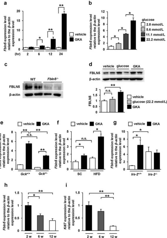
At irst, we identiied by gene expression microarray analysis (GSE41248), that stimulation of mouse pancreatic islets with a GKA for 24 hours induced Fbln5 expression in the islets (12.6-fold enhanced expression as compared to that in the vehicle control; p= 0.0043)12. To validate this upregulation of Fbln5 expression by treatment with a GKA in mousepan-creatic islets, we investigated Fbln5 mRNA expression in isolated islets from C57BL/6 J mice. Consistent with the results of the microarray analysis, the Fbln5 mRNA expression in the isolated islets was signiicantly increased, in a time-dependent manner, by treatment with a GKA (Fig. 1a). Ambient glucose also induced Fbln5 expression in the islets in a concentration-dependent manner (Fig. 1b). We detected FBLN5 protein expression in the wild-type mouse islets, as well as in INS-1 rat insulinoma cell line (Fig. 1c and d) but not in the Fbln5-deicient (Fbln5−/−) islets (Fig. 1c). he treatment with a GKA also increased FBLN5 protein expression levels in INS-1 cells (Fig. 1d). Moreover, in glucokinase hetero-deicient (Gck+/−) mouse islets, GKA-stimulated Fbln5 mRNA expression levels were reduced as compared to those in the islets from wild-type mice (Fig. 1e). No diference was detected in Fbln5 mRNA expression levels between vehicle-treated Gck+/− islets and the wild-type islets (p= 0.357) (Fig. 1e). hese results suggest that Fbln5 expression is induced by glucokinase activation in the pancreatic islets. Furthermore, the GKA-induced increase in Fbln5 expression was more pronounced in the islets of mice reared on a high-fat diet for 20 weeks than in the islets of standard chow-fed mice (Fig. 1f), although there were no signiicant difer-ences between the vehicle-treated islets from standard chow-fed and high-fat diet-fed mice (p= 0.24), consistent with the report that glucokinase-mediated signaling in the β-cells is activated by a high-fat diet8, 29. In contrast,
in insulin receptor substrate 2 (IRS-2)-deicient (Irs-2−/−) mouse islets, basal Fbln5 expression was signiicantly increased compared with those of wild-type mice (Fig. 1g). However, the response of Fbln5 induction to GKA was almost abolished in Irs-2−/− mouse islets (Fig. 1g). It may also explain the more pronounced upregulation of islet Fbln5 expression in high-fat diet-fed mice than in normal chow-fed mice, as GKA is known to induce IRS-2 expression in the β-cells of mice reared on a high-fat diet8. he lack of Fbln5 induction in Irs-2−/− islets suggests that IRS-2 is involved in the GKA-induced upregulation of islet Fbln5 expression. Moreover, we found that Fbln5 was strongly expressed in the islets of 2-week-old pre-weaning mice, the expression level decreasing by 6 or 12 weeks of age (Fig. 1h). his expression pattern of Fbln5 is consistent with the expression of the proliferation marker Ki67 in the islets (Fig. 1i).
Glucokinase/K
ATPchannel/calcineurin/NFAT signaling is required for glucose-mediated
Fbln5
expression in islets.
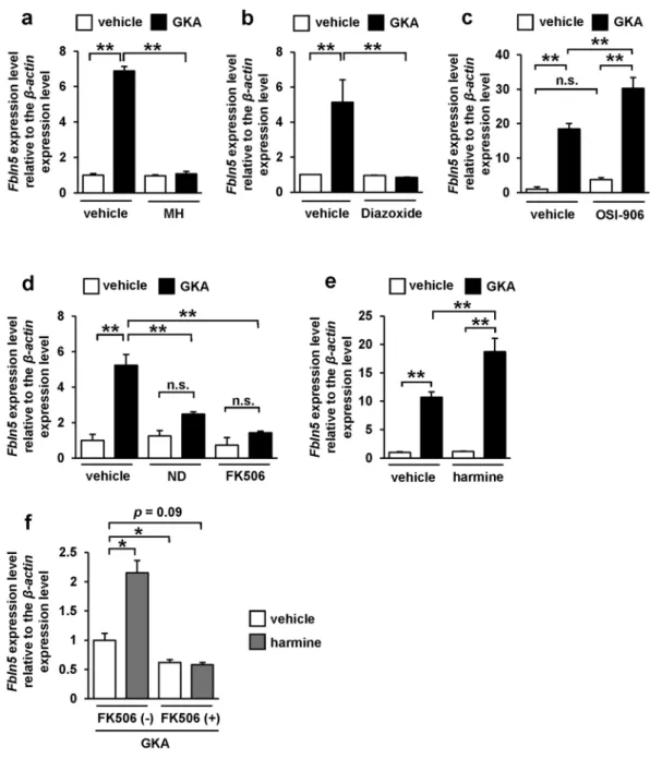
We next assessed the signaling pathways underlying the GKA-induced upregulation of Fbln5 in the pancreatic islets. Treatment with D-mannoheptulose, a speciic inhibitor of glucokinase, completely abolished the GKA-induced upregulation of Fbln5 in the pancreatic islets (Fig. 2a). In addition, treatment with diazoxide, a KATP channel (ATP-sensitive potassium channel) opener, also suppressed the GKA-induced elevation of Fbln5 expression in the islets (Fig. 2b). Treatment with OSI-906, a dual insulin and IGF-1 receptor inhibitor, did not reduce the Fbln5 induction by GKA, but enhanced it (Fig. 2c). hese results imply that an inlux of Ca2+ into the β-cells via depolarization of the plasma membrane accompanied by the closure of KATP channel, and not the autocrine action of insulin, is involved in the GKA-induced upregulation of Fbln5 in the pancreatic islets.Calcineurin is activated in an intracellular Ca2+-dependent manner30, leading to NFAT activation by
dephos-phorylation and subsequent translocation of NFAT from the cytosol to the nucleus31. Glucose-induced regulation
of Irs-2 expression has been reported to be mediated via this Ca2+/calcineurin/NFAT signaling in the pancreatic
β-cells32. Hence, we evaluated the efects of a Ca2+ channel blocker, a calcineurin inhibitor, and a DYRK1A inhib-itor on the upregulation of Fbln5 in the islets treated with a GKA. Blockade of the L-type voltage-dependent Ca2+ channels (L-type VDCCs) with nifedipine in isolated mouse islets abrogated the GKA-induced increase in Fbln5 expression in the islets (Fig. 2d). Moreover, treatment with FK506, which speciically inhibits calcineurin activity, also almost completely abolished the GKA-induced increase in Fbln5 expression in the pancreatic islets (Fig. 2d). Dual-speciicity tyrosine phosphorylation-regulated kinases (DYRKs), including DYRK1A, inactivate the NFAT1 proteins by phosphorylating its SP-3 motif33. Notably, harmine, a DYRK1A inhibitor, enhanced the Fbln5
www.nature.com/scientificreports/
islets was blunted in the presence of FK506 (Fig. 2f). hese results suggest that the transcriptional regulation of Fbln5 in the islets is mediated by glucose signaling and downstream Ca2+/calcineurin/NFAT signaling.
Fbln5
−/−mice exhibited normal glucose tolerance and normal glucose-stimulated insulin
secre-tion and
β
-cell proliferation evoked by GKA.
To investigate the role of Fbln5 in glucose metabolism and insulin secretion, we used 8- to 12-week-old Fbln5 knockout (Fbln5−/−) mice16 to evaluate whether Fbln5 deletionmay inluence glucose homeostasis in vivo. Fbln5−/− mice showed normal glucose tolerance and comparable insu-lin secretion during an oral glucose tolerance test (Fig. 3a and b). No signiicant diference in glucose-stimulated insulin secretion was observed between islets isolated from Fbln5−/− mice and wild-type mice (Fig. 3c). hese results imply that Fbln5 has no efect on insulin secretion in healthy young adult mice.
We next assessed the β-cell mass in the 8- to 12-week-old Fbln5−/− mice. No signiicant diferences in the islet morphology and the β-cells area relative to the total pancreatic area were observed between the wild-type mice and Fbln5−/− mice (Fig. 3d and e). Furthermore, we evaluated the GKA-induced β-cell proliferation activity
Figure 2. Glucose-signal induced Fbln5 upregulation via the glucokinase/KATP channel/calcineurin/NFAT signaling pathway in pancreatic islets. (a–f) Fbln5 mRNA expression. Islets from C57BL/6 J mice were incubated with 10 nmol/L of D-mannoheptulose (MH) (n = 4) (a), 200 µmol/L of diazoxide (n = 4) (b), 200 nmol/L of OSI-906 (n = 4) (c), 50 µmol/L of nifedipine (ND), 10 µmol/L of FK506 (n = 3) (d), 10 µmol/L of harmine (n = 3) (e), or a combination of 10 µmol/L of FK506 and 10 µmol/L of harmine (n = 4) (f) for 24 hours in the presence or absence of 30 µmol/L of GKA CpdA (vehicle; DMSO). Data are represented as means ± SEM.
www.nature.com/scientificreports/
in Fbln5−/− and wild-type islets. Treatment with GKA for 48 hours markedly increased the EdU-incorporated proliferating insulin-positive β-cells to a similar extent in the islets isolated from both genotypes of mice (Fig. 3f and g). On the other hand, the luorescent intensity of insulin was signiicantly increased in the GKA-treated islets compared with the vehicle-treated islets in wild-type mice, but not in Fbln5−/− mice (see Supplementary Fig. S1a). his result in islets from wild-type mice is consistent with the observation that glucokinase activation enhances insulin gene expression and insulin secretion in β-cells12, 34. However, GKA-induced insulin secretion
was not decreased in Fbln5−/− islets compared with wild-type islets (see Supplementary Fig. S1b). Insulin content
in GKA-treated Fbln5−/− islets showed a tendency to be decreased compared to wild-type islets, but it did not reach statistical signiicance (see Supplementary Fig. S1b). hus, Fbln5 is not required for the development and maintenance of β-cell function or proliferation.
In immunohistochemical analysis of paraffin-embedded endocrine pancreatic tissue from 8-week-old wild-type mice and Fbln5−/− mice, FBLN5 was seemed to be around CD34 (endothelial marker) -positive inter-stitial tissue, but not in β-cells, α-cells, or δ-cells in the islets (see Supplementary Fig. S2). Next, fetal pancreatic tissue parain sections from wild-type mice at the age of embryonic day 15 were immunostained for FBLN5. he area and intensity of the FBLN5 signal in the islets seemed to be more abundant compared with those in adult mice (see Supplementary Fig. S3a). hen, we used non-parainized cultured islets from 8-week-old wild-type mice for immunostaining. Notably, FBLN5-positive β-cells were detectable in non-parainized adult wild-type islets (see Supplementary Fig. S3b). In addition, FBLN5 is observed at cytoplasmic granular structures in INS-1 cells, as shown in Supplementary Fig. S3c, suggesting that FBLN5 is also expressed in β-cells.
Adenovirus-mediated
Fbln5
overexpression increased glucose-stimulated insulin secretion and
inhibited cell proliferation in INS-1 cells.
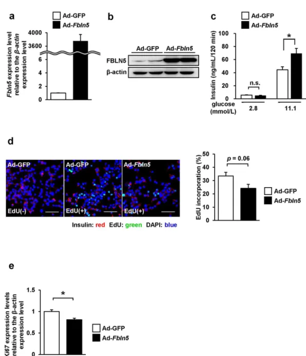
Next, we evaluated the properties of Fbln5 by forced expression of Fbln5 gene expression using an adenovirus vector (Ad-Fbln5) in INS-1 cells. Following adenovirus-mediated infection of Ad-Fbln5 in INS-1 cells, overexpression of Fbln5 was conirmed by measuring the mRNA and pro-tein expression levels (Fig. 4a and b). he cells overexpressing Fbln5 showed enhanced insulin secretion in the presence of 11.1 mmol/L glucose as compared to the control cells (1.6-fold, p= 0.017), although basal insulin secretion was not signiicantly diferent between the Ad-Fbln5- and Ad-GFP- infected INS-1 cells (Fig. 4c). he efects of Fbln5 overexpression on the cell proliferation activity was evaluated by measuring the EdU incorpo-ration and Ki67 expression in Ad-Fbln5-infected INS-1 cells. We were almost able to ignore the GFP-signals when we adjusted the gain of signals according to the luorescent intensity of EdU. (Fig. 4d let panel). he ratio of EdU-incorporated proliferating INS-1 cells to the total count of INS-1 cells tended to be decreased in the Ad-Fbln5-infected cells as compared with that in the control cells (Fig. 4d). In addition, Ad-Fbln5-infected INS-1 cells showed signiicant reduction in the Ki67 expression (Fig. 4e). hese results indicate that overexpression of Fbln5 enhances insulin secretion whereas decreases cell proliferation in β-cells.Discussion
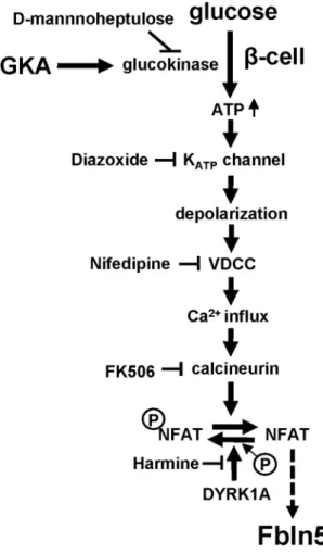
In this study, we showed the glucose signaling-induced transcriptional regulation of Fbln5 in pancreatic islets, which is mediated by glucose metabolism via glucokinase and downstream Ca2+/calcineurin/NFAT signaling pathway (Fig. 5).
Because β-cells are exposed to high ambient glucose concentrations under the diabetic condition, glucokinase, which acts as a glucose sensor, transmits the impact of the hyperglycemia to the β-cells. In the pancreatic islets, glucokinase is mainly expressed in the β-cells, with very low levels of expression observed in the α-cells (unpub-lished data). We conirmed GKA-induced increase in FBLN5 expression in INS-1 cells. Immunohistochemical analysis of INS-1 cells also supported that FBLN5 is expressed in the β-cells. However, further investigation using more speciic antibody is needed to clarify the localization of FBLN5 since we observed FBLN5 signal not only in cytoplasm but also in the nucleus in INS-1 cells. FBLN5 immunoluorescence from parain-embedded tissue was mainly detected in non-β-cells tissue in the islets. FBLN5 is reported to be deposited on microibrils during the development of mature elastic iber26. Co-staining FBLN5 with CD34 in islets from parain-embedded specimens
indicated that FBLN5 is strongly expressed in endothelial cells or small vessels in islets, consistent with the previ-ous reports, which showed FBLN5 secretion from vascular smooth muscle cells or endothelial cells35, 36. FBLN5
was also detectable in β-cells in the non-parainized cultured islets. It is therefore possible that FBLN5 in β-cells was lost or masked in the process of parain embedding or deparainization. Since dual inhibition of insulin and IGF-1 receptor with OSI-906 did not abrogate GKA-induced Fbln5 upregulation in the islets, it is unlikely that this upregulation is mediated by an autocrine action of insulin on the insulin receptor. Because FBLN5 is a secreted protein, islet-derived FBLN5 might be deposited outside of the islets and play a role in non-islet tissue functions.
Our data showed that GKA-induced upregulation of Fbln5 was more pronounced in islets isolated from obese mice reared on a high-fat diet than in the islets of control mice fed normal chow. his could be explained by the involvement of glucokinase in the compensatory β-cell hyperplasia induced by a high-fat diet8, 29. Consistent
with this notion, we found that Irs-2 deletion increased basal Fbln5 expression and attenuated the GKA-induced upregulation of Fbln5 in the isolated islets. Chronic hyperglycemia in Irs-2−/− mouse may cause the elevation in Fbln5 in the islets at the basal state. Other factors that are related to insulin resistance with Irs-2 deletion in mice can possibly be involved in this basal elevation. Glucose-induced transcriptional regulation of Irs-2 gene expression in the β-cells is mediated by the Ca2+/calcineurin/NFAT pathway32, which is involved in β-cell
pro-liferation in mice and humans37, 38. In addition, the DYRK1A inhibitor has been demonstrated to enhance β-cell
proliferation in mice39, 40. It is also reported that GKA-induced increase of the mRNA expressions of Nfatc1 and
its downstream genes are involved in β-cell maturation and β-cell proliferation in neonatal islets38. A recent study
showed that glucose-induced mouse pancreatic β-cell proliferation is mediated via IRS-2, MTOR and cyclin D2, but not by the insulin receptor41. We also found that the Fbln5 expression was higher in the islets harvested from
pre-weaning mice, which showed robust β-cell proliferation as conirmed by the high Ki67 expression. Fbln5 is strongly expressed during embryogenesis and plays a role in tissue remodeling25. herefore, Fbln5 could be a
predictor for compensatory β-cell proliferation and remodeling of β-cell mass induced by activation of IRS-2 expression.
How does NFAT signaling regulate the transcriptional activity of Fbln5 ? Fbln5 expression is positively regu-lated via transforming growth factor β1 (TGF-β1) in ibroblasts or epithelial cells25. Calcineurin inhibitors induce
www.nature.com/scientificreports/
(HIF-1α) is also a Fbln5-inducible factor in the endothelial cells43. HIF-1α expression is also reportedly regulated
through calcineurin activity or dephosphorylation of RACK1 in mast cells44, 45. We have identiied NFAT
consen-sus sequences in the 5-upstream region of the mouse Fbln5 gene at: −698 to −693 (AGGAAA), +386 to +391 (TGGAAA), +428 to +433 (TGGAAA), +591 to +596 (TGGAAA), and 4 other sites from the irst transcription initiation site. Further analysis, including of the TGF-β and HIF-1α pathways, are needed to clarify the precise mechanism of Fbln5 transcription via NFAT in pancreatic islets.
Loss of systemic Fbln5 expression had no signiicant efects on the insulin secretion from the pancreatic islets or β-cell proliferation/expansion in young adult mice, suggesting that Fbln5 does not seem to be involved in β-cell
Figure 4. Fbln5 overexpression in INS-1 cells enhanced glucose-stimulated insulin secretion, but inhibited
development or functions at this stage of life. However, the efects of Fbln5 on pancreatic β-cell functions under diabetic- or insulin-resistant conditions remain unclear. In addition, we showed that Fbln5 expression is abun-dant in the islets from fetal or pre-weaning mice. Testing juvenile mice, therefore, is required for further investi-gation into the physiological role of Fbln5 in the context of developmental process. Fbln5 overexpression in INS-1 cells revealed that Fbln5 could positively regulate glucose-stimulated insulin secretion from the pancreatic β-cells. By contrast, Fbln5 overexpression possibly suppress cell proliferation in the INS-1 cells. In fact, Fbln5 overexpres-sion decreased Ki67 expression in INS-1 cells, although Fbln5 and Ki67 expression were increased in proliferat-ing juvenile islets. Fbln5 is reported to promote cell proliferation or tumor growth in mouse 3T3-L1 ibroblasts or human HT1080 ibrosarcoma cells25, mouse pancreatic ductal adenocarcinoma46, and human gastric cancer
MGC-803 cells47. On the other hand, several previous studies have demonstrated inhibition of cell proliferation
by Fbln5 overexpression in mouse vascular smooth muscle cells18, human breast cancer cells48, mink lu Mv1Lu
epithelial cells25, primary human saphenous vein endothelial cells49, and rat retinal pigment epithelial cells50.
hus, further investigation of the pathway that mediate Fbln5 action on β-cell proliferation is required. hese efects of Fbln5 on β-cell functions and β-cell proliferation might be explained by the distinct proliferative and functional state of the β-cells. A previous study showed that a high rate of insulin production suppressed β-cell proliferation because of increased ER stress, in a cell-autonomous manner51. On the other hand, genes involved
in β-cell functions were suppressed when proliferation-related genes were upregulated in replicating β-cells52.
here is a report in the literature which suggests that another matricellular protein, SPARC, which is expressed in stromal cells within the islets, can regulate β-cell growth and survival by inhibiting growth factor responses53.
hus, the interactions between Fbln5 and pancreatic β-cell functions, which are still poorly understood, may rep-resent novel molecular mechanisms involved in glucose metabolism and provide new insights for the treatment in diabetes.
www.nature.com/scientificreports/
In summary, we demonstrated that expression of the matricellular protein Fbln5 is upregulated by high ambi-ent glucose concambi-entrations in the pancreatic islets though glucokinase-dependambi-ent glucose and downstream Ca2+/ calcineurin/NFAT signaling. Further study of the regulation of islet Fbln5 expression is warranted, especially in relation to glucose signaling and proliferation of β-cells.
Methods
Animals and Animal Care.
All the animal procedures were performed in accordance with the guidelines of the Animal Care Committee of Yokohama City University. he protocol was approved by the Yokohama City University Institutional Animal Care and Use Committee (IACUC) (Permit Number: F-A-16-026). C57BL/6 J mice were purchased from Jackson. We backcrossed Fbln5 knockout (Fbln5−/−) mice16, 19 with C57BL/6 J micemore than 10 times. Both Fbln5−/− mice and wild-type littermates were fed a standard chow (MF, Oriental Yeast, Tokyo, Japan) or a high-fat diet (Clea Japan, Tokyo, Japan). All the experiments were conducted on male lit-termates. Animal housing rooms were maintained at a constant room temperature (25 °C) and a 12-hour light (7:00 a.m.) /dark (7:00 p.m.) cycle.
Adenovirus.
Fbln5-overexpressing recombinant adenovirus (Ad-Fbln5)18 and GFP-expressing controlade-novirus (Ad-GFP) were used for the experiments at a multiplicity of infection of 50 viruses per cell. In brief, the FLAG-tagged full-length rat Fbln5 was inserted in an adenoviral vector (pACCMVpLpA(−) loxP-SSP). Viruses were generated by transfection into the Human Embryo Kidney 293 (HEK293) cell line.
Islet isolation and culture.
Isolation of islets from mice was conducted using collagenase, as described in a previous report54. he isolated islets were cultured in RPMI 1640 medium (Wako Pure Chemical Industries)containing 5.6 mmol/L glucose supplemented with 10% FCS, 100 units/mL of penicillin, and 100 µg/mL of strep-tomycin. he islets were treated with 30 µmol/L of GKA Cpd A, 50 µmol/L of nifedipine, 10 µmol/L of FK506, 10 µmol/L of D-mannoheptulose (Toronto Research Chemicals), 200 µmol/L of diazoxide (Wako Pure Chemical Industries), 200 nmol/L of OSI-906 (Selleck Chemicals). All the reagents were added concomitantly to the medium in each experiment.
Oral glucose tolerance test.
All the mice were denied access to food for 14–16 hours before the oral glu-cose tolerance test (OGTT) and then orally loaded with gluglu-cose at 1.5 mg/g body weight. Blood gluglu-cose levels and serum insulin levels were determined using Glutest Neo Super (Sanwa Chemical Co. Kanagawa, Japan) and an insulin ELISA kit (Morinaga Institute of Biological Science, Yokohama, Japan), respectively.Glucose-stimulated insulin secretion in isolated islets and INS-1 cells.
Ten islets isolated from Fbln5−/− mice and wild-type mice were incubated at 37 °C for 1.5 hours in Krebs-Ringer bicarbonate bufer con-taining 2.8, 11.1 or 22.2 mmol/L of glucose. When examining the efect of Fbln5 deiciency on GKA-induced insulin secretion, islets were incubated at 37 °C for 1.5 hours in Krebs-Ringer bicarbonate bufer containing 2.8 mmol/L glucose with or without 30 µmol/L of GKA CpdA, or 11.1 mmol/L glucose without GKA CpdA. For measuring insulin content, islets were extracted with acid ethanol. INS-1 cells were infected with adenovirus (Ad-GFP or Ad-Fbln5) and cultured for 48 hours. Subsequently, the cells were incubated at 37 °C for 2 hours in Krebs-Ringer bicarbonate bufer containing 2.8, 11.1 or 22.2 mmol/L of glucose. hen, the insulin concentration in the assay bufer and insulin content was measured with an insulin ELISA kit.Cell culture.
INS-1 (832/13) cells55 were cultured in RPMI 1640 containing 10 mmol/L of HEPES,11.1 mmol/L of glucose, 10% FBS, 1 mmol/L of sodium pyruvate, 2 mmol/L of L-glutamine, 50 µmol/L of 2-mercaptoethanol, 100 units/mL of penicillin and 100 µg/mL of streptomycin. he cells were maintained at 37 °C in humidiied air containing 5% CO2. Before the experiments, the INS-1 cells were starved by incubation in RPMI1640 medium containing 2.8 mmol/L of glucose, 100 units/mL of penicillin, and 100 µg/mL of streptomycin for 16 hours.
Histological analysis.
Pancreatic tissue sections from embryonic day15 and 8-week-old Fbln5−/− mice and wild-type mice were analyzed ater formalin ixation and parain embedding. For non-parainized tissue staining, isolated islets from 8-week-old wild-type mice attached to 0.1%-gelatin-coated coverslips (Falcon) were analyzed ater ixation with paraformaldehyde. Pancreatic islets isolated from 8-week-old wild-type mice were analyzed ater ixation without parain embedding. he sections or attached islets on coverslips were immu-nostained with antibody directed against insulin (Santa Cruz Biotechnology), glucagon (Abcam), somatosta-tin (GeneTex), CD34 (Santa Cruz Biotechnology), or rabbit polyclonal anti-ibulin-5 (BSYN 1923; 1:100)16.Proliferation Assay.
Isolated islets from Fbln5−/− mice and wild-type mice were incubated with a modiied thymidine analog, EdU (5-ethynyl-2’-deoxyuridine; Click-iT EdU; Invitrogen Cat. No. C10637) in the presence or absence of 30 µmol/L of GKA. Ater the treatment for 48 hours, the islets were ixed and sections were prepared of the embedded islets in 1% agarose-gel.INS-1 cells were infected with an adenovirus vector (Ad-GFP or Ad-Fbln5) and cultured for 48 hours. hen, the cells were incubated with 10 µM of EdU for 3 hours and ixed. EdU incorporation and detection were per-formed as described in the manufacturer’s protocol. he images were taken using the FluoView FV1000-D confocal laser scanning microscope. We counted EdU-positive proliferative cells ater adjusting the gain of luo-rescence. At that condition there were no signiicant changes in the luorescent intensity between GFP-positive cells and GFP-negative cells.
Real-time PCR.
Total RNA was isolated from the pancreatic islets using an RNase-free DNase and RNeasy Kit (Qiagen, Valencia, CA). cDNA was prepared using High Capacity cDNA Reverse-Transcription Kits (Applied Biosystems). Quantitative PCR was performed by using TaqMan Gene Expression Assays (7900 Real-Time PCR System; Applied Biosystems) with the THUNDERBIRD qPCR Master Mix (TOYOBO). All the probes were pur-chased from Applied Biosystems (mouse Fbln5; Mm00488601_m1, mouse β-actin; Mm00607939_s1, mouse Ki67; Mm01278617_m1, rat Fbln5; Rn0069712_m1, rat β-actin; Rn00667869_m1, rat Ki67; Rn01451446_m1). Data were normalized to the expression level of β-actin.Immunoblotting.
For immunoblotting, isolated mouse islets and INS-1 cells were lysed in ice-cold RIPA bufer with protease and phosphatase inhibitor cocktail. he islets and cell extracts were subjected to immu-noblotting. The primary antibodies used were rabbit anti-FBLN5 (BSYN1923) at the dilution of 1:100, or Anti-Fibulin-5-Antibody (Millipore) at the dilution of 1:5000, and β-actin (Sigma-Aldrich). Densitometry was performed using Image J sotware.Statistical analysis.
All the data are expressed as the means ± SEM, and were analyzed using the Student’s t test or ANOVA. Differences between two groups were analyzed by Student’s t test (Figs 1a, 3b-c and e, 4, Supplementary Fig. S1b-c). For comparisons among more than two groups, we used the one-way ANOVA fol-lowed by the Tukey HSD post hoc test (Figs 1b and d–i, 2a and d, 3g, Supplementary Fig. S1a). When the data had unequal variance, we used Welch’s one-way ANOVA followed by the Games-Howell post hoc test (Fig. 2b,c,e,f). Diferences were considered signiicant if the p value was <0.05 (*) or <0.01 (**).References
1. Otonkoski, T., Andersson, S., Knip, M. & Simell, O. Maturation of insulin response to glucose during human fetal and neonatal development. Studies with perifusion of pancreatic isletlike cell clusters. Diabetes37, 286–291 (1988).
2. Soleimanpour, S. A. et al. Calcineurin signaling regulates human islet {beta}-cell survival. J Biol Chem285, 40050–40059, doi:10.1074/jbc.M110.154955 (2010).
3. Alonso, L. C. et al. Glucose infusion in mice: a new model to induce beta-cell replication. Diabetes56, 1792–1801, doi:10.2337/db06-1513 (2007).
4. Bonner-Weir, S., Deery, D., Leahy, J. L. & Weir, G. C. Compensatory growth of pancreatic beta-cells in adult rats ater short-term glucose infusion. Diabetes38, 49–53, doi:10.2337/diab.38.1.49 (1989).
5. Kwon, G., Marshall, C. A., Pappan, K. L., Remedi, M. S. & McDaniel, M. L. Signaling elements involved in the metabolic regulation of mTOR by nutrients, incretins, and growth factors in islets. Diabetes53(Suppl 3), S225–232, doi:10.2337/diabetes.53.suppl_3.S225 (2004).
6. Porat, S. et al. Control of pancreatic beta cell regeneration by glucose metabolism. Cell Metab13, 440–449, doi:10.1016/j. cmet.2011.02.012 (2011).
7. Matschinsky, F. M. Banting Lecture 1995. A lesson in metabolic regulation inspired by the glucokinase glucose sensor paradigm. Diabetes45, 223–241, doi:10.2337/diab.45.2.223 (1996).
8. Terauchi, Y. et al. Glucokinase and IRS-2 are required for compensatory beta cell hyperplasia in response to high-fat diet-induced insulin resistance. J Clin Invest117, 246–257, doi:10.1172/jci17645 (2007).
9. Njolstad, P. R. et al. Neonatal diabetes mellitus due to complete glucokinase deiciency. he New England journal of medicine344, 1588–1592, doi:10.1056/nejm200105243442104 (2001).
10. Osbak, K. K. et al. Update on mutations in glucokinase (GCK), which cause maturity-onset diabetes of the young, permanent neonatal diabetes, and hyperinsulinemic hypoglycemia. Human mutation30, 1512–1526, doi:10.1002/humu.21110 (2009). 11. Kassem, S. et al. Large islets, beta-cell proliferation, and a glucokinase mutation. The New England journal of medicine362,
1348–1350, doi:10.1056/NEJMc0909845 (2010).
12. Shirakawa, J. et al. Glucokinase activation ameliorates ER stress-induced apoptosis in pancreatic beta-cells. Diabetes62, 3448–3458, doi:10.2337/db13-0052 (2013).
13. Tornovsky-Babeay, S. et al. Type 2 diabetes and congenital hyperinsulinism cause DNA double-strand breaks and p53 activity in beta cells. Cell metabolism19, 109–121, doi:10.1016/j.cmet.2013.11.007 (2014).
14. Grimsby, J. et al. Allosteric activators of glucokinase: potential role in diabetes therapy. Science (New York, N.Y.)301, 370–373, doi:10.1126/science.1084073 (2003).
15. Nakamura, T. et al. Fibulin-5/DANCE is essential for elastogenesis in vivo. Nature415, 171–175, doi:10.1038/415171a (2002). 16. Yanagisawa, H. et al. Fibulin-5 is an elastin-binding protein essential for elastic ibre development in vivo. Nature415, 168–171,
doi:10.1038/415168a (2002).
17. Kowal, R. C., Richardson, J. A., Miano, J. M. & Olson, E. N. EVEC, a novel epidermal growth factor-like repeat-containing protein upregulated in embryonic and diseased adult vasculature. Circ Res84, 1166–1176, doi:10.1161/01.RES.84.10.1166 (1999). 18. Spencer, J. A. et al. Altered vascular remodeling in fibulin-5-deficient mice reveals a role of fibulin-5 in smooth muscle cell
proliferation and migration. Proc Natl Acad Sci USA102, 2946–2951, doi:10.1073/pnas.0500058102 (2005).
19. Budatha, M. et al. Extracellular matrix proteases contribute to progression of pelvic organ prolapse in mice and humans. J Clin Invest
121, 2048–2059, doi:10.1172/JCI45636 (2011).
20. Kapustin, A. et al. Fibulin-5 binds urokinase-type plasminogen activator and mediates urokinase-stimulated beta1-integrin-dependent cell migration. Biochem J443, 491–503, doi:10.1042/BJ20110348 (2012).
www.nature.com/scientificreports/
22. Yue, W. et al. Fibulin-5 suppresses lung cancer invasion by inhibiting matrix metalloproteinase-7 expression. Cancer research69, 6339–6346, doi:10.1158/0008-5472.can-09-0398 (2009).
23. Lomas, A. C. et al. Fibulin-5 binds human smooth-muscle cells through alpha5beta1 and alpha4beta1 integrins, but does not support receptor activation. Biochem J405, 417–428, doi:10.1042/BJ20070400 (2007).
24. Tang, J. C., Xie, A. Y. & Cai, X. J. [Diverse functions of ibulin-5 in tumors]. Molekuliarnaia biologiia48, 875–880 (2014). 25. Schiemann, W. P., Blobe, G. C., Kalume, D. E., Pandey, A. & Lodish, H. F. Context-speciic efects of ibulin-5 (DANCE/EVEC) on
cell proliferation, motility, and invasion. Fibulin-5 is induced by transforming growth factor-beta and affects protein kinase cascades. J Biol Chem277, 27367–27377, doi:10.1074/jbc.M200148200 (2002).
26. Hirai, M. et al. Fibulin-5/DANCE has an elastogenic organizer activity that is abrogated by proteolytic cleavage in vivo. J Cell Biol
176, 1061–1071, doi:10.1083/jcb.200611026 (2007).
27. Hammar, E. et al. Extracellular matrix protects pancreatic beta-cells against apoptosis: role of short- and long-term signaling pathways. Diabetes53, 2034–2041, doi:10.2337/diabetes.53.8.2034 (2004).
28. Weber, L. M., Hayda, K. N. & Anseth, K. S. Cell-matrix interactions improve beta-cell survival and insulin secretion in three-dimensional culture. Tissue engineering. Part A14, 1959–1968, doi:10.1089/ten.tea.2007.0238 (2008).
29. Takamoto, I. et al. Crucial role of insulin receptor substrate-2 in compensatory beta-cell hyperplasia in response to high fat diet-induced insulin resistance. Diabetes, obesity & metabolism10(Suppl 4), 147–156, doi:10.1111/j.1463-1326.2008.00951.x (2008). 30. Rusnak, F. & Mertz, P. Calcineurin: form and function. Physiological reviews80, 1483–1521 (2000).
31. Hogan, P. G., Chen, L., Nardone, J. & Rao, A. Transcriptional regulation by calcium, calcineurin, and NFAT. Genes & development
17, 2205–2232, doi:10.1101/gad.1102703 (2003).
32. Demozay, D., Tsunekawa, S., Briaud, I., Shah, R. & Rhodes, C. J. Speciic glucose-induced control of insulin receptor substrate-2 expression is mediated via Ca2+-dependent calcineurin/NFAT signaling in primary pancreatic islet beta-cells. Diabetes60, 2892–2902, doi:10.2337/db11-0341 (2011).
33. Gwack, Y. et al. A genome-wide Drosophila RNAi screen identiies DYRK-family kinases as regulators of NFAT. Nature441, 646–650, doi:10.1038/nature04631 (2006).
34. Lu, M. et al. Characterization of a novel glucokinase activator in rat and mouse models. PLoS One9, e88431, doi:10.1371/journal. pone.0088431 (2014).
35. Williamson, M. R., Shuttleworth, A., Canield, A. E., Black, R. A. & Kielty, C. M. he role of endothelial cell attachment to elastic ibre molecules in the enhancement of monolayer formation and retention, and the inhibition of smooth muscle cell recruitment. Biomaterials28, 5307–5318, doi:10.1016/j.biomaterials.2007.08.019 (2007).
36. Yanagisawa, H., Schluterman, M. K. & Brekken, R. A. Fibulin-5, an integrin-binding matricellular protein: its function in development and disease. J Cell Commun Signal3, 337–347, doi:10.1007/s12079-009-0065-3 (2009).
37. Heit, J. J. et al. Calcineurin/NFAT signalling regulates pancreatic beta-cell growth and function. Nature443, 345–349, doi:10.1038/ nature05097 (2006).
38. Goodyer, W. R. et al. Neonatal beta cell development in mice and humans is regulated by calcineurin/NFAT. Developmental cell23, 21–34, doi:10.1016/j.devcel.2012.05.014 (2012).
39. Dirice, E. et al. Inhibition of DYRK1A Stimulates Human beta-Cell Proliferation. Diabetes65, 1660–1671, doi:10.2337/db15-1127 (2016).
40. Wang, P. et al. A high-throughput chemical screen reveals that harmine-mediated inhibition of DYRK1A increases human pancreatic beta cell replication. Nat Med21, 383–388, doi:10.1038/nm.3820 (2015).
41. Stamateris, R. E. et al. Glucose Induces Mouse beta-Cell Proliferation via IRS2, MTOR, and Cyclin D2 but Not the Insulin Receptor. Diabetes65, 981–995, doi:10.2337/db15-0529 (2016).
42. Akool el, S. et al. Molecular mechanisms of TGF beta receptor-triggered signaling cascades rapidly induced by the calcineurin inhibitors cyclosporin A and FK506. Journal of immunology (Baltimore, Md.: 1950) 181, 2831-2845 (2008).
43. Guadall, A. et al. Fibulin-5 is up-regulated by hypoxia in endothelial cells through a hypoxia-inducible factor-1 (HIF-1alpha)-dependent mechanism. J Biol Chem286, 7093–7103, doi:10.1074/jbc.M110.162917 (2011).
44. Liu, Y. V. et al. Calcineurin promotes hypoxia-inducible factor 1alpha expression by dephosphorylating RACK1 and blocking RACK1 dimerization. J Biol Chem282, 37064–37073, doi:10.1074/jbc.M705015200 (2007).
45. Walczak-Drzewiecka, A., Ratajewski, M., Wagner, W. & Dastych, J. HIF-1alpha is up-regulated in activated mast cells by a process that involves calcineurin and NFAT. Journal of immunology (Baltimore, Md.: 1950)181, 1665–1672, doi:10.4049/ jimmunol.181.3.1665 (2008).
46. Topalovski, M., Hagopian, M., Wang, M. & Brekken, R. A. Hypoxia and Transforming Growth Factor beta Cooperate to Induce Fibulin-5 Expression in Pancreatic Cancer. J Biol Chem291, 22244–22252, doi:10.1074/jbc.M116.730945 (2016).
47. Shi, X. Y. et al. Efect of Fibulin-5 on cell proliferation and invasion in human gastric cancer patients. Asian Paciic journal of tropical medicine7, 787–791, doi:10.1016/s1995-7645(14)60137-1 (2014).
48. Mohamedi, Y. et al. Fibulin-5 downregulates Ki-67 and inhibits proliferation and invasion of breast cancer cells. International journal of oncology48, 1447–1456, doi:10.3892/ijo.2016.3394 (2016).
49. Preis, M. et al. Efects of ibulin-5 on attachment, adhesion, and proliferation of primary human endothelial cells. Biochem Biophys Res Commun348, 1024–1033, doi:10.1016/j.bbrc.2006.07.156 (2006).
50. Li, F., Xu, H., Zeng, Y. & Yin, Z. Q. Overexpression of ibulin-5 in retinal pigment epithelial cells inhibits cell proliferation and migration and downregulates VEGF, CXCR4, and TGFB1 expression in cocultured choroidal endothelial cells. Current eye research
37, 540–548, doi:10.3109/02713683.2012.665561 (2012).
51. Szabat, M. et al. Reduced Insulin Production Relieves Endoplasmic Reticulum Stress and Induces beta Cell Proliferation. Cell Metab
23, 179–193, doi:10.1016/j.cmet.2015.10.016 (2016).
52. Klochendler, A. et al. he Genetic Program of Pancreatic beta-Cell Replication In Vivo. Diabetes65, 2081–2093, doi:10.2337/db16-0003 (2016).
53. Ryall, C. L. et al. Novel role for matricellular proteins in the regulation of islet beta cell survival: the efect of SPARC on survival, proliferation, and signaling. J Biol Chem289, 30614–30624, doi:10.1074/jbc.M114.573980 (2014).
54. Shirakawa, J. et al. Protective efects of dipeptidyl peptidase-4 (DPP-4) inhibitor against increased beta cell apoptosis induced by dietary sucrose and linoleic acid in mice with diabetes. J Biol Chem286, 25467–25476, doi:10.1074/jbc.M110.217216 (2011). 55. Hohmeier, H. E. et al. Isolation of INS-1-derived cell lines with robust ATP-sensitive K+ channel-dependent and -independent
glucose-stimulated insulin secretion. Diabetes49, 424–430, doi:10.2337/diabetes.49.3.424 (2000).
56. Shirakawa, J. et al. Efects of liraglutide on beta-cell-speciic glucokinase-deicient neonatal mice. Endocrinology153, 3066–3075, doi:10.1210/en.2012-1165 (2012).
Acknowledgements
Author Contributions
T.O., J.S., and Y. Te. designed the study. T.O. and J.S. performed the experiments, analyzed the data and wrote the manuscript. H.Y. provided us the Fbln5KO mice and contributed to the discussion. M.K., S.Y., K.T., and Y. To. assisted in the experiments. All authors gave inal approval of the version to be published.
Additional Information
Supplementary information accompanies this paper at doi:10.1038/s41598-017-02535-0
Competing Interests: he authors declare that they have no competing interests.
Publisher's note: Springer Nature remains neutral with regard to jurisdictional claims in published maps and institutional ailiations.
Open Access This article is licensed under a Creative Commons Attribution 4.0 International License, which permits use, sharing, adaptation, distribution and reproduction in any medium or format, as long as you give appropriate credit to the original author(s) and the source, provide a link to the Cre-ative Commons license, and indicate if changes were made. he images or other third party material in this article are included in the article’s Creative Commons license, unless indicated otherwise in a credit line to the material. If material is not included in the article’s Creative Commons license and your intended use is not per-mitted by statutory regulation or exceeds the perper-mitted use, you will need to obtain permission directly from the copyright holder. To view a copy of this license, visit http://creativecommons.org/licenses/by/4.0/.