Introduction
Early diagnosis and appropriate treatment strategies, such
as endoscopic resection and surgical management, have made gastric cancer a curable disease. However, approxi- mately half of newly diagnosed patients bear more advanced- stage disease [1] and often experience recurrence or metas- tasis even after undergoing curative surgery. A sustained pursuit for the pathogenesis of gastric cancer has revealed that various factors, including diets high in salt, a low intake of fruits and vegetables and chronic inflammation induced by Helicobacter pylori infection, are associated with car- cinogenesis [2] [3] [4]. In addition to these etiologic factors,
multiple genetic and epigenetic alterations are implicated in the multistep process of the development and spread of gas- tric cancer, including lymph node metastasis and peritoneal dissemination [5, 6]. However, even with the advent of mo- lecular biology, the precise mechanisms involved in these processes remain to be elucidated.
Recently, cancer stem cells, defined as cancer cells having
the capacity for self-renewal, thereby giving rise to a hetero- geneous lineage of cancer cells that eventually constitute a tumor, have been reported to be involved in the carcinogen- esis of a variety of malignancies [7]. In this concept, called the cancer stem cell (CSC) theory, CSCs play a crucial role in metastasis. Failure to eliminate CSCs may be attributed
MS#AMN 07159
Positivity for cancer stem cell markers, CD44 and CD133, is a useful biomarker for predicting the outcomes of patients with advanced gastric cancer
Kengo K
anetaka1), Mitsuhisa T
akatsuki1), Tamotsu K
uroki1), Tomayoshi H
ayashi2), Junya F
ukuoka2), Susumu E
guchi1)1 Department of Surgery, Nagasaki University Graduate School of Biomedical Sciences
2 Department of Pathology, Nagasaki University Hospital
Background; The CD44 and CD133 expressions have been identified to be putative cancer stem cell (CSC) markers. Some reports have revealed a correlation between these CSC markers and a poor prognosis in the clinical setting. However, clinical impact of combined use of these markers has not been determinated in advanced gastric cancer.
Methods; Specimens were obtained from 73 patients with gastric cancer with invasion beyond the muscularis (T3/4a) who underwent curative gastrectomy. Immunohistochemically, tumors with more than 5% CD44-positive cells or at least one CD133- positive cancer cell were regard as being CSC marker-positive.
Results; Forty patients were CSC marker-positive. There were significant correlations between the CSC marker expression and the extent of lymphatic (p=0.04) and vessel invasion (p<0.001). The CSC marker-positive patients exhibited poor prognoses in both the overall (p=0.006) and disease-free survival analyses (p=0.019). Based on the results of the univariate analysis, the peritoneal CEA value, the extent of lymph node metastasis and CSC marker positivity were analyzed in the multivariate analysis.
The results revealed that the extent of lymph node metastasis (p<0.001) and CSC marker positivity (p=0.04) were significant risk factors.
Conclusion; CSC marker positivity is an independent prognostic factor in patients with T3/4a gastric cancer.
ACTA MEDICA NAGASAKIENSIA 59: 83−89, 2015 Key words: Cancer stem cell, gastric cancer
Address correspondence: Kengo Kanetaka, Nagasaki University Graduate School of Biomedical Sciences,1-7-1 Sakamoto, Nagasaki 852-8501 Japan
Tel: +81-95-819-7316, Fax: +81-95-819-7319, E-mail: k
anetaka
@nagasaki-u.ac.jp Received May 12, 2014; Accepted July 10, 2014to recurrence following curative treatment [8]. This theory was first described in relation to hematopoietic malignancy;
however, the concept has achieved popularity with respect to the exploration of carcinogenesis in patients with solid tu- mors, such as those of the breast [9], brain [10], colon [11, 12], prostate [13], head and neck [14] and liver [15]. In this context, CSCs may also be involved in the establishment of secondary metastatic tumors in distant organs due to their tumor-initiating capacity [16]. Many putative cell surface markers (CSC markers) are reported to identify CSC popula- tion so far. In gastric cancer, some authors also have re- vealed a correlation between these CSC markers such as CD44 and CD133 and poor prognoses using immunohis- tochemistry[17-20].
To improve prognosis of gastric cancer, it is important to
select the patients with worse prognosis for candidate of ad- juvant treatment. However, it is unknown which CSCs marker could be more effective to select the patients with worse prognosis. In addition, immunohistochemical stain sometimes show false positive. To pick these patients up thoroughly, we performed combined detection of two repre- sentative CSC markers, CD44 and CD133, which may be expressed in cancer stem cells in various types of tumors.
We considered patients with gastric cancer deeply penetrat- ing the gastric wall (T3/4a) to be good candidates for an analysis to elucidate the significance of the expressions of CSC markers in tumor spread. Hence, we selected patients with T3a/4 gastric cancer for this study. Our results indicate that positivity for CSC markers is a useful biomarker for pre- dicting the outcome of advanced gastric cancer.
Materials and Methods
PatientsAmong the 256 patients who underwent gastrectomy at
Nagasaki University Hospital between 2002 and 2006, 73 patients who were treated with curative surgery and had pathologically proven T3/4a gastric adenocarcinoma were enrolled in this study.
The demographic data of all patients were recorded, and
TNM staging was performed according to the Japanese Clas- sification of Gastric Carcinoma issued by the Japanese Re- search Society for Gastric Cancer [21]. The histological type was classified as differentiated or undifferentiated. Well- and moderately-differentiated tubular or papillary adenocarcino- mas were classified as differentiated. The undifferentiated group consisted of poorly-differentiated adenocarcinoma, signet-ring cell carcinoma and mucinous adenocarcinoma.
Immunohistochemistry
Formalin-fixed and paraffin wax-embedded gastric cancer
specimens obtained from 73 patients were investigated. He- matoxylin and eosin staining was used for the histological study, and one block with maximal section of tumoral tissue was chosen for immunostaining. The specimens were cut into 4-microm-thick sections, which were dewaxed and stained using the streptavidin-biotin-peroxidase complex method. For antigen retrieval, the slides were heated at 95c for 20 minutes in a microwave oven for CD44 staining or 125c for 10 minutes for CD133 staining. The sections were incubated with antibodies at the following dilutions: primary anti-CD44 antibodies (Novacastra; Newcastle, UK) at 1:100 and anti-CD133 antibodies (Cell Signaling Technology, Danvers, MA, USA) at 1:100. The sections were incubated with secondary antibodies for 60 minutes at room tempera- ture. The slides were washed three times with Tris-Buffered saline (TBS), followed by incubation with Envision kit/HRP (Dako, Glostrup, Denmark) for 30 minutes (CD44) or CSAII (Dako, Glostrup, Denmark) for 15 minutes (CD133). After being washed three times with TBS, each slide was incu- bated for 5 minutes in 2% 3,3ʼ-diaminobenzidine tetrahydro- chloride 50 mM tris-buffer (pH 7.6) containing 0.3% hydro- gen peroxidase as a chromogen, then counterstained with hematoxylin. Tumors with more than 5% membranous stained cancer cells were regard as being positive for CD44 [17]. Tumors with at least one cell with membranous or cy- toplasmic staining per 10 high power fields were designated as being CD133-positive. All sections were evaluated by a pathologist who was unaware of the clinical outcomes of the patients.
Statistical analysis
The correlations between the expressions of CD44 and
CD133 and the clinicopathologic factors were analyzed us- ing Studentʼs t-test and the chi-square test. A survival analy- sis was performed using the Kaplan-Meier method, and sur- vival differences were analyzed using the log-rank test. A Cox regression model was used to evaluate the associations between the clinical covariates and cancer-specific survival.
Hazard ratios (HRs) and 95% confidence intervals (CIs) were estimated from the Cox proportional hazard models.
For the multivariable analysis, all variables that were moder-
ately associated with cancer-specific survival were included
(p<0.10). Differences at p<0.05 were considered to be sig-
nificant. All statistical analyses were performed using the
Statview software program.
The CD44 and CD133 expressions in gastric cancer The patients in this study included 52 males and 21 fe-
males 35 to 90 years of age (median: 70.0 years of age). All patients were followed up for survival, and 63 patients whose data were available were also followed up for recurrence- free survival. Recurrence was defined as initial tumor recur- rence, and only deaths from cancer were considered in the analysis of survival in this study.
Forty-six percent of the patients were CD44-positive and
54% were CD44-negative. The CD44 expression was intense primarily on the cell membranes of the cancer tissues, and none of the patients exhibited CD44 staining in the cyto- plasm (Fig.1A). In contrast, CD133 staining was observed in either the cytoplasm or apical membrane of the tumor cells. Of the 73 gastric cancer specimens, 23 tumor tissues (31.5%) exhibited positive immunoreactivity for CD133 (Fig.1B). A majority of the CD44-positive cases demon- strated large distribution of positive cells. On the other hand, CD133-positive cells were rare. In both CD44 and CD133 staining, there were no differences in the distribution of pos- itive cells between the tumor surface and more deeply inva- sive areas.
CD44 and CD133 expressions associated with clinicopatho- logical features
The relationships between CSC marker staining and clini-
copathological characteristics were examined (Table 1).
There were no significant correlations between the CD44 ex- pression and clinicopathological factors such as tumor his- tology and lymph node metastasis. The CD133-positive gastric cancer patients exhibited larger tumor sizes and more lymphatic invasion than the CD133-negative patients with marginal statistical significance.
The CSC marker expression and survival
To investigate the clinical impact of the expressions of
CSC markers in patients with gastric cancer, we performed combined detection of CD44 and CD133. We classified the 73 patients into CSC marker-positive and -negative groups.
The CSC marker-positive group included CD44- and/or CD133-positive patients, for a total of 40 patients. The CSC marker-negative group included patients with both CD44- and CD133-negative staining, consisting of 33 patients.
There were 12 patients with both CD44- and CD133-posi- tive staining; however, the distribution of each CD44 and CD133 positive cells was quite different.
There were no correlations between CSC marker positiv-
ity and the extent of lymph node metastasis, although we did find a significant correlation between the CSC marker ex- pression and factors such as lymphatic and vessel invasion.
Next, we investigated the correlations between the ex-
pressions of CSC markers and the prognoses of the patients.
The median follow-up time was 928 days (range, 14 to 3,582 days). Among these patients, the date of recurrence was un- certain in 10 patients who died from cancer. Therefore, re- currence-free survival was investigated in 63 patients.
Table 2 lists the site of first tumor recurrence in the two
groups. The most frequent site of recurrence in both groups was the peritoneum, and the pattern of recurrence was simi- lar between the two groups.
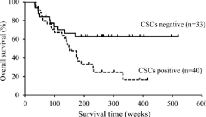
Fig.2 shows the Kaplan-Meier curves for survival in the
two groups of gastric cancer patients subdivided according to the expressions of the CSC markers. The patients in the CSC marker-positive group exhibited poorer prognoses (log- rank test, p=0.006) than those in the CSC marker-negative group. The recurrence-free survival of the CSC marker-pos- itive group was also significantly worse than that of the CSC marker-negative group (log-rank test, p=0.019) (Fig.3).
In order to identify significant risk factors for cancer-spe-
cific survival in the 73 patients with T3/4a gastric cancer Results
Figure 1. Representative images of immunohistochemical staining for CD44 and CD133. (A) CD44 was expressed prominently in the membranes of the cancer cells (x400). (B) CD133 positivity was found in the cytoplasm of the cancer cells (x400).
who underwent curative surgery, a multivariate analysis was performed (Table 3). First, a univariate analysis of the six clinicopathological factors was carried out. Based on the results of the univariate analysis, lymph node metastasis (p<0.001), lymphatic invasion (p=0.09) and CSC marker positivity (p<0.01) were included as covariates in the multi- variate regression analysis. The results revealed that second to lymph node metastasis, CSC marker positivity was a sta- tistically significant risk factor for overall survival. The CSC marker expression was found to be a statistically significant prognostic factor in patients with T3/4a gastric cancer.
Recently, cancer stem cell (CSC) theory was introduced
into the field of cancer research. Due to their tumor-initiat- ing capacity, CSCs are involved in the establishment of sec- ondary metastatic tumors in distant organs. With regard to peritoneal dissemination, Nishii et al. reported that CSC markers, such as CD44 and Sox2, are significantly overex- pressed in tumor cells constituting the bulk of peritoneal dis- semination in animal models [22]. Their results imply the existence of a correlation between stemness and the devel- opment of peritoneal dissemination; however, few clinical studies using immunohistochemistry to evaluate the signifi- cance of the CSC marker expression have been reported thus far.
In order to investigate the role of CSC markers in gastriccancer metastasis and the possibility to pick patients with worse prognosis up in clinical practice, we performed com- bined detection of two representative CSC markers in pa- tients with advanced gastric cancer, i.e., tumors invading the subserosa or penetrating the serosa (T3/4a). To minimize bias from the operative procedures, we excluded patients with tumor invasion to adjacent organs because such tumors require combined resection of the involved organs.
Various CSC markers have been reported thus far, includ- Table 1. Association of CSCs marker staining with clinicopathological features of gastric cancer
Variables
CD44 expression CD133 expression CSC marker expression
Positive Negative P-value Positive Negative P-value Positive Negative P-value
(n=29) (n=44) (n=23) (n=50) (n=40) (n=33)
Age (yr) 69.3 +/- 1.88 67.4 +/- 1.65 0.43 70.1 +/- 1.59 67.2 +/- 1.65 0.28 70.0 +/- 1.46 66.0 +/- 2.06 0.11
Sex 0.65 <0.05 0.44
Male 22 30 22 30 33 19
Female 7 14 1 20 7 14
Tumor size (mm) 61.7 +/- 4.70 62.9 +/- 5.71 0.89 72.8 +/- 7.59 57.6 +/- 4.37 0.07 66.8 +/- 5.22 57.1 +/- 5.79 0.22
Histology >0.99 0.21 0.48
Differentiated 12 18 12 18 18 12
Undifferentiated 17 26 11 32 22 21
Lymphatic invasion 0.44 0.05 0.04
ly0-1 7 15 3 19 8 14
ly2-3 22 29 20 31 32 19
Vessel invasion 0.08 0.08 <0.01
v0-1 7 20 5 22 8 14
v2-3 22 24 18 28 32 19
Lymph node metastasis 0.62 0.80 >0.99
N0-1 17 29 14 32 25 21
N2-3 12 15 9 18 15 12
Table 2. The site of first recurrence
CSCs marker expression
Positive Negative P-value
(n=24) (n=10)
Site of first recurrence 0.33
Peritoneal 12 6
Lymph nodes 6 1
Liver 3 0
Others 3 3
Discussion
ing CD44 and CD133. CD44, a transmembrane glycopro- tein, is involved in cellular adhesion and motility in response to the microenvironment [23]. Furthermore, CD44 is known to be a determinant of metastatic and invasive behavior in different malignancies, including breast [24] [25] and col- orectal [26, 27] cancers. Among patients with gastric can- cer, Ghaffazadehgan et al. reported that the CD44 expression is correlated with a poor prognosis, especially in those with intestinal type of gastric cancer [17]. In an animal model,
Takaishi et al. reported that only CD44-positive subpopula- tions of malignant gastric cell lines give rise to xenograft tumors in SCID mice and demonstrated that CD44 positivity suggests the stem cell properties of self-renewal and differ- entiation [28].
CD133 is another putative CSCs marker. This molecule is
a pentaspan transmembrane glycoprotein that has been iden- tified to be a CSC marker in pancreatic [29], ovarian [30], and liver cancers [31]. In addition, many reports have shown
Figure 3. The recurrent-free survival curves for the CSC marker- positive and -negative gastric cancer patients. The CSC marker- positive patients exhibited significantly shorter survival times than the CSC marker-negative patients according to the log-rank test (p=0.019).Figure 2. The overall survival curves for CSC marker-positive and -negative gastric cancer patients. The CSC marker-positive patients exhibited significantly shorter survival times than the CSC marker-negative patients according to the log-rank test (p=0.006).
Table 3. Univariate and multivariate analysis of CSCs marker expression and overall survival Univariate analysis Multivariate analysis
HR (95% CI) P-value HR (95% CI) P-value
Histology 0.91
Differentiated referrence
Undifferentiated 0.96 (0.47-1.95)
Lymphatic invasion 0.09 0.85
ly0-1 referrence referrence
ly2-3 1.95 (0.88-4.32) 0.92 (0.38-2.24)
Vessel invasion 0.42
v0-1 referrence
v2-3 1.35 (0.65-2.77)
Lymph node metastasis 0.001> 0.001>
N0-1 referrence referrence
N2-3 4.34 (2.12-8.88) 3.90 (1.75-8.67)
CSCs marker 0.01> 0.04
negative referrence referrence
positive 2.73 (1.29-5.73) 2.22 (1.04-4.76)
Adjuvant chemotherapy 0.60
no referrence
yes 1.20 (0.61-2.37)
that the presence of CD133-positive cells is a significant prognostic factor in patients with glioma [32] and colon can- cer [33, 34]. In gastric cancer patients, previous reports have demonstrated that CD133 is positively correlated with tumor size, the depth of tumor invasion and the presence of lymph node metastasis [19, 20]. Furthermore, multivariate analy- ses have revealed that CD133 is an independent prognostic factor [18, 19]. In an animal study, OʼBrien et al. reported that CD133-positive cells share the characteristics of CSCs, in which CD133-positive cells are able to initiate tumor growth in a minor quantity of cells when transplanted into mice [11].
Our results revealed a correlation between combined de-
tection of CSC positivity and clinicopathological features, such as lymphatic and vessel invasion. Moreover, the CSC marker expression was, second to lymph node metastasis, found to be a statistically significant prognostic factor in pa- tients with T3/4a gastric cancer. Immunohistochemical studies using resected specimens provide morphological in- formation, such as the amount and distribution of CSC marker-positive cells. Theoretically, CSCs should comprise a small population of cancer cells in specimens; however, our results demonstrated that relatively large areas of the specimens exhibited CSC marker positivity and an equal distribution of positive cells between the tumor surface and more deeply invasive areas.
Moreover, contrary to our expectations, we found a quite
different distribution of CD44 and CD133 positive cells even in the same section of the tumor. This implied the pos- sibility that several CSC clones coexist and compete within single primary tumors, as recently indicated in hematopoi- etic malignancies [35, 36]. An alternative possibility is that CD44 and/or CD133 may not represent the CSC population itself, but rather a downstream population such as progenitor cells. Wang T et al. also investigated these CSCs marker expression in gastric carcinogenesis, as characterized by the Correa pathway [37]. The expression of CD44 was found to encompass intestinal metaplasia to cancer tissue, whereas CD133 was expressed considerably in the latter. Similar re- sults were obtained by Wakamatsu et al., who investigated the expressions of ALDH, CD44 and CD133 in non-neo- plastic gastric mucosa and cancer tissue [38].
Our present results demonstrate several important points
for clinical practice. First, this method is easy to adapt in hospitals that are not specialized because only one or two specimens of the primary part of the tumor are used for the immunohistochemical analysis. Second, the combined de- tection of CSC markers is useful for selecting T3/4a gastric cancer patients with worse prognoses, and our results showed
that evaluating only two CSC markers is sufficient for such selection.
In conclusion, the expressions of CSC markers can be
used to detect gastric cancer patients with poorer prognoses, even those in the advanced stage.
Acknowledgment
This work was supported by KAKENHI (21890191).
References
1. Isobe Y, Nashimoto A, Akazawa K et al.: Gastric cancer treatment in Japan: 2008 annual report of the JGCA nationwide registry. Gastric Cancer 2011;14:301-316.
2. Ushijima T, Sasako M: Focus on gastric cancer. Cancer cell 2004;5:121- 3. Milne AN, Carneiro F, OʼMorain C et al.: Nature meets nurture: mo-125.
lecular genetics of gastric cancer. Human genetics 2009;126:615-628.
4. Suerbaum S, Michetti P: Helicobacter pylori infection. The New Eng- land journal of medicine 2002;347:1175-1186.
5. Nguyen DX, Bos PD, Massague J: Metastasis: from dissemination to organ-specific colonization. Nature reviews Cancer 2009;9:274-284.
6. Talmadge JE, Fidler IJ: AACR centennial series: the biology of cancer metastasis: historical perspective. Cancer research 2010;70:5649-5669.
7. Clarke MF, Dick JE, Dirks PB et al.: Cancer stem cells--perspectives on current status and future directions: AACR Workshop on cancer stem cells. Cancer research 2006;66:9339-9344.
8. Jones RJ, Matsui WH, Smith BD: Cancer stem cells: are we missing the target? Journal of the National Cancer Institute 2004;96:583-585.
9. Al-Hajj M, Wicha MS, Benito-Hernandez A et al.: Prospective identi- fication of tumorigenic breast cancer cells. Proc Natl Acad Sci U S A 2003;100:3983-3988.
10. Singh SK, Hawkins C, Clarke ID et al.: Identification of human brain tumour initiating cells. Nature 2004;432:396-401.
11. OʼBrien CA, Pollett A, Gallinger S et al.: A human colon cancer cell capable of initiating tumour growth in immunodeficient mice. Nature 2007;445:106-110.
12. Dalerba P, Dylla SJ, Park IK et al.: Phenotypic characterization of hu- man colorectal cancer stem cells. Proc Natl Acad Sci U S A 2007;104:10158-10163.
13. Collins AT, Berry PA, Hyde C et al.: Prospective identification of tu- morigenic prostate cancer stem cells. Cancer research 2005;65:10946- 10951.
14. Prince ME, Sivanandan R, Kaczorowski A et al.: Identification of a subpopulation of cells with cancer stem cell properties in head and neck squamous cell carcinoma. Proc Natl Acad Sci U S A 2007;104:973- 15. Ma S, Chan KW, Hu L 978. et al.: Identification and characterization of
tumorigenic liver cancer stem/progenitor cells. Gastroenterology 2007;132:2542-2556.
16. Baccelli I, Trumpp A: The evolving concept of cancer and metastasis stem cells. The Journal of cell biology 2012;198:281-293.
17. Ghaffarzadehgan K, Jafarzadeh M, Raziee HR et al.: Expression of cell adhesion molecule CD44 in gastric adenocarcinoma and its prog- nostic importance. World journal of gastroenterology : WJG 2008;14:6376- 6381.
18. Ishigami S, Ueno S, Arigami T et al.: Prognostic impact of CD133 ex- pression in gastric carcinoma. Anticancer research 2010;30:2453-2457.
30. Ferrandina G, Martinelli E, Petrillo M et al.: CD133 antigen expres- sion in ovarian cancer. BMC cancer 2009;9:221.
31. Song W, Li H, Tao K et al.: Expression and clinical significance of the stem cell marker CD133 in hepatocellular carcinoma. International journal of clinical practice 2008;62:1212-1218.
32. Zeppernick F, Ahmadi R, Campos B et al.: Stem cell marker CD133 affects clinical outcome in glioma patients. Clinical cancer research : an official journal of the American Association for Cancer Research 2008;14:123-129.
33. Horst D, Kriegl L, Engel J et al.: CD133 expression is an independent prognostic marker for low survival in colorectal cancer. British jour- nal of cancer 2008;99:1285-1289.
34. Kojima M, Ishii G, Atsumi N et al.: Immunohistochemical detection of CD133 expression in colorectal cancer: a clinicopathological study.
Cancer science 2008;99:1578-1583.
35. Anderson K, Lutz C, van Delft FW et al.: Genetic variegation of clon- al architecture and propagating cells in leukaemia. Nature 2011;469:356- 36. Notta F, Mullighan CG, Wang JC et al.: Evolution of human BCR-361.
ABL1 lymphoblastic leukaemia-initiating cells. Nature 2011;469:362- 37. Wang T, Ong CW, Shi J et al.: Sequential expression of putative stem 367.
cell markers in gastric carcinogenesis. British journal of cancer 2011;105:658-665.
38. Wakamatsu Y, Sakamoto N, Oo HZ et al.: Expression of cancer stem cell markers ALDH1, CD44 and CD133 in primary tumor and lymph node metastasis of gastric cancer. Pathology international 2012;62:112- 119.
19. Yu JW, Zhang P, Wu JG et al.: Expressions and clinical significances of CD133 protein and CD133 mRNA in primary lesion of gastric ade- nocacinoma. Journal of experimental & clinical cancer research : CR 2010;29:141.
20. Zhao P, Li Y, Lu Y: Aberrant expression of CD133 protein correlates with Ki-67 expression and is a prognostic marker in gastric adenocar- cinoma. BMC cancer 2010;10:218.
21. Association JGC: Japanese classification of gastric carcinoma: 3rd English edition. Gastric Cancer 2011;14:101-112.
22. Nishii T, Yashiro M, Shinto O et al.: Cancer stem cell-like SP cells have a high adhesion ability to the peritoneum in gastric carcinoma.
Cancer science 2009;100:1397-1402.
23. Jang BI, Li Y, Graham DY et al.: The Role of CD44 in the Pathogen- esis, Diagnosis, and Therapy of Gastric Cancer. Gut and liver 2011;5:397-405.
24. Joensuu H, Klemi PJ, Toikkanen S et al.: Glycoprotein CD44 expres- sion and its association with survival in breast cancer. The American journal of pathology 1993;143:867-874.
25. Friedrichs K, Kugler G, Franke F et al.: CD44 isoforms in prognosis of breast cancer. Lancet 1995;345:1237.
26. Mulder JW, Kruyt PM, Sewnath M et al.: Colorectal cancer prognosis and expression of exon-v6-containing CD44 proteins. Lancet 1994;344:1470-1472.
27. Wielenga VJ, Heider KH, Offerhaus GJ et al.: Expression of CD44 variant proteins in human colorectal cancer is related to tumor pro- gression. Cancer research 1993;53:4754-4756.
28. Takaishi S, Okumura T, Tu S et al.: Identification of gastric cancer stem cells using the cell surface marker CD44. Stem cells 2009;27:1006- 1020.
29. Maeda S, Shinchi H, Kurahara H et al.: CD133 expression is correlat- ed with lymph node metastasis and vascular endothelial growth fac- tor-C expression in pancreatic cancer. British journal of cancer 2008;98:1389-1397.