著者
仲田 真理子
内容記述
この博士論文は内容の要約のみの公開(または一部
非公開)になっています
year
2016
その他のタイトル
オスマウスの社会行動制御におけ るエストロゲン
受容体βの役割
学位授与大学
筑波大学 (University of Tsukuba)
学位授与年度
2016
報告番号
12102甲第7901号
URL
http://hdl.handle.net/2241/00145483
Roles of estrogen receptor β in the regulation of
social behaviors in male mice
(オスマウスの社会行動制御におけるエストロゲン受容体β の役割)
筑波大学 人間総合科学研究科 感性認知脳科学専攻 Kansei, Behavioral & Brain Sciences
Graduate School of Comprehensive Human Sciences at the University of Tsukuba
仲田 真理子
Chapter 1: General Introduction ... 1
1.1. Introductory remarks ...2
1.2. Social behaviors in male rodents ...3
1.2.1. Assessment of social information processing of male rodents ... 3
1.2.2. Assessment of male sexual behavior ... 5
1.2.3. Assessment of social behavior between male mice ... 6
1.2.3.1. Aggressive behavior ... 6
1.2.3.2. Establishment of dominance hierarchy ... 6
1.3. Testosterone action and estrogen receptors ...7
1.3.1. Testosterone and its metabolites ... 7
1.3.2. Subtypes of estrogen receptors ... 8
1.3.3. Organizational and activational actions of testosterone ... 8
1.4. Regulation of male social behaviors by testosterone...10
1.4.1.Regulation of male social behaviors by activational action of testosterone ... 10
1.4.2.Regulation of male social behaviors by organizational action of testosterone ... 11
1.5. Neural network for male social behaviors ...12
1.5.1.Neural network for social information processing ... 12
1.5.2.Neural network for sexual behavior ... 13
1.5.3.Neural network for aggressive behavior ... 14
1.6. Regulation of male social behavior by estrogenic signaling in the brain ...14
1.6.1. Distribution of estrogen receptors in the neural network for male social behaviors ... 14
1.6.2.Regulation of male social behaviors by testosterone via estrogen receptors ... 15
1.6.3.Site-specific regulation of male social behaviors by estrogenic signaling ... 16
1.7. Possible regulation of male social behavior by ERβ ...17
1.8. Site-specific knockdown of ERs with RNA interference (RNAi) ...19
1.8.1. Development and mechanisms of RNAi methods ... 19
2.1. Experimental animals ...25
2.1.1. Mice ... 25
2.1.2. Tail DNA extraction and PCR genotyping of βERKO mice ... 25
2.2. Estrogen receptor β silencing using small hairpin RNA ...26
2.3. Behavioral tests ...27
2.3.1. Sexual behavior test ... 27
2.3.2. Aggressive behavior test ... 28
2.3.3. Sexual preference tests ... 28
2.3.3.1. Olfactory sexual preference test ... 29
2.3.3.2. Sexual preference test with freely moving two females (2F Sex test) ... 30
2.3.4. Social investigation test ... 31
2.3.5. Agonistic behavior test ... 32
2.3.6. Tube test... 33
2.3.6.1. Training... 33
2.3.6.2. Testing ... 33
2.3.7. Quantitative analysis of behavioral data ... 34
2.4. Histological analysis ...35
2.4.1. Preparation of brain tissues for immunohistochemistry ... 35
2.4.2. Immunohistochemistry ... 36
2.4.3. Analysis of immunopositive cells ... 36
Chapter 3: Experiment 1- Effects of Pre-Pubertal ERβ Knockdown in
the MPOA and MeA ... 38
3.1. Introduction ...39
3.2. Methods ...40
3.3. Results ...41
3.3.1 Effects of pre-pubertal ERβ knockdown in the MPOA ... 41
3.3.2. Effects of pre-pubertal ERβ knockdown in the MeA ... 43
3.3.3. Examination of bilateral hit of AAV vector within targeted brain site ... 45
3.2. Methods ...54
3.3. Results ...54
3.4. Discussion ...58
Chapter 5: Effects of Adult ERβ Knockdown in the MeA ... 60
5.1. Introduction ...61
5.2. Experiment 3: Effects of adult ERβ knockdown in the MeA on male-type sexual preference, sexual and aggressive behavior ...63
5.2.1. Methods ... 63
5.2.2. Results ... 64
5.2.3. Conclusions ...71
5.3. Experiment 4: Effects of adult ERβ knockdown in the MeA on sexual preference test with freely moving two females (2F Sex test) ...72
5.3.1. Methods ... 72
5.3.2. Results ... 72
5.3.3. Conclusions ... 75
5.4. Discussion ...76
Chapter 6: Experiment 5- Influence of Systemic Deletion of ERβ Gene
on Repeated Social Interaction ... 79
Chapter 7: General Discussion ... 80
7.1. Site-specific regulation of male social behavioral mediated by ERβ ...82
7.1.1. MPOA... 84
7.1.2. MeA ... 87
7.2. Contribution of ERβ to the establishment of inter-male social relationship ...89
7.3. Future directions ...92
7.3.1. Identification of type of ERβ-expressing neurons ... 92
7.3.2.Investigation of role of pubertal ERβ in social information processing in the MeA and MPOA ... 93
7.3.3.Investigation of precise role of ERβ in establishment of social relationship ... 93
-Chapter 1-
1. General Introduction
1.1. Introductory remarks
Social interaction is an essential component of our life. Interaction with other individuals is profitable for survival. For instance, living in a group is advantageous for efficient foraging and detection of predators. Moreover, interaction with an individual of same species is crucial for reproductive success. Adult animals mate with opposite-sex conspecifics, and take care of their pups. Defense of the territory, choice of an appropriate partner, and appropriate behavior toward the partner and pups are necessary for successful reproduction. Furthermore, individuals of social species including rodents, monkeys, and human, establish social relationship with conspecifics through repeated episodes of social interaction. Choosing suitable behavioral repertoire to each situation and performance of these behaviors in appropriate way are necessary to establish social relationship. Living within the social relationship is essential for survival in social animals.
I focused on social behaviors in male mice in this study. Previous studies using rodents including mice, rats, and hamsters elucidated essential roles of testosterone, one of gonadal steroid hormones, in the regulation of male social behaviors. However, underlying neural mechanism of behavioral regulation by testosterone is not completely understood. Notably, little is known about the roles of estrogen receptor β (ERβ), a subtype of estrogen receptor and one of the major mediators of testosterone action. Although several lines of evidence suggested importance of ERβ in “fine-tuning” of components of male social behaviors including aggressive behavior, social reactivity, and social information processing (Reviewed in Weiser et al., 2008; Handa et al., 2012), precise role and relative importance of ERβ in the regulation of social interaction and its neural mechanism are not well understood. In this study, I focused to investigate (1)
site-specific role of ERβ in the regulation of essential components of male social behaviors, i.e. social information processing, sexual and aggressive behaviors. In addition to these “behavioral components”, I intended to examine (2) whether ERβ is necessary for establishment of social relationship in male mice.
1.2. Social behaviors in male rodents
Typically, male mice behave differently toward same- and opposite-sex conspecifics. When a male rodent encounters another male, it intensively sniffs body, face and anogenital region of the opponent. In a laboratory setting such as a resident-intruder paradigm (see 1.2.3.), aggressive behavior is often observed following to social investigation. After repeated and/or a long-term social interaction, male rodents often establish dominance hierarchy (Ginsburg and Allee, 1942). On the other hand, males show sexual behavior toward a female. In mice, females spontaneously ovulate every fourth or fifth day. Behavioral estrus, in which females show sexual receptivity, lasts about 24 hours during an estrous cycle (Tomihara, 2010). Receptive posture of a female is critical for completion of male sexual behavior even though males are able to mount to a receptive female (McGill, 1962). Thus, males prefer a receptive female over a non-receptive female when two females are presented simultaneously (Kondo and Sachs, 2002).
To respond properly to each of different types of opponents, males mainly use olfactory information. They judge sex, age, and reproductive status of an opponent and whether the opponent is familiar one or not. Auditory information is also used for social interaction. In rats and mice, ultrasonic vocalization is utilized during copulatory interaction, juvenile play behavior, and nursing (Portfors, 2007).
1.2.1. Assessment of social information processing of male rodents
Olfactory information from an opponent is necessary for performance of social behaviors in rodent species (Rowe and Edwards, 1971, 1972). Olfactory system in rodents consists of main and accessory olfactory systems. Traditionally, the main olfactory system (MOS) has been implicated in detection of volatile odorant molecules and the accessory olfactory system (AOS) has been implicated in pheromonal communication mediated by non-volatile chemicals. Although previous studies suggested that this classification is not definite (Tucker, 1963; Meredith, 1998), relative importance of AOS for male sexual and aggressive behavior is well established (e.g. Clancy et al., 1984).
These two olfactory systems are known to converge in some brain regions including medial amygdala (MeA) (Meredith, 1998). To investigate underlying mechanism of social information processing in these brain regions, several behavioral testing paradigms have been developed. Among them, in sexual preference tests, subject animals are allowed to investigate odors of urine, or soiled bedding from two types of stimulus animal and preferential investigation toward one of these stimuli is assessed. Sexually active males preferentially investigate the odor from receptive females compared to that from non-receptive females or males. Moreover, they show preference to a gonadectomized male over an intact male rat (Xiao et al., 2004). In sexual preference tests, abilities to discriminate two stimuli and respond to intrinsically attractive stimuli (i.e. receptive females or gonadectomized males) are also assessed. In addition, total investigation duration can be used as an index of social interest to the stimuli.
Furthermore, rodents can discriminate and memorize other conspecific individuals using olfactory information. Information whether an opponent is familiar or novel is necessary for territory defense, partner choice, and parental care. Not only recognition of a same-sex individual, but also that of an opposite-sex individual plays an important role
for successful reproduction. For instance, rats and mice prefer a novel female odor than a familiar female odor (Carr et al., 1980). Moreover, it is reported that, after exposure to a novel female odor, male mice show increased levels of risk-taking behaviors (Kavaliers et al., 2008).
Ability of social recognition and social memory has been assessed using a habituation-dishabituation paradigm (Ferguson et al., 2002). In this paradigm, subject animal is exposed to a same stimulus animal (stimulus A) repeatedly with a fixed inter-trial interval. Decreases of investigation along a repeated exposure (habituation) indicate that the subject animal is able to keep the memory of the stimulus animal A. Restoration of investigation duration upon an exposure to a novel stimulus animal (stimulus B) (dishabituation) indicates that the subject animal is able to discriminate the stimulus A and B. These social information processing is crucial for subsequent social behaviors and establishment of social relationship.
1.2.2. Assessment of male sexual behavior
Male mice show sexual behaviors when they encounter a female mouse. At first, precopulatory behaviors including sniffing of facial and anogenital region and emission of 50kHz ultrasonic vocalizations are observed. Then, the male mouse shows stereotypical copulatory behaviors such as mount, intromission, and ejaculation (McGill, 1962; Hull and Dominguez, 2007). If the female mouse is sexually receptive, she shows receptive posture called “lordosis”. Lordosis posture is helpful for males to successfully ejaculate although males occasionally show ejaculation to non-receptive female (McGill, 1962).
After completion of the ejaculation, male mice rarely copulate again for 24h, unlike male rats that show ejaculation several time in a single testing day. In addition, mounting
behavior toward another male is sometimes observed as a part of dominance behavior in mice and rats (Wang et al., 2011).
1.2.3. Assessment of social behavior between male mice
1.2.3.1. Aggressive behavior
In the article by Nelson and Trainor (2007), aggression is defined as “overt behavior that has the intention of inflicting physical damage on another individual, and the potential for aggressive behavior exists whenever the interests of two or more individuals conflict.” To assess aggressive behavior in male mice, individual housing and/or co-habitation with female conspecifics are general procedure to potentiate aggression toward other males (Siegfried et al., 1981). Experimental paradigm called “resident-intruder paradigm” has been widely used. A stimulus male mouse (intruder) is introduced into a home cage of subject male mouse (resident). Aggressive behaviors by the resident are then observed and recorded. To minimize the levels of fight-back by intruder mice, they are often group-housed and/or olfactory bulbectomized. Behavioral acts such as chasing, boxing, wrestling, tail rattling, biting, and offensive lateral attack are defined as main components of aggressive behavior. Aggressive behavior can also be assessed in neutral cages. In this method, experimental mice are placed in each sides of a divider placed in a neutral cage and allowed to habituate for a few minutes. The divider is then removed and aggressive behavior between two mice is observed.
1.2.3.2. Establishment of dominance hierarchy
Male mice establish hierarchical social relationship through social interaction. Wang et al. (2011) tested social behavior of group-housed (four mice) male C57BL/6J mice and reported that a linear hierarchy was observed in 89% of the cases although a non-linear
hierarchy was occasionally observed, i.e., if mouse A was dominant over B, and B was dominant over C, then A was dominant over C. In this study, it is also reported that the hierarchy was not very stable, since the rank of each mouse often (about 41%) changed between days during 7 days testing period.
Establishment of a dominance hierarchy is not necessarily accompanied with intensive aggressive behavior. The term of “agonistic behavior” includes all interactive behavior among conspecific animals such as sniffing, grooming, and submissive behavior in addition to aggressive behavior (Scott, 1966) and is often used in analysis of hierarchical social relationship. Social dominance among multiple animals can be assessed using various testing paradigms. In some cases, agonistic behavior with direct physical contact observed during behavioral tests is used for an assessment of social dominance. In other cases, social dominance is assessed by a comparison of territorial or courtship behaviors between males. Furthermore, in the tube test developed by Lindzey et al. (1966), mice are forced to compete for occupation of a narrow tube. Social rank in the tube test is reported to be consistent with the rank measured using other test paradigms (Wang et al., 2011).
1.3. Testosterone action and estrogen receptors
1.3.1. Testosterone and its metabolites
Testosterone is one of gonadal steroid hormones classified as androgen and plays an essential role in the regulation of a series of male social behaviors. In males, testosterone is synthesized from cholesterol mainly in Leydig cells of testes. Gonadotropin releasing hormone (GnRH) induces secretion of follicle-stimulating hormone (FSH) and luteinizing hormone (LH) from the anterior pituitary. Thereafter, FSH and LH induce secretion of testosterone from the testes. Testosterone acts on androgen receptors (AR) as
its original form or as dihydrotestosterone (DHT) after the conversion by 5α-reductase. Moreover, testosterone is converted into estradiol by aromatase, a metabolic enzyme belonging to Cytochrome P450 superfamily (Simpson et al., 1994). After aromatization to estradiol, testosterone regulates organization and activation of male-type neural circuitry via estrogen receptors (ER).
1.3.2. Subtypes of estrogen receptors
Two subtypes of ERs, estrogen receptor α (ERα) and estrogen receptor β (ERβ), are well known as key mediators of behavioral regulation by testosterone. They are members of a nuclear receptor subfamily, and are ligand-dependent transcription factors (Fawell et al., 1990; Tremblay et al., 1997). After ligand binding, ERs are transported to cell nuclei and bind to target sites of DNA (Kumar and McEwan, 2012). ERα and ERβ are known to regulate a wide variety of target genes including progesterone receptor, oxytocin receptor (Young et al., 1998; Lindberg et al., 2003). Moreover, rapid, non-genomic action of estradiol via ERα, ERβ and G-protein coupled ER has been focused on in recent studies (Björnström and Sjöberg, 2005).
1.3.3. Organizational and activational actions of testosterone
Two types of actions of testosterone are well documented. One is called “organizational action” and the other is called “activational action”. The organizational action is permanent and is involved in formation and development of neural networks. It occurs during the critical period in lifetime, such as the perinatal and pubertal period.
Figure 1 illustrates changes of circulating testosterone levels in lifetime of male mice. From the embryonic day 18 to the neonatal period, there is a drastic increase of circulating testosterone levels, which is called “androgen surge” (blue arrow in Figure 1). During this
period, the neural network controlling male social behaviors is masculinized and defeminized. Although both AR and ERs are implicated in this process, a pivotal role of estrogenic signaling has been demonstrated (Arnold and Breedlove, 1985). During the androgen surge, aromatized testosterone enables activation of ERs only in male (MacLusky and Naftolin, 1981) since ovaries are not active and do not secrete estradiol. Furthermore, in the pre-natal period, alpha-fetoprotein binds to estradiol originated from a mother and prevents masculinization and defeminization of female brains (Bakker et al., 2006).
At the onset of puberty, testosterone levels start to increases again and reach the adult level by the end of the pubertal period (red arrow in Figure 1). Recent studies demonstrated that testosterone during the pubertal period is also necessary for full masculinization of the central nervous system (Romeo, 2003; Sisk 2015). Male hamsters with depletion of pubertal testosterone by pre-pubertal castration failed to show restoration of aggressive behavior in response to testosterone implant in adult (Schulz et al., 2004).
Figure 1. Illustration of the change of circulating testosterone level in lifetime of male mice. Blue arrow indicates perinatal period. Red arrow indicates pubertal period.
In contrast, the activational action is transient and occurs throughout life. Testosterone is able to fully regulate male social behavior by acting on the neural circuitry masculinized/defeminized by its organizational action. Testosterone induces behavioral and/or physiological changes through genomic and/or non-genomic action. Genomic action occurs through DNA binding of dimerized receptors and occurs after a time-lag of hours or days, is well documented as classical mechanisms. On the other hand, rapid non-genomic action, which occurs in seconds or minutes without direct binding to DNA, is also noted in recent years (Björnström and Sjöberg, 2005).
1.4. Regulation of male social behaviors by testosterone
1.4.1. Regulation of male social behaviors by activational action of testosterone
It is well established that testosterone is necessary for expression of male-type social behaviors. Gonadectomy disrupted sexual behavior of male rats and simultaneous implant of a silastic capsule filled with testosterone or estradiol but not DHT, non-aromatized androgen, can reverse the effect of gonadectomy (Meisel et al., 1984). Estrogen replacement is also effective to restoration of sexual behavior in male mice (Edwards and Burge, 1971). Thus, estrogenic signaling is important in the performance of male sexual behavior. For aggressive behavior, estrogenic signaling is also necessary (Ogawa et al., 1997) although both signaling via estrogen and androgen receptors are necessary for full expression of aggressive behavior (Nelson and Trainor, 2007). Deletion of testosterone by gonadectomy disrupts not only aggressive behavior, but also dominance hierarchy between male mice. Albert et al. (1986) reported that a gonadectomized dominant male rat without testosterone replacement lost his dominance over subordinate males. Relationship between male social dominance and testosterone level was reported in
previous studies with human (Mazur and Booth, 1998) and rhesus monkeys (Rose et al., 1971). In male mice, Zielinski and Vandenbergh (1993) revealed that treatment with physiological doses of testosterone successfully restored dominance status of males over subordinate mice treated with lower doses of testosterone.
Activational action of testosterone is also necessary for social information processing in male rodents. Xiao et al. (2004) reported that gonadectomy of adult male rats disrupted their male-type sexual preference.
1.4.2. Regulation of male social behaviors by organizational action of testosterone
The organizational action of testosterone is also necessary for expression of male social behaviors. As described in 1.3.3., estrogenic signaling in perinatal period contributes to masculinization of the nervous system. Neonatal treatment of female rats with estradiol enabled them to express male-type sexual behavior in response to testosterone injection in adulthood (Christensen and Gorski, 1978). Severe deficits of male-type sexual and aggressive behaviors and sexual preference were reported in male aromatase knockout (AromKO) mice, which cannot synthesize estradiol. Neonatal treatment with estradiol to AromKO male mice restored male sexual and aggressive behaviors in adulthood (Toda et al., 2001a, b; Harada et al., 2009). These findings demonstrate relative importance of estrogenic signaling in the perinatal period.
Importance of pubertal testosterone in the organization of the neural network for male-type social behaviors is also documented in male rodents (Romeo, 2003; Sisk and Foster, 2004; Sisk, 2015). Depletion of testosterone during puberty by gonadectomy at postnatal day (PND) 21 disrupted adult male sexual and aggressive behaviors even with testosterone complement after the end of puberty (Schulz et al., 2004). However, precise underlying mechanisms of pubertal organizational action of testosterone is not well
understood. Contribution of ERα in pubertal formation and/or development of neural network for male sexual and aggressive behavior is demonstrated recently (Sano et al., 2016) and described below (see 1.6.3.).
1.5. Neural network for male social behaviors
1.5.1 Neural network for social information processing
Processing of social information of other individual is essential to choose appropriate social behavior. As described above, there are dual olfactory systems. In the MOS, main olfactory epithelium sends olfactory information to the main olfactory bulb. On the other hand, vomelonasal organ is a receptive organ of AOS and sends information to the accessory olfactory bulb. Both main and accessory olfactory bulbs project to the MeA (Baum, 2009). The MeA has been considered to integrate olfactory information. From the MeA, the information is sent to other brain sites in the hypothalamic and limbic areas regulating male social behaviors, either directly or via the bed nucleus of the stria terminalis (BNST) (Ferguson et al., 2002). Lesions of the MeA disrupt male-type sexual preference (Kondo and Sachs, 2002). Recently, Dhungel et al. (2011) also reported disruption of sexual preference by MeA lesions. They proposed that the MeA might be important for preference exhibited by male rats toward a receptive female rat over a non-receptive female rat, but not over a gonadally intact male rat. The medial preoptic area (MPOA), a hypothalamic nucleus responsible for the performance male sexual behavior, is also implicated in male-type sexual preference. Unlike the MeA, lesions of the MPOA suppressed preference of male rats toward receptive female rats over not only a non-receptive female rat but also a gonadally intact male rat (Dhungel et al., 2011). These findings suggest that these two brain areas may be involved differently in the regulation of male-type sexual preference. The MPOA is implicated in the control of sexual
motivation (Hull et al., 1995) whereas the MeA plays an important role in processing of odor information of other individuals. It should be noted that the MeA processes not only information relevant to sexual preference but also individual discrimination. For instance, it is reported that oxytocin in the MeA is necessary for social recognition (Ferguson et al., 2001) and ERα expressed in the MeA may be involved in the control of social recognition by regulating the levels of oxytocin receptors (Choleris et al., 2003).
1.5.2. Neural network for sexual behavior
Several lines of evidence indicate the involvement of the MPOA, MeA and BNST in the regulation of male sexual behavior. Among those, the MPOA is considered to play the most critical role. Lesions of the MPOA greatly reduced male sexual behavior in rats and mice (Paredes, 2003; Hull and Rodoriguez-Manzo, 2009). An increased number of Fos immunoreactive cells were observed in the MPOA in male rats after sexual behavior (Veening et al., 2005). The MPOA receives innervations from the MeA and BNST, which are supposed to mediate information of sexually receptive female. In the MeA and BNST, increased Fos immunoreactivity was also observed after sexual behavior (Veening et al., 2005; Hull and Rodoriguez-Manzo, 2009). Ventromedial nucleus of hypothalamus (VMN) is also indicated in male sexual behavior. Involvement of ERα positive neurons in the VMN in sexual behavior is revealed by recent studies (Sano et al., 2013; Lee et al., 2014).
1.5.3. Neural network for aggressive behavior
The MeA, BNST, lateral septum (LS), anterior hypothalamic area (AHA) and VMN are implicated in the regulation of aggressive behavior. Neuronal activation indicated by an increase of Fos immunoreactivity after aggressive encounter was reported in these brain sites (Veening et al., 2005). Social odor information is integrated in the MeA and sent to the BNST, LS, and AHA. These brain sites send innervation to the periaqueductal gray (PAG), which is responsible for execution of cooperated body movement for aggressive behavior (Nelson and Trainor, 2007). Recently, an essential role of the VMN in aggressive behavior was revealed using optogenetics (Lin et al., 2011). Similar to the findings in sexual behavior, ERα positive neurons in the VMN may play a significant role in the regulation of aggressive behavior in male mice (Sano et al., 2013; Lee et al., 2014). On the other hand, involvement of the MPOA in the regulation of male aggressive behavior is still controversial. Newman (1999) proposed a relatively minor role of the MPOA in aggressive behavior since Fos expression was unaffected after aggressive encounter in male Syrian hamsters (Kollack-Walker and Newman, 1995). However, other studies using male rats and mice indicated that the MPOA might also be involved in the regulation of male aggressive behavior (Patil and Brid, 2010; Wu et al., 2014).
1.6. Regulation of male social behavior by estrogenic signaling in the brain
1.6.1. Distribution of estrogen receptors in the neural network for male social behaviors
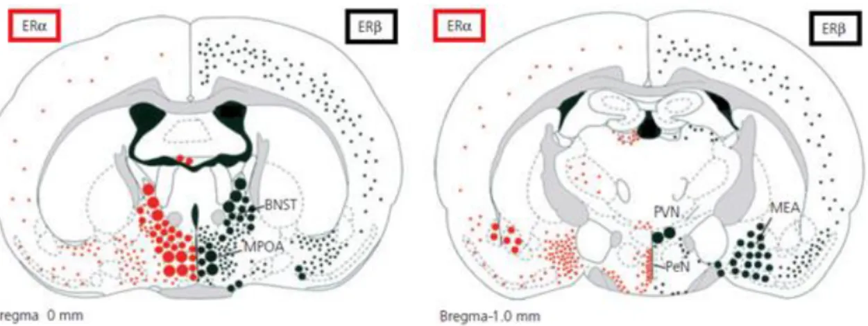
Both ERα and ERβ are widely expressed in the brain sites of the neural network for male social behaviors. In the hypothalamic and limbic areas, distribution of ERα and ERβ is often overlapped, but in some of areas, either ERα or ERβ is predominantly expressed. Figure 2 shows representative expression sites of ERα and ERβ in the mouse brain (modified from Mitra et al., 2003 and Handa et al., 2012). Among expression sites, the
MPOA, BNST (left panel) and the MeA (right panel) express both ERα and ERβ abundantly. In the MeA, co-localization of ERα protein and ERβ mRNA is reported (Shughrue et al., 1998). On the other hand, ERβ but not ERα is expressed in the PVN (right panel). Moreover, in the VMN, ERα but not ERβ is abundantly expressed (Shughrue et al., 1997; Mitra et al., 2003; Merchenthaler et al., 2004).
Figure 2. Differential distribution of ERα and ERβ in the mouse brain. Two coronal planes through the brain (left panel: at bregma, right panel: at -1mm to bregma) show the anatomical distribution of ERα (left, red dots) and ERβ (right, black dots). BNST, bed nucleus of the stria terminalis; MPOA, medial preoptic area; MEA, medial amygdala; PVN, paraventricular nucleus of the hypothalamus; PeN, periventricular nucleus. Gray shading shows white matter tracts (modified from Mitra et al., 2003 and Handa et al., 2012)
1.6.2. Regulation of male social behaviors by testosterone via estrogen receptors
The exact receptor type(s) and their expression site(s) those mediate activational and organizational actions by testosterone for the regulation of male social behavior are still not completely understood. Studies using knockout mice indicate that ERα and ERβ may play different roles. ERα is necessary for performance of male social behaviors since
αERKO male mice showed severe deficits in sexual and aggressive behaviors (Ogawa et al., 1997, 1998, 2000). On the other hand, the role played by ERβ in the regulation of male social behaviors is still unclear. Survival of sexual behavior and partially increased aggressive behavior in βERKO male mice suggest that ERβ may play a role in fine-tuning rather than induction of male social behavior (Ogawa et al., 1999; Nomura et al., 2002). Moreover, both ERα and ERβ are implicated in social information processing, but responsible brain site(s) may be different (Imwalle et al., 2002; Choleris et al., 2003; Kavaliers et al., 2004, 2008).
1.6.3 Site-specific regulation of male social behaviors by estrogenic signaling
It is considered that each brain site in the neural circuitry for male social behavior may be differently involved in the regulation of behaviors (Newman, 1999). Abundant but somewhat differential expression of ERs and ARs in this neural network (Simerly et al., 1990; Shughrue et al., 1997; Mitra et al., 2003; Merchenthaler et al., 2004) indicated that male social behaviors are site-specifically regulated by testosterone in these brain regions via ERs and/or ARs. Among these brain areas, the MPOA and MeA, in which both ERα and ERβ are abundantly expressed, have been focused as regulatory sites of male social behaviors via ERs. Local testosterone implants into the MPOA or MeA restored sexual behavior of castrated male hamsters (Wood and Newman, 1995). Several lines of evidence demonstrated an importance of estrogenic signaling. For instance, Wood (1996) reported that local administration of estradiol but not DHT in the MeA could restore male sexual behavior after in castrated hamsters. Similarly, in the MPOA, local administration of estradiol was more effective than DHT in restoring sexual behavior in castrated male rats (Hull and Rodoriguez-Manzo, 2009).
Effects of site-specific knockdown of ERα (αERKD) in the MPOA, MeA, or VMN on male sexual and aggressive behavior were examined in adult male mice. As a result, αERKD in the MPOA decreased sexual behavior without affecting aggressive behavior, whereas αERKD in the MeA affected neither sexual nor aggressive behavior. On the other hand, αERKD in the VMN reduced both of sexual and aggressive behavior. Recently, Lee et al. (2014) also provided evidence of importance of ERα expressing neurons in the VMN. They reported that activation of ERα positive neuron in the VMN induced social investigation toward the opponent, sexual behavior, and aggressive behavior in scalable manner.
Sano et al. (2016) revealed that ERα might also be site-specifically involved in the formation and/or development of neural networks for male social behaviors in pubertal period. Site-specific αERKD at postnatal day (PND) 21 in the MeA, which continuously suppressed ERα expression from pubertal period to adult, reduced sexual and aggressive behaviors in adulthood. Considering the finding of negative effects of αERKD in the MeA only in adulthood discussed above (Sano et al., 2013), these findings indicate that ERα in the MeA may be necessary for the pubertal organization of neural network for male sexual and aggressive behaviors.
1.7. Possible regulation of male social behavior by ERβ
Compared with ERα, the precise role of ERβ in the regulation of male social behavior still remains unclear. Behavioral alteration by βERKO in male mice has been investigated in previous studies. Ogawa et al. (1999) reported survival of sexual behavior and partially increased aggressive behavior in βERKO male mice. Adult male βERKO mice showed longer duration of and shorter latency to aggressive behavior than wild-type (WT) mice in the first test of three repeated aggressive behavior tests. Increased levels of aggressive
behavior in pubertal and adolescent periods in βERKO males (Nomura et al., 2002) also suggested an inhibitory role of ERβ in the regulation of male aggressive behavior. Moreover, it is hypothesized that ERβ modulates aggressive behavior induced by activation of ERα at an adequate level. Increased level of aggressive behavior induced by estrogen treatment in gonadectomized βERKO suggested potentiation of estrogen-inducible aggression by disruption of ERβ gene (Nomura et al., 2006). Not only the performance of typical aggressive behavior, but also the reaction to social stimuli may be modulated by ERβ. In the situation of encounter to another mouse without direct physical contact, hyper-reactivity has been reported in both male (Handa et al., 2012) and female (Tsuda et al., 2014) βERKO mice.
Moreover, ERβ is implicated in social information processing. In social recognition test using habituation-dishabituation paradigm (see 1.2.1 for details of the paradigm), βERKO female (Choleris et al., 2003) but not male (Sánchez-Andrade and Kendrick, 2011) mice showed disrupted social recognition of same-sex stimulus animals. Although ERβ may play a minor role in social recognition of same-sex conspecifics, ERβ might have a role in recognition of opposite-sex individual in male mice. Kavaliers et al., (2008) revealed that risk-taking behavior was altered in WT, but not in βERKO, male mice by the degree of familiarity of female exposed before the test. Thus, βERKO males possibly are unable to distinguish familiar and novel females.
Additionally, it is known that ERβ possibly mediates anxiolytic effect of estradiol in rodents. Selective ERβ agonist reduced anxiety-related behavior in contrast to anxiogenic effects of ERα agonist in gonadectomized female rats (Lund et al., 2005). Similar behavioral effects of ERβ agonist treatment were reported in gonadectomized males in social and non-social situation (Weiser et al., 2008). It is hypothesized that ERβ may be involved in the maintenance of adequate expression levels of male social behaviors,
which is necessary for animal’s survival, not only by direct regulation of stereotypical social behaviors but also by the regulation of emotional aspect.
Previous studies also have suggested ERβ-mediated organizational action. Female-type sexual behavior in hormonally treated βERKO male mice suggests that ERβ may be involved in defeminization of male brains (Kudwa et al., 2005). On the other hand, combined with increased levels of aggression, higher testosterone levels reported in βERKO male at 5 weeks of age (Nomura et al., 2002) suggest that ERβ may play a role in the regulation of puberty onset. Although these studies have not identified the exact period(s) of the organizational action via ERβ, it is possible that ERβ is involved in the formation and development of male-type neural network in neonatal and/or pubertal period.
In a previous study investigating possible neonatal organizational action via ERβ, it is reported that neonatal treatment of male rats with selective ERβ agonist increased aggressive behavior in adulthood (Patisaul and Bateman, 2008). To elucidate complicated role of ERβ in the regulation of male social behaviors, it is necessary to further investigate its organizational and activational action in different stages in lifetime.
1.8. Site-specific knockdown of ERs with RNA interference (RNAi)
1.8.1. Development and mechanisms of RNAi methods
Invention and development of RNAi method enabled us to suppress the expression of a targeted gene. This technique originated from the finding that hairpin-shape short RNA interfered gene expression (Lee et al., 1993). Subsequently, Fire et al. (1998) succeeded to inhibit gene expression by introduction of double-strand RNA into cell. These findings and subsequent development of RNAi methods enabled site- or cell type- specific knockdown of a targeted gene by introduction of small double-strand RNA.
Small hairpin RNAs (shRNA) incorporated to adeno-associated virus (AAV) or lenti virus in plasmid vector are often used for introduction of the RNA. After introduction into the cell, double-strand RNA is converted to siRNA which is single-strand RNA with about 21 base long. Conversion to siRNA enabled introduced RNA to inhibit expression of a targeted RNA (Ghildiyal and Zamore, 2009; Kim et al., 2009; Siomi, H. and Siomi, M.C., 2009). Double-strand RNA is processed by dicer and Argonaute proteins. After the processing, siRNA and Argonaute protein form RISC complex. Targeted RNA with RISC binding is cut by slicer activity of Argonaute protein and decomposed by other RNAase. Thus, this process selectively inhibits translation of the targeted RNA and expression of the targeted gene.
1.8.2. Site-specific knockdown of ERs using RNAi method
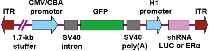
AAV vector for brain site-specific knockdown of ERα was constructed by Dr. Sergei Musatov. Musatov et al. (2006) for the first time, succeeded in site-specific knockdown of ERα in the VMN of female mice and provided definitive evidence that ERα in the VMN play an essential role in female sexual behavior. As described above, Sano et al. (2013) investigated the site-specific regulation of male sexual and aggressive behavior by ERα in male mice using the same method. Likewise, Cushing et al. (2008) demonstrated that ERα in the MeA play a role in male prosocial behavior in adult male prairie voles. Figure 3 illustrates the construct of a viral vector for ERα knockdown (shRNA ERα) or a control vector (shRNA LUC) used in these studies. Viral infection induces simultaneous expression of Green Fluorescent Protein (GFP), which enables to visualize injection site and spread of the virus in the targeted brain site. Recently, AAV vector for knockdown of ERβ was constructed by a research team of Dr. Sergei Musatov at the Cornell Medical School and Dr. Sonoko Ogawa at the University of Tsukuba. Construct of the viral vector
for ERβ knockdown is similar to that described in Figure 3.
Figure 3. Schematic representation of the AAV vector construct for ERα knockdown or control (modified from Sano et al, 2013)
1.9. Thesis objectives
In this thesis, I aimed to investigate the role of ERβ in the regulation of male social behaviors. Precise role of ERβ in male social behavior is not completely understood. As described above, ERβ may play an important role in modulation of social behaviors, which are possibly turned on by ERα. Investigation of relative importance of ERα vs ERβ, and possible differences in mechanisms of action must contribute greatly to better understanding of precise mechanisms of behavioral regulation by testosterone. Furthermore, ERβ-mediated social information processing may play a role in establishment of social relationship in male mice. However, responsible brain site(s) of ERβ action is virtually unknown. Thus, in this thesis, I aimed to investigate the role of ERβ in social behavior regulation from two aspects. The first question was how, where
and when each component of male social behaviors, such as social information processing, sexual and aggressive behavior, is regulated by ERβ? Site-specific
knockdown using RNAi methods enabled us to investigate site- and age- specific role of ERβ in the regulation of social behaviors in male mice. Secondly, in addition to the regulation of each component of social interaction, relative importance of ERβ in actual
social relationship to other males was examined.
In experiments 1-4, I examined the effects of pre-pubertal or adult site-specific knockdown ERβ in the MPOA and MeA on male social behaviors. Among several expression sites, the MPOA and MeA express both ERα and ERβ abundantly, and are known to be responsible for male social behaviors (Kondo, 1992; Paredes et al., 1993; Hull et al., 1999, Kondo and Sachs, 2002; Patil and Brid, 2010; Wang et al., 2013). I first tested whether ERβ in the MPOA or the MeA is necessary for male sexual and aggressive behaviors by site-specific knockdown of ERβ (βERKD) before puberty. I then examined the influence of site-specific βERKD only in adulthood in the MPOA. In the MeA, I intended to examine the influence of adult βERKD on social information processing including male-type sexual preference and social recognition in addition to sexual and aggressive behaviors since the MeA is known to play a pivotal role in social information processing (Ferguson et al, 2002; Baum, 2009). I also assessed an effect of βERKD in the MeA on partner choice during actual sexual behavior tests in Experiment 4. By comparing the influence of pre-pubertal and adult βERKD, I aimed to elucidate the roles of pubertal and adult ERβ in the MPOA and MeA.
In Experiment 5, I aimed to investigate whether ERβ plays a significant role not only in the regulation of stereotypical social behaviors in a single encounter, but also repeated social interaction and establishment of social relationship. The role of ERβ in the establishment of hierarchical inter-male social relationship between two males was examined using adult βERKO mice.
Summary of Objective
1) Investigate the effects of pre-pubertal site-specific knockdown ERβ in the MPOA and MeA on social behaviors of male mice.
2) Investigate the effects of adult site-specific knockdown ERβ in the MPOA and MeA on social behaviors of male mice.
3) Investigate the effects of deletion of ERβ gene on establishment of social relationships in male mice.
-Chapter 2-
General Methods
2. General Methods
2.1. Experimental animals
2.1.1. Mice
Gonadally intact ICR/Jcl male mice were used as experimental animals in Experiments 1-4. They were originally purchased from a commercial breeder (CLEA Japan Inc., Japan) and maintained in a breeding colony at the University of Tsukuba. In Experiment 5, βERKO male mice were used. βERKO mice were originally created in C57BL/6J and 129 background by Dr. Kenneth S. Korach’s group at the National Institute of Environmental Health Sciences (Krege et al., 1998). Heterozygous breeding pairs completely backcrossed to C57BL/6J were then gifted to Dr. Sonoko Ogawa at the University of Tsukuba. They were also maintained in a breeding colony at the University of Tsukuba. All mice were kept under standard housing conditions (23±2°C, 12:12 light/dark cycle with lights off at 12:00) in polypropylene clear plastic cages (19x29x12 cm; Allentown Inc., USA) with corncob bedding (Greentrue, Purina PetCare Co., USA). Food (Rodents Diet MF, Oriental Yeast Co., Ltd., Japan) and water were provided ad
libitum. All procedures were conducted in accordance with the National Institutes of
Health guidelines and were approved by the Animal Care and Use Committee and the Recombinant DNA Use Committee at the University of Tsukuba. All efforts were made to minimize the number of animals and their suffering.
2.1.2. Tail DNA extraction and PCR genotyping of βERKO mice
To identify genotype of each subject animal, mouse genomic DNA from tail tip samples of βERKO mice (used in Experiment 5) were collected on the day of weaning (PND21). Samples for PCR genotyping were prepared with a Hot Sodium Hydroxide and
Tris (HotSHOT) method (Truett et al., 2000). Tail samples were incubated in an alkaline lysis reagent (25 mM NaOH, 0.2 mM EDTA·2Na; pH 12) for 1hr at 95°C. Thereafter, samples were cooled to 4°C and a neutralizing reagent (40 mM Tris-HCl; pH 5) was then added for pH adjustment. DNA was stored at 4°C until used for PCR amplification.
Genotyping of tail DNA was performed by PCR amplifications of ER gene fragments as previously described (Krege et al., 1998). Intron 2 (5’-TGGACTC- ACCACGTAGGCTC-3’), exon 3 (5’-CATCCTTCACAG GACCAGACAC-3’) and the 3’ end of Neo (5’-GCAGCCTCTGTTCC ACATACAC-3’) primers were used. Each tail DNA sample was blended with the above three primers, a standard PCR cocktail mix (10x PCR buffer and 2mM dNTP), and Taq DNA polymerase. All samples were run in the following PCR conditions: denaturation at 94°C for 30s, annealing at 56°C for 30s, elongation at 72°C for 60 s and each cycle was repeated for a total of 36 cycles. PCR samples were run in a 2% agarose gel at 130 V for 30 min. A 1,435bp band (intron 2 and exon 3 primers) is amplified for homozygous wild-type (+/+) mice, 1,479bp band (intron 2 and Neo primers) for homozygous mutant (-/-) mice, and both bands for heterozygous (+/-) mice. In Experiment 5, homozygous wild-type (WT) and homozygous mutant (βERKO) animals were used as experimental mice.
2.2. Estrogen receptor β silencing using small hairpin RNA
In Experiments 1-4, experimental animals were stereotaxically injected with shRNA expressing AAV vectors either on PND 21 (Experiment 1) or in adulthood (Experiments 2-4). AAV-shRNA against the sequence specific for the ERβ gene
(AAV-shERβ: 5-GATCCCCGCCACGAATCAGTGTACCATCTTCCTGTCAATGGT
ACACTGATT CGTGGCTTTTTTGGAAT-3 and 5-CTAGAGCCACGAATCAGTG
AAV-shRNA against the sequence specific for luciferase (LUC) (AAV-shLUC: 5
-GATCCCCCCGCTGGAGA GCAACTGCATCTTCCTGTCAATGCAGTTGCTCT CCAGCGGTTTTTGGAA-3 and 5
-CTAGTTCCAAAAACCGCTGGAGAGCAACTGCATGAGCAACTGCATTG ACAGGAAGATGCAGTTGCTCTCCAGCGGGGG-3) was also used as control. The nucleotides specific for ERβ and LUC are underlined. These vectors also express enhanced GFP as a reporter to visually detect transfected cells.
Mice were anesthetized with sodium pentobarbital (60mg/kg; Kyouritsu Seiyaku Co. Ltd., Japan) and placed in a stereotaxic frame (Model 900, David Kopf Instruments, USA). A 26G injection needle attached to a 10 µl Hamilton syringe was inserted by aiming either at the MeA or MPOA (coordinates were determined for each experiment separately). Each animal was bilaterally injected with 1 µl of either AAV-shERβ or AAV-shLUC (1012 packaged genomic particles, 0.5 µl/hemisphere) over 5 min. The needle was left in place for an additional 10 min following the end of the infusion.
2.3. Behavioral tests
All mice were individually housed in the plastic cages starting at least 7 days before the first behavior test. Time course of behavioral assay in each experiment is described in each chapter.
2.3.1. Sexual behavior test
Each experimental animal was tested for sexual behavior against a receptive female mouse in its home cage. Each trial was 30 min and conducted under red light illumination during the dark phase of the light/dark cycle. At the beginning of each trial, a hormonally primed ovariectomized (OVX) ICR/Jcl female stimulus mouse was introduced. All
stimulus animals were obtained from the breeding colony maintained at the University of Tsukuba. To ensure high sexual receptivity, all females were subcutaneously (s.c.) injected with 10 µg estradiol benzoate (EB) in 0.1 ml sesame oil at 48 and 24 h and 500µg progesterone (P) in 0.1 ml sesame oil at 4-6 h before testing. Each male was tested against a different female mouse in each of the repeated trials. The cumulative number of mounts and intromissions, and the latency to the first mount or intromission were recorded.
2.3.2. Aggressive behavior test
Aggressive behavior was assessed in a resident-intruder paradigm for 15 min under red light illumination during the dark phase of the light/dark cycle. One test consisted of three trials conducted in three consecutive days. At the beginning of the test, an age-matched gonadally intact ICR/Jcl male mouse (intruder) was introduced into a home cage of an experimental animal (resident). All intruder mice were olfactory bulbectomized and group-housed (3-5 animals per cage). OBX was conducted to inhibit offensive aggression by intruders. Each resident mouse was tested against a different intruder mouse in each of the repeated aggression tests. An aggressive bout was defined as a series of behavioral interactions consisting of at least one of the following: chasing, boxing, tail rattling, wrestling, biting, and offensive lateral attack (often accompanied by biting). The cumulative number and duration of aggressive bouts were recorded. A maximum of three seconds could elapse between two aggressive bouts to be considered as one aggressive bout. If the interval exceeded three seconds, the two bouts were scored as two separate aggressive bouts.
2.3.3. Sexual preference tests
In olfactory sexual preference test (2.3.3.1.), experimental animals were prevented from direct interaction with stimulus animals. In two-female sexual behavior test (2.3.3.2.), males were allowed direct physical contact with females.
The testing apparatus consisted of a white plastic testing cage (31x35x17 cm) placed centrally in a white polyvinyl chloride box (46x51x25 cm). Testing cage was covered with a clear acrylic board during tests and a video camera was placed 57 cm from the bottom of the testing cage.
2.3.3.1. Olfactory sexual preference test
In Experiments 2 and 3, each experimental mouse was tested for sexual preference of a receptive female over a non-receptive female (PTFF) and a receptive female over an intact male (PTFM). In Experiment 3, each experimental mouse was tested for preference of a gonadectomized male over intact male (PTMM) in addition to PTFF and PTFM. In PTFF, a hormonally primed (see 2.3.1.) OVX C57BL/6J female mouse (receptive female: RF) and an OVX C57BL/6J female without hormonal priming (non-receptive female: XF) were used as stimulus animals. In PTFM, a RF and a gonadally intact C57BL/6J male (IM) mouse were used. In PTMM, a gonadectomized C57BL/6J male (XM) mouse and an IM were used. Each test was 15 min and conducted under white light illumination (26 lux) during the dark phase of the light/dark cycle. Clear sectoral Plexiglas cylinders (7 cm in radius, 16 cm in height) with 13 holes (6 mm diameter) near the bottom 3 cm (Mouse Cylinder SIOT3, OʼHara & Co., Ltd., Japan) were used to present stimulus mice. Experimental mice were able to sniff olfactory cues from stimulus mice through perforated parts of the cylinders.
At least two days before testing, each experimental mouse was transferred to a testing cage with clean bedding and allowed to establish its own home territory. On the day of
the testing, they were first habituated to two empty cylinders for one hour. The cylinders were placed at diagonal corners of the testing cage. At the beginning of the test, empty cylinders were removed and two cylinders with stimulus animals were placed at the same two diagonal corners. After completion of each test, cylinders were thoroughly washed, wiped with 70% ethanol, and then air-dried.
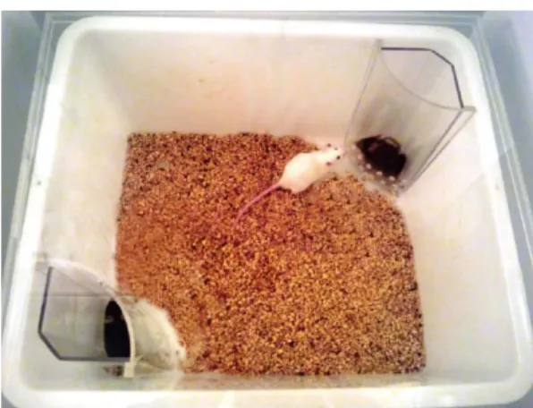
Social investigation (SI) was defined as sniffing toward each stimulus animal through the holes of the cylinder (Figure 4). The cumulative duration of SI to each stimulus mouse was recorded separately. A maximum of one second could elapse between two SIs to be considered as one bout. If the interval exceeded one second, they were recorded as two bouts.
Figure 4. Social investigation of an experimental mouse (white mouse) in olfactory sexual preference test.
2.3.3.2. Sexual preference test with freely moving two females (2F Sex test)
At least one week before testing, subject mice was transferred to a testing cage and allowed to establish home territory. Each trial was 30 min and conducted under red light illumination during the dark phase of the light/dark cycle. At the beginning of the test, two ovariectomized ICR/Jcl female stimulus mice were introduced into subject’s cage. One of the females was hormonally primed as described in 2.3.1. to ensure high sexual
receptivity (RF). On the other hand, another female was not hormonally primed (XF). In this test, latency to first mount and intromission to each stimulus mouse was recorded separately to evaluate which stimulus female was chosen as a partner for sexual behavior.
2.3.4. Social recognition test
Each experimental mouse was tested for social recognition with RF, XF, and IM mice. Each test was conducted under white light illumination (26 lux) during the dark phase of the light/dark cycle. Test apparatus other than the cylinder was same as sexual preference tests. One empty round cylinder (7 cm in diameter at the bottom and 4.4 cm in diameter at the top, 16cm in height) with 28 holes (6 mm diameter) near the bottom 3cm (Tsuda and Ogawa, 2012; Mouse Cylinder SIOT1, O’Hara & Co., Ltd.) was introduced in the center of testing cages 1 h before the first trial. Experimental mice were tested four times, 4 min each, with 17 min inter-trial intervals (Figure 5). In the first three trials, each experimental mouse was tested against the same stimulus mouse (Stimulus A) whereas in the fourth trial, he was tested against a different (novel) stimulus mouse (Stimulus B). Same types of mice (i.e., RF, XF or IM) were used for Stimuli A and B. The cumulative duration of SI was recorded in each trial. Definition of SI was the same as that in olfactory sexual preference test (see 2.3.3.1.).
2.3.5. Agonistic behavior test
Agonistic behavior between two experimental male mice was assessed in a neutral testing cage (a plastic cage of the same type as animals’ home cage) for 15 min under red light illumination during the dark of the light/dark cycle. Before each testing trial, testing cage was separated into two compartments by a black Plexiglas board (divider). Each experimental animal was placed in each compartment and was habituated to the testing environment for 5 min. At the beginning of the test, the divider was removed and agonistic behaviors were observed. Number and duration of following behaviors were recorded. The cumulative number and duration of aggressive behavior (definition is described in 2.3.2.), fleeing, approaching, sniffing, huddling, and grooming, and the cumulative number of tail rattling were recorded. These behavioral indices were classified into either agonistic or prosocial interaction. Agonistic interaction was defined as a series of behavioral interactions consisting of at least one of the following: aggressive behavior, fleeing, and tail rattling. For calculation of the cumulative duration of agonistic interaction, cumulative duration of aggressive behavior and fleeing were added. Prosocial interaction was defined as a series of behavioral interactions consisting of at least one of the following: approach, sniffing, huddling and grooming.
2.3.6. Tube test
Tube test was conducted using testing arena (70x50 cm) surrounded by black wall. A transparent Plexiglas tube (length: 45 cm, inner diameter: 3 cm) was set on the center of the testing arena.
2.3.6.1. Training
from one end to the other end. A black plastic escape box (13x14x13 cm) was attached at the end of the tube in some of the training trials. Each experimental animal experienced 8 trials per day for two consecutive days. On the first day of the training, mice experienced initial 4 training trials with the escape box and 4 trials thereafter without the escape box. On the second day, initial 2 trials were conducted with the escape box and the rest of the trials were without the escape box. At the begging, each animal was gently held and put into one end of the tube. Starting side in initial trial was counterbalanced. When a mouse stopped in the tube, an experimenter gently pushed animal’s back with a plastic pole. After a mouse reached the end, he was trained to run in an opposite direction. At the end of training of each animal, all apparatuses were wiped with 70% ethanol and air-dried.
2.3.6.2. Testing
Before the testing trial on each day, two training trials without escape box were conducted. At the begging of the testing trial, a pair of experimental animals was set on each end of the tube and an experimenter released the mice to let them run into the tube (Figure 6, left). Starting side of each animal was counterbalanced. The test trial ended when one of the mice was ejected from the end where he first entered (Figure 6, right). A mouse stayed inside of the tube at the end of the trial was called as a “winner” and an ejected mouse was called as a “loser”. Alternatively, if two minutes elapsed without ejection of either mouse, the trial ended as a “tie”. At the end of each test trial, all apparatuses were wiped with 70% ethanol and air-dried.
Winner’s animal ID and the latency to the end of trial were recorded in each trial. Furthermore, occurrence of “invasion” by a winner was recorded. When both hind paws of a winner crossed mid-point of the tube to loser’s side, invasion was recorded (Figure 6, right). The loser was able to walk back spontaneously and exit the tube even if it was
not pushed by the winner.
Figure 6. Schema of experimental procedure for tube test. At the beginning of the test, mice were released and run into the tube (left panel). The test ended after ejection of a loser (right panel). Invasion: both hind paws of a winner crossed mid-point of the tube (red line of the right panel) to loser’s side.
2.3.7. Quantitative analysis of behavioral data
All behavioral tests were recorded using digital video cameras. All video recordings were scored by an experimenter unaware of animals’ experimental group using a digital event recorder program (Recordia 1.0b, O’Hara & Co., Ltd.).
Behavioral data from sexual and aggressive behavior tests was analyzed by a two-way analysis of variance (ANOVA) for repeated measurements for the main effects of vector treatment, tests and their interactions. The data from sexual preference tests except for total SI duration in olfactory sexual preference test was analyzed in each vector treatment group separately by a paired t-test between two stimulus mice. Total SI duration in olfactory sexual preference test was analyzed by an unpaired t-test between vector treatment groups. Average SI duration in social investigation test was analyzed in each vector treatment group separately by one-way ANOVA for repeated measurements for three types of stimulus mouse. Behavioral data from agonistic behavior tests and tube test was analyzed by a two-way ANOVA for repeated measurements for the main effects of genotype, days, and their interaction. Post hoc analysis was conducted with Bonferroni correction when interaction was significant. All these data were analyzed using the SPSS ver. 21.0 (SPSS Inc., USA). Proportion difference in the test trials in Experiment 5 was
analyzed in Fischer’s Exact Test. Fischer’s Exact Test was conducted using the R (The R Project for Statistical Computing). Statistically significant differences were considered when p<0.05.
2.4. Histological analysis
In Experiments 1-4, histological analysis was conducted after behavioral tests.
2.4.1. Preparation of brain tissues for immunohistochemistry
After the completion of the last behavioral tests, all experimental animals were deeply anesthetized with heparin-containing pentobarbital sodium solution (60 mg/kg body weight, i.p.). They were then perfused through the left cardiac ventricle with 40 ml of 100 mM phosphate buffered saline (PBS; pH 7.2) for blood removal, followed by 40 ml of 4% paraformaldehyde-containing 100mM phosphate buffer (PB; pH 7.2) for fixation with the use of a peristaltic pump. Brains were removed and post-fixed in the same fixative at 4℃ for 24h. After cryoprotection in 30% sucrose in 100 mM PB at 4℃, coronal sections (30 µm thickness) were prepared using a freezing microtome. Serial sections were collected in four sets with 120 µm intervals, and stored in anti-freezing buffer (30% ethylene glycol and 30% glycerol in 0.05 M Tris-buffered saline (TBS), pH 7.2) at -20°C until use.
2.4.2. Immunohistochemistry
Freely floating sections were incubated in PBS containing 0.2% triton X (PBS-X) with 0.3% H2O2 for 20 min at room temperature (RT) for blocking. After washing, sections were pretreated with 5% bovine serum albumin (BSA) in PBS–X (blocking buffer) for 2 h at RT. The sections were then incubated with goat polyclonal anti-GFP
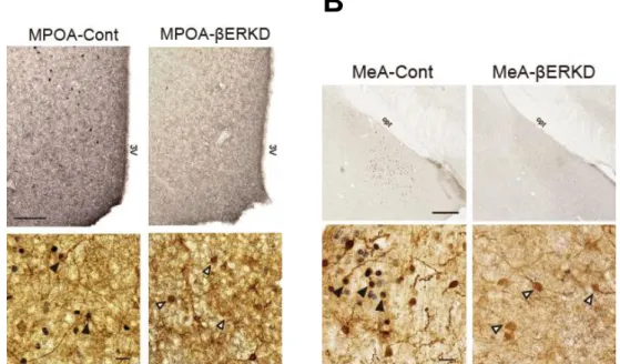
antiserum (1:5,000; ab6673, Abcam, USA) in blocking buffer for one night at 4℃. They were washed and incubated with biotinylated rabbit anti-goat secondary antiserum (1:250; Vector Laboratories) in blocking buffer for 2 h at RT. After washing, sections were reacted to avidin-biotin complex (Vectastain ABC Elite kit; Vector Laboratories) PBS for 1 h at RT, and washed. They were then incubated in 0.02% (DAB) and 0.003% H2O2 in PBS for 2 min, followed by wash with PBS. A few sections from each group were also processed for double immunohistochemical staining for GFP and ERβ. Prior to immunohistochemistry for GFP, they were incubated with rabbit polyclonal ERβ antiserum (1:1000; Z8P, lot 10766190, Zymed Laboratories, USA) for 3 days at 4°C followed by biotinylated goat anti-rabbit secondary antiserum (1:250; Vector Laboratories) for 2 h and visualized in 0.03% diaminobenzidine (DAB), 0.15% NiNH4SO4, and 0.003% H2O2 in TBS for 12-14 min, followed by wash with TBS (pH 7.2).
All sections were mounted on gelatin-coated slides, air-dried, dehydrated through ascending series of ethanol, cleaned with xylene, and coverslipped with Permount (Fisher Scientific, USA).
2.4.3. Analysis of immunopositive cells
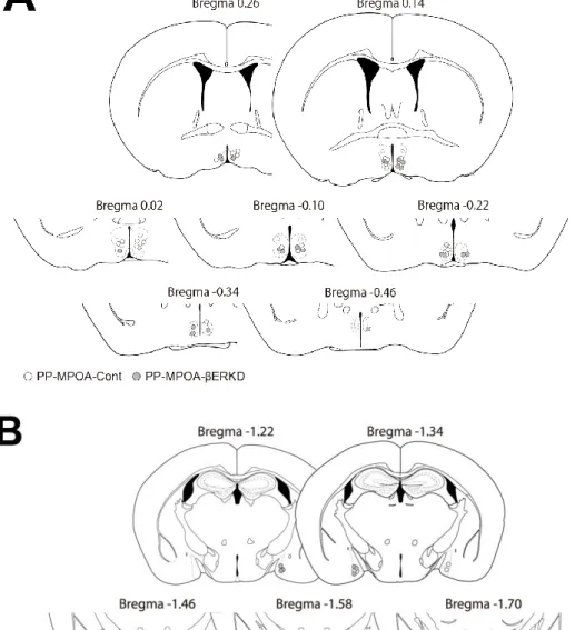
Nine sections containing the MPOA (Bregma 0.38 to -0.58) and nine sections containing the MeA (Bregma -1.10 to -2.06) were selected for histological analysis of immunopositive cells for GFP. Each brain area was photographed at 20x magnification with a digital camera mounted on a microscope (BZ-X710, KEYENCE Corporation, Japan). Spread of GFP immunopositive cells were recorded for confirmation of AAV infection in the targeted area. We also selected three double-immunostained sections in the MPOA (Bregma 0.02, 0.10, and 0.22) and in the MeA (Bregma 1.82, 1.94, and
-2.06) where most intensive ERβ expression was observed in the control groups. In these sections, we counted (3 mice per group) number of ERβ-immunopositive cells and double-labeled cells for ERβ and GFP in each side of the hemisphere within the targeted site. The data was analyzed in each section separately by a Welch’s t-test between two vector treatment groups using the SPSS ver. 21.0 (SPSS Inc., USA). Statistically significant differences were considered at p < 0.05.
-Chapter 3-
Experiment 1:
Effects of Pre-Pubertal ERβ Knockdown
in the MPOA and MeA
3. Experiment 1: Effects of Pre-Pubertal ERβ Knockdown
in the MPOA and MeA
3.1. Introduction
It is still unknown when, where in the brain, and how testosterone regulates male social behavior via ERβ. In addition to androgen surge in perinatal period, circulating testosterone level starts to increase from the beginning of pubertal period and reaches to adult level at the end of puberty. Thereafter, activation of adult neural network, which is formed and developed by perinatal and pubertal organizational action of testosterone, induces a variety of male social behaviors.
Although it is known that ERβ is involved in the regulation of male social behavior (Ogawa et al., 1999; Nomura et al., 2002, 2006; Kavaliers et al., 2008), precise time course of its action is still unclear. Previous study using selective ERβ agonist has reported that ERβ activation in the perinatal period can facilitate aggressive behavior in adulthood (Patisaul and Bateman, 2008). Thus, it is possible that ERβ mediates not only activational, but also organizational action of testosterone. However, the role of ERβ in pubertal period and adulthood in different brain sites remains to be elucidated.
To investigate relative importance of ERβ in pubertal period and adulthood, effects of pre-pubertal site-specific knockdown ERβ in the MPOA and MeA on the performance of sexual and aggressive behavior in adulthood was examined. It is well documented that both MPOA and MeA, play an important role in the regulation of sexual and aggressive behavior (Paredes et al., 1993; Hull et al., 1999; Patil and Brid, 2010). Since knockdown of ERβ in pre-pubertal period suppresses ERβ gene expression permanently after AAV injection, it can be tested whether pubertal and adult ERβ in the MPOA and MeA is necessary for the performance of sexual and aggressive behavior. Moreover, the MPOA
and MeA express high levels of both ERα and ERβ (Shughrue et al., 1997; Mitra et al., 2003). It is intriguing to clarify whether ERβ in these target sites have similar roles as ERα reported by Sano et al. (2013, 2016).
3.2. Methods
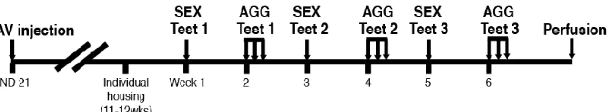
A total of 12 litters of ICR/Jcl male mice were assigned to either MPOA or MeA groups on PND 21 after being weaned. Mice from each litter were further divided into two shRNA injection groups of either AAV-shERβ or AAV-shLUC. Those four groups were designated as pre-pubertal treatment (PP)-MPOA-βERKD (n=11), PP-MPOA-Cont (n=13), PP-MeA-βERKD (n=9), and PP-MeA-Cont (n=9). Coordinates for the MPOA group were AP +0.02, ML ±0.5, DV-5.2, and those for the MeA group were AP -1.25, ML ±2.2, DV -5.15. All coordinates were determined based on The Mouse Brain Stereotaxic Coordinates (Paxinos and Franklin, 2001) with an adjustment for the brain size on PND 21. All mice were then group housed with their littermates (4~5 mice per cage) until they were tested for sexual and aggressive behavior in adult as gonadally intact (11.9±0.21 wks old at the first behavioral test). Starting one week before the first behavioral test, all mice were individually housed. Three sexual behavior tests (SEX) and three sets of aggressive behavior tests (AGG) were done in alternate weeks for a total of six weeks (Figure 7). After the completion of the last behavioral test, brain tissues were collected and processed for immunohistochemistry for GFP and ERβ.
Figure 7. Schema of experimental procedures. Tick marks under the horizontal bar indicate one week. SEX, sexual behavior; AGG, aggressive behavior.
3.3. Results
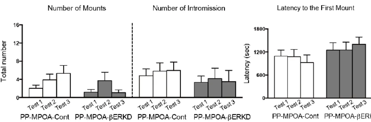
3.3.1 Effects of pre-pubertal ERβ knockdown in the MPOA
There was no difference in male sexual behaviors between the PP-MPOA-βERKD and PP-MPOA-Cont groups in sexual behavior tests (Figure 8). Statistical analysis revealed that there was no significant main effects of treatment and test, and interaction of treatment and test in any of number of mounts (treatment: F1,18 = 1.117, n.s.; test: F2,36 = 2.631, p = 0.086; treatment x test: F2,36 = 1.770, n.s.) and intromissions (treatment: F1,18 = 0.396, n.s.; test: F2,36 = 0.030, n.s.; treatment x test: F2,36 = 0.302, n.s.), and latency to the first mount (treatment: F1,18 = 0.860, n.s.; test: F2,36 = 0.078, n.s.; treatment x test: F2,36 =2.161, n.s.). These results indicated that pre-pubertal ERβ knockdown in the MPOA has minimal effects on sexual behavior of adult male mice.