Abbreviations: BHI, brain-heart infusion; CA, cholic acid; GCA, glycocholic acid; GLC, gas-liquid chromatography; 7α-HSDH, 7α-hydroxysteroid dehydrogenase; Me-DMES, methyl ester dimethylethylsilyl ether; PHP GEL, piperidino-hydroxypropyl dextran gel; 3α12α7=O, 3α,12α-dihydroxy-7-oxo-5β-cholanoic acid; ββ, 7β,12β-dihydroxy-5β-cholanoic acid
Aerobic and Anaerobic Biotransformation of Bile Acids by
Escherichia coli (III)
Yoshio Ogura, Tsuyoshi Takei, Nobuo Yamaga, Kazuo Yamada and Kiyohisa Uchida
Division of Medical Biochemistry, Department of Pathophysiological and Therapeutic Science, School of Medicine, Tottori University Faculty of Medicine, Yonago 683-8503 Japan
The oxidation/reduction reactions of bile acids by Escherichia coli (E. coli) K-12 were examined in both Davis and brain-heart infusion (BHI) media under aerobic and an-aerobic conditions. The pH in the Davis medium changed by almost the same amount, around pH 6.5–7.0 in both aerobic and anaerobic cultures, but the pH in the BHI me-dium was different in both cultures, that is, about pH 9.0 in the aerobic culture but only about 6.5 in the anaerobic culture. The growth curve of E. coli in the Davis medium showed a similar pattern in both conditions. Cholic acid (CA) was oxidized to 3
α12α-dihydroxy-7-oxo-5β-cholanoic acid (3α12α7=O) in both cultures, but the reaction in the
anaerobic culture was somewhat slower than that in the aerobic culture. On the other hand, reduction of 3α12α7=O to CA did not occur in the aerobic culture, but about 10%
reduction was observed in the anaerobic culture after 4 days. These data suggest that the oxidation/reduction reaction of E. coli was oxidative in aerobic culture but reduc-tive in anaerobic culture and these characteristics were not due to the changes in the pH of the medium. The reactions of CA and glycocholic acid to crude 7α-HSDH prepared
from E. coli were examined and it was found that both free and conjugated CA as a sub-strate for the 7α-HSDH showed similar Km values.
Key words: Escherichia coli; cholic acid; glycocholic acid; 3α,12α-dihydroxy-7-oxo-5β-cholanoic acid; in vitro biotransformation
Escherichia coli (E. coli) possesses 7α-hydroxy- steroid dehydrogenase (7α-HSDH) and oxidizes 7α-hydroxyl bile acids to the corresponding 7-oxo-bile acids, or reduces 7-oxo-bile acids to the corresponding 7α-hydroxyl bile acids (Aries and Hill, 1970; Haslewood and Haslwood, 1976; Mac-donald et al., 1973). In a previous paper (Ogura et al., 2003), we reported that E. coli in aerobic cul-ture transformed a large proportion of cholic acid (CA) to 3α,12α-dihydroxy-7-oxo-5β-cholanoic acid (3α12α7=O) but hardly transformed
7-oxo-bile acid to CA, whereas in anaerobic culture about 50% of CA was oxidized and about 50% of 7-oxo-bile acid was reduced. A similar result was also obtained with chenodeoxycholic acid. There-fore, we speculated that the oxidation/reduction reaction of E. coli attained different equilibrium values depending on the presence or absence of oxygen. In other words, the reaction equilibrium shifted to the oxidative side in aerobic culture but was almost equally balanced in anaerobic culture. In previous experiments with brain-heart
infusion (BHI) medium, the pH of the cultured medium increased to about 9 in aerobic culture but decreased to around 6.5 in anaerobic culture (Ogura et al., 2003). Since it has been reported that the reaction of 7α-HSDH is oxidative in alka-line conditions but reductive in acidic conditions (Groh et al., 1993; Macdonald et al., 1983), we suspected our previous results were dependent on the difference of pH of the medium rather than on the difference between aerobic and anaerobic conditions.
Meanwhile, we found that when E. coli was cultured in the Davis medium (Davis and Min-gioli, 1950) containing 0.2% glucose, the pH of the medium did not differ significantly between aerobic and anaerobic conditions. Therefore, we examined the effect of E. coli on bile acids in both aerobic and anaerobic cultures in the Davis medium and compared it with that in the BHI me-dium.
In addition, E. coli dehydrogenates taurine- or glycine-conjugated CA to the corresponding taurine- or glycine-conjugated 3α12α7=O with-out deconjugation, but the conversion rates for conjugated CA were lower than for free CA (Ogura et al., 2003). In the present experiments, we ana-lyzed the effect of concentrations of conjugated and free CA on crude 7α-HSDH prepared from E.
coli to clarify the difference mentioned above. Materials and Methods
Chemicals
CA was purchased from Sigma Chemical, St. Louis, MO. 3α,12α-Dihydroxy-7-oxo-5β-cholanoic acid (3α12α7=O) was prepared by oxidation of CA with N-bromosuccinimide (Fie-ser and Rajagopalan, 1949). 7β,12β-Dihydroxy-5β-cholanoic acid (ββ), an internal standard for analysis by capillary gas-liquid chromatography (GLC), was synthesized as described previously (Arimoto et al., 1982). Piperidinohydroxypro-pyl dextran gel (PHP GEL) was purchased from
Shimadzu, Kyoto, Japan, and dimethylethylsily-limidazole from Tokyo Kasei Kogyo, Tokyo, Ja-pan. The other reagents and solvents of analytical grade were obtained from Wako Pure Chemical Industries, Osaka, Japan. If not otherwise stated, the solvents were distilled once before use.
Bacteriological procedures
E. coli K-12 was obtained from the American Type Culture Collection (Manassas, VA). The BHI medium (Becton Dickinson, Sparks, MD) and the Davis medium (pH 7.2) were used for the culture experiments. The Davis medium contains 0.7% K2HPO4, 0.2% KH2PO4, 0.047% sodium
citrate, 0.005% MgSO4, 0.1% (NH4)2SO4 and 0.2%
glucose. The pH was adjusted to 7.2. E. coli was precultured aerobically in the BHI medium at 37˚C for 1 day. Aliquots of the precultured E.
coli (3 × 10⁷ in 10 µL medium) was added each
to 2 mL of the fresh BHI medium, or to the fresh Davis medium, containing 1 mM of bile acids and cultured at 37˚C for 4 days. The aerobic culture was performed in the open air with an incubator (PCI-100; Iuchi, Osaka, Japan) adjusted at 37˚C, and the anaerobic culture was performed with the use of a gas-pack (BBL Gas Pak Pouch System; Becton Dickinson). Viable cells of E. coli in both media were counted by colony formation on an agar plate after seeding the diluted culture me-dium.
Analytical methods
Bile acids were analyzed according to the method reported previously (Ogura et al., 2003). A por-tion of the medium cultured, 100 µL, was heated with 8 volumes of ethanol at 80˚C for 10 min and then filtered through a glass microfiber filter (GF/ C; Whatman, Maidstone, United Kingdom). The filtrate was evaporated to dryness under a stream of nitrogen and the residue was acidified with diluted hydrochloric acid. The bile acids were ex-tracted with ethylether, converted to methyl ester dimethylethylsilyl ether (Me-DMES) derivatives
and quantified by gas-liquid chromatography with a Hicap CBP-1 capillary column (25 m × 0.25 mm i.d.; Shimadzu). Conversions were expressed as % of bile acids converted in the culture me-dium.
Enzyme activity of cell-free 7α-HSDH
E. coli K-12 was cultured aerobically for one day in the BHI medium. Crude 7α-HSDH was pre-pared according to the method of Macdonald et al. (1973). The enzyme activity was assayed by the method of Macdonald et al. (1974, 1975) with a slight modification. Briefly, the reaction was performed at 37˚C for 10 min in 0.18 M glycine-NaOH buffer (pH 9.5) containing 7 mM NAD+, 0
to 4 mM bile acid and 45 µL of the crude enzyme preparation in a final volume of 1 mL.
Results and Discussion
Major conversions of bile acids by intestinal bac-teria include deconjugation, oxidation of hydroxy groups at C-3, C-7 and/or C-12 positions, and 7α/ β-dehydroxylation. E. coli possesses 7α-HSDH for the oxidation, but has neither deconjugation nor dehydroxylation activity. In the present study, we examined the effect of E. coli on bile acids in
both Davis and BHI media, which showed dif-ferent pH after cultures in aerobic and anaerobic conditions.
Changes in the pH of the BHI and Davis media cultured with E. coli are given in Fig. 1. The pH in the Davis medium changed almost by a similar value under both aerobic and anaerobic conditions; it decreased slightly to pH 6.5 on the first day of incubation and remained almost con-stant or very slightly increased thereafter, while the pH in the BHI medium markedly increased to about pH 9.0 in the aerobic condition but de-creased to about pH 6.5 in the anaerobic condi-tion. The changes in the Davis medium were almost the same to the change in the BHI medium in the anaerobic condition.
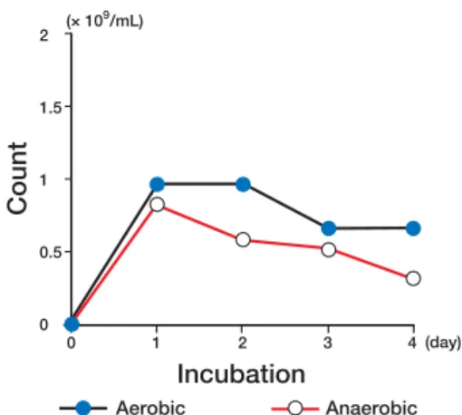
Figure 2 shows changes in viable cell counts during aerobic and anaerobic cultures of E. coli K-12 in the Davis medium. Although the viable cell counts in the anaerobic culture were higher than those in the aerobic culture, they changed almost by the same amount in both conditions.
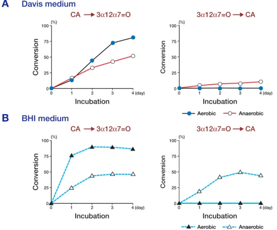
Figure 3 shows the time course of changes in 7α-dehydrogenation of CA and 7α-hydrogenation of 3α12α7=O by E. coli in the Davis and BHI media. In the Davis medium, CA was oxidized to 3α12α7=O in both aerobic and anaerobic cul-tures, but the reaction in anaerobic culture was somewhat slower than that in aerobic culture. On
Fig. 1. Changes in pH of the Davis medium and the
brain-heart infusion (BHI) medium during aerobic and anaerobic cultures of Escherichia coli K-12.
Fig. 2. Changes in viable cell counts during aerobic and
anaerobic cultures of Escherichia coli K-12 in the Davis medium. ���������� ��������������� ������������� ����������������� ��������������� ����� �� �� �� �� � ��� �� �� �� �� �� �� �� � � � �� �� ���������� ���������� ����� ������� ��������� ����� �� �� �� �� � ��� � � � � ��� �� ��� ��
the other hand, the reduction of 3α12α7=O to CA did not occur in aerobic culture, but about 10% re-duction was observed in anaerobic culture after 4 days. In the BHI medium, the oxidation of CA in aerobic culture was much higher than that in an-aerobic culture and the reduction of 3α12α7=O, though it was not observed in aerobic culture, oc-curred obviously in anaerobic culture.
These changes in the Davis medium were less marked than those in the BHI medium, but nevertheless showed the same characteristics for CA oxidation and 7-oxo-bile acid reduction, sug-gesting that this 7α-HSDH reaction was oxidative in aerobic culture but reductive in anaerobic cul-ture.
Therefore, these data indicate that the oxida-tion/reduction reaction of 7α-HSDH is not affect-ed by extracellular pH. In addition, it has been
Fig. 3. Conversion of cholic acid (CA) and 3α,12α-dihydroxy-7-oxo-5β-cholanoic acid (3α12α7=O) by Escherichia
coli K-12 in the Davis medium and brain-heart infusion (BHI) medium.
reported that the intercellular pH of E. coli is maintained at around 7.6 in a wide range of extra-cellular pH (Padan et al., 1976; Shimamoto et al., 1994). Therefore, we conclude that the 7α-HSDH activity of E. coli is not affected by changes in pH of the culture medium.
NAD+ is a cofactor required for the oxidative
activity of 7α-HSDH. The content of NAD+ in E. coli was reported to increase in aerobic culture (Leonardo et al., 1996). This would explain the shift to the oxidative side in the transformation of bile acids by 7α-HSDH.
In our previous paper, we found that conju-gated CA was dehydrogenated by E. coli, but to a lesser extent than free CA (Ogura et al., 2003). The reactions of CA and glycocholic acid (GCA) by the crude enzyme preparation were then exam-ined in vitro. As shown in Fig. 4, CA and GCA
�%� ���������� �� �� �� �� � ����� ���������� �� �� �� �� � ����� ���������� �� �� �� �� � ����� ���������� �� �� �� �� � ����� ���������� ��� �� ��� �� ��� �%� ���������� ��� �� ��� �� ��� �%� ���������� ��� �� ��� �� ��� �%� ���������� ��� �� ��� �� ��� ��������� ����������������� ��������� ����������������� � ���� � ��������� ��������� � ��� � ���� � ��������� ��������� � ���
A
B
Davis medium BHI mediumFig. 4. Dehydrogenation of cholic acid (CA) and
glyco-cholic acid (GCA) in vitro by 7α-hydroxysteroid dehy-drogenase prepared from Escherichia coli K-12. showed almost the same reactions, suggesting that
both free and conjugated CA have similar affini-ties for the enzyme. This result coincides with the data demonstrated by Haslewood and Haslewood (1976). Therefore, the data with the crude en-zyme preparation suggest that the lower reaction for conjugated CA is due to a slower penetration rate of conjugated CA into living cells than free CA.
In conclusion, the oxidation/reduction reac-tion of E. coli was oxidative in aerobic culture but rather reductive in anaerobic culture. These re-sults were not due to the changes in the pH of the medium. In addition, the transport step through the cell membrane will be one of the rate limits for the transformation of bile acids in whole cells.
Acknowledgments: This work was supported by a Grant-in-Aid for Scientific Research from the Yakult Bio-Science Foundation (2005).
References
1 Aries V, Hill MJ. Degradation of steroids by intesti-nal bacteria II. Enzymes catalysing the oxidoreduc-tion of the 3α-, 7α- and 12α-hydroxyl groups in cho-lic acid, and the dehydroxylation of the 7-hydroxyl group. Biochim Biophys Acta 1970;202:535–543. 2 A r i moto K, Adach i K, Ya maga N.
7β,12β-Dihydroxy-5-cholan-24-oic acid as an internal stan-dard for quantitative determination of bile acids by gas chromatography. Steroids 1982;39:631–641. 3 Davis BD, Mingioli ES. Mutants of Escherichia coli
requiring methionine or vitamin B12. J Bacteriol
1950;60:17–28.
4 Fieser LF, Rajagopalan S. Selective oxidation with
N-bromosuccinimide. I. Cholic acid. J Am Chem Soc 1949;71:3935–3938.
5 Groh H, Schade K, Hörhold-Schubert C. Steroid metabolism with intestinal microorganisms. J Basic Microbiol 1993;33:59–72.
6 Haslewood ES, Haslewood GAD. The specificity of a 7α-hydroxysteroid dehydrogenase from
Escherich-ia coli. Biochem J 1976;157:207–210.
7 Leonardo MR, Dailly Y, Clark DP. Role of NAD in regulating the adhE gene of Escherichia coli. J Bac-teriol 1996;178:6013–6018.
8 Macdonald IA, Williams CN, Mahony DE. 7α-
Hydroxysteroid dehydrogenase from Escherichia
coli B: preliminary studies. Biochim Biophys Acta 1973;309:243–253.
9 Macdonald IA, Williams CN, Mahony DE. A 3α- and 7α-hydroxysteroid dehydrogenase assay for conjugated dihydroxy-bile acid mixtures. Anal Bio-chem 1974;57:127–136.
10 Macdona ld I A, Wi l l ia m s C N, Ma hony DE , Christie WM. NAD- and NADP-dependent 7α- hydroxysteroid dehydrogenases from Bacteroides
fragilis. Biochim Biophys Acta 1975;384:12–24. 11 Macdonald IA, Bok kenheuser VD, Winter J,
McLernon AM, Mosbach EH. Degradation of ste-roids in the human gut. J Lipid Res 1983;24:675–700. 12 Ogura Y, Yamaga N, Kido Y, Katayama R, Yamada
K, Uchida K. Aerobic and anaerobic biotransforma-tion of bile acids by Eschirichia coli (I). Bioscience Microflora 2003;22:133–137.
13 Padan E, Zilberstein D, Rottenberg H. The proton electro-chemical gradient in Escherichia coli cells. Eur J Biochem 1976;63:533–541.
14 Shimamoto T, Inaba K, Thelen P, Ishikawa T, Goldberg EB, Tsuda M, Tsuchiya T. The NhaB Na+/
H+ antiporter is essential for intracellular pH
regula-tion under alkaline condiregula-tions in Escherichia coli. J Biochem 1994;116:285–290.
Received January 11, 2006; accepted February 24, 2006 Corresponding author: Yoshio Ogura, PhD
���������������������� ��������� �� �������������� ��� �� ���� �� �� �� �� � �� �� �� �� � ��� �� �� �� � � � � � ��� � � � � ���� ���� ���� �� �������������������������� ������