INTRODUCTION
The lung is a vulnerable target in collagen vascu-lar diseases (CVDs) represented by rheumatoid ar-thritis (RA) because of its abundant vasculature and large content of connective tissue (1). Pulmonary involvements of CVD are various, for example, pleu-ritis, interstitial pneumonia (IP) and vasculitis. IP especially is common to various CVDs, and has a great influence on patients’ prognosis. The frequency
of IP associated with CVD (IP- CVD) varies depend-ing on the histological pattern : usual IP (UIP), non-specific IP (NSIP), organizing pneumonia (OP), diffuse alveolar damage (DAD), lymphoid IP (LIP), as well as on the underlying CVD. Patients with pul-monary fibrosis associated with CVD have an im-proved prognosis compared to idiopathic pulmonary fibrosis (IPF) (2, 3). However, in CVD patients, other histological patterns of IP are often observed. Al-though such patients have been considered to have more favorable prognosis than patients with idiopathic interstitial pneumonia (IIP) who have the same his-tological patterns, there is currently no clear proof. To the best of our knowledge, there is no report of detail pulmonary findings of RA without the in-fluence of therapy. Here, we report a case with
natu-CASE REPORT
Pulmonary findings without the influence of therapy in a
patient with rheumatoid arthritis : an autopsy case
Kiyoshi Hashimoto
1, Hirofumi Nakanishi
1, Akira Yamasaki
1, Hiroki Chikumi
1,
Yasuyuki Hasegawa
1, Masanari Watanabe
1, Hisao Ito
2, and Eiji Shimizu
1 1Division of Medical Oncology and Molecular Respirology, Department of Multidisciplinary Inter-nal Medicine, Faculty of Medicine, Tottori University, Tottori, Japan, 2
Division of Organ Pathology, Department of Microbiology and Pathology, Faculty of Medicine, Tottori University, Tottori, Japan Abstract : We report the autopsy findings of a 40- year- old woman with lung complica-tions of rheumatoid arthritis. She has been suffering from rheumatoid arthritis and interstitial pneumonia without satisfactory therapies because of her poor compliance. At autopsy, diffuse pleural adhesions and many protruding cysts were observed. The cut sur-faces had rich fibrous changes and honey-comb like appearances dominantly in the left lower lobe. Microscopically, remarkable fibrous changes were observed with destruction of the alveolar structure. These fibroses were temporally homogeneous and lacked promi-nent fibroblastic foci. The histological pattern was consistent with fibrous non- specific interstitial pneumonia. In peripheral pulmonary arterioles, some thrombi were detected with much recanalization. Systemic amyloidosis was observed in the submandibular gland, thyroid, heart, and arterioles of the lung, kidney, and digestive tract. In the left pulmo-nary artery, a large embolus was detected. This embolism was the direct cause of death. Her pulmonary findings, except for the embolism, were considered sober states of lung complications of rheumatoid arthritis without the influence of therapy. J. Med. Invest. 54 : 340-344, August, 2007
Keywords : pulmonary findings, rheumatoid arthritis, interstitial pneumonia, autopsy
Received for publication January 10, 2007 ; accepted February 8, 2007.
Address correspondence and reprint requests to Kiyoshi Hashimoto, M.D., Ph.D., Division of Medical Oncology and Molecular Respi-rology, Department of Multidisciplinary Internal Medicine, Fac-ulty of Medicine, Tottori University 36-1, Nishi-cho, Yonago, Tottori 683-8504, Japan and Fax : +81-859-38-6539.
ral pulmonary findings of RA.
CASE REPORT
A 40- year- old woman was admitted to the hos-pital because of her mental derangement attendant on hypoxemia. When she was 30- year- old, she was diagnosed as RA by orthopedists. Moreover, she had been suffering from IP for 12 years. Although she was recommended to take disease modifying antirheumatic drugs (DMARDs) or internal steroids several times, they had not been taken continuously because of her poor compliance. Lung transplanta-tion was also impossible because of her mental and circumstantial problems. Pulmonary hypetension (PH) was very severe. Two years before her death, pulmonary pressure was 40 mmHg, rising to 100 mmHg just before her death. Figure 1A and 1B show
the findings of chest high resolution computed to-mography (HRCT) just before her death. Multiple cystic lesions and fibrous changes were observed in the bilateral lung fields. Honey-comb like appear-ances were found dominantly in the left and back side. Figure 1A also shows the dilatation of the right pulmonary artery. These HRCTs presented no defi-nite rheumatoid nodules.
Recovering her mental derangement and begin-ning rehabilitation, she suddenly complained of dyspnea and lost her consciousness, breathing, and heart beat, leading to death without avail of cardio-pulmonary resuscitation. The direct cause of death was clinically regarded as pulmonary embolism. We performed an autopsy with the permission of her family.
AUTOPSY FINDINGS
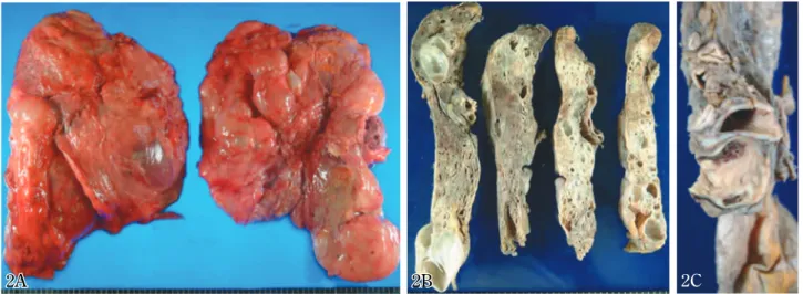
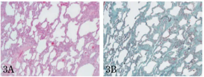
Extensive and hard pleural adhesions were de-tected in the bilateral thoracic cavities (Figure 2A). Many protruding cysts (max : 7.5 cm in diameter) were also observed. The cut surfaces (Figure 2B) had the pleural thickness, many cysts of various size, remarkable proliferation of fibrous tissue, and honey-comb like appearances dominantly in the left lower lobe. Microscopically, comparatively loose in-terstitial fibroses that were temporally homogene-ous and lacked prominent fibroblastic foci were dem-onstrated (Figure 3A and 3B). This histological pat-tern was NSIP, because no temporal heterogene-ity of fibrosis was seen despite the lack of steroidal or immunosuppressive therapy (4). Dense fibro-ses were also observed in the alveoli and alveolar
Figure 1. Chest high resolution computed tomography (HRCT) shows multiple cystic lesions and interstitial fibrotic changes in the bilateral lung fields. Honey-comb like appearance is observed in the left and back side.
Figure 2A. Pleural adhesions and many cysts are seen on the surface of the bilateral lungs.
Figure 2B. The cut surfaces of the left lung have the pleural thickness, many cystic lesions and remarkable fibrous changes.
Figure 2C. The left pulmonary artery is filled up by an embolus.
1A 1B
2C 2
walls with moderate infiltration of lymphoid cells es-pecially around the small airways (Figure 4A and 4B). The original structures of alveolar walls were de-stroyed with fragmentation of elastic fibers (Figure 4B). Samples from the left lower lobe showed the pleural thickness and honeycomb-like appearance with dense fibrosis histologically (Figure 5). This
fibrosis was temporally homogeneous and lacked fibroblastic foci. Although the lumens of airspaces were partially covered by bronchiolar metaplastic epi-thelial cells, complete bronchiolization was never ob-served. Smooth muscle filaments were not increased so much. In the peripheral arterioles, fibrous wall thicknesses with poor infiltration of lymphoid cells were noted especially in the intima. Some arterioles were filled up by organized thrombi with much re-canalization (Figure 6A and 6B).
Autopsy also revealed remarkable thickness of right ventricular wall (8 mm) (not shown). Phe-nomenons of right ventricular dysfunction were also observed, for example, subcutaneous edema, peri-cardial effusion (80 ml), ascitis (850 ml), and con-gestive changes of the liver, kidney, and spleen.
Systemic amyloidosis was detected in the subman-dibular gland, thyroid, coronary arteries and arteri-oles of the lung, kidney and digestive tract, showing immunoreactions against only AA protein, but not against Aλ, Aκ, transthyretin, and beta2-microglobulin in immunohistochemistry (not shown).
A large embolus was found in the left pulmonary artery (Figure 2C), filling up its lumen. It consisted of reddish coagulations and white organizations.
DISCUSSION
The pulmonary manifestation of RA has tradition-ally been considered under the following 5 catego-ries : pleuritis with or without effusion, necrobiotic
Figure 3A. Comparatively loose interstitial fibrosis that is tem-porally homogeneous and lacks prominent fibroblastic foci is dem-onstrated in the sample from the right lower lobe (hematoxylin-eosin, original magnification
!
40).Figure 3B. Proliferated collagen fibers are greenly stained in the same sample as Figure 3A (the combination Verhoeff’s elas-tic and Masson’s trichrome : Elaselas-tica-Masson, original magnifi-cation
!
40).Figure 4A. The collagen fibers are remarkably increased in the left upper lobe with moderate infiltration of lymphoid cells around the small airways (hematoxylin-eosin, original magnifi-cation
!
200).Figure 4B. The alveoli are replaced by fibroses (green stain-ing) with fragmentations of elastic fiber of the alveolar walls (black staining) in the same sample as Figure 4A (Elastica-Masson, original magnification
!
200).Figure 5. A sample from the left lower lobe shows the pleu-ral thickness and honeycomb-like appearance with dense fibro-sis. The fibrosis is temporally homogeneous (hematoxylin-eosin, original magnification
!
40).Figure 6A. Thrombus with many recanalizations is observed in a lung peripheral arteriole of the right lower lobe (hematoxylin-eosin, original magnification
!
100).Figure 6B. This indicates that the lumen is filled up by the thrombus with organizations (green staining) and many reca-nalizations in the same sample as Figure 6A (Elastica-Masson, original magnification
!
100).3A 3B
4A 4B
6A 6B
nodules (nonpneumoconiotic intrapulmonary rheu-matoid nodules), Caplan’s syndrome (rheurheu-matoid pneumoconiosis), pulmonary arteritis and hyperten-sion, and IP (1). Although the rheumatoid necrobi-otic nodule is comparatively specific, the others are not so specific to RA (5). Therefore, we should di-agnose the case without rheumatoid nodule as RA lung disease, being supported by the clinical sys-temic diagnosis of RA. Pulmonary findings of this case corresponded to lung complications of RA, show-ing pleuritis, IP, arteriolar thrombi probably caused by the arteriolitis, but not the rheumatoid nodule. We finally diagnosed her lung as RA lung disease, being supported by her clinical diagnosis.
Kim et al. summarized previous reports, showing that IP- CVD was diverse and included UIP, NSIP, OP, DAD, LIP, and apical fibrosis (6). They also described that the histopathologic findings of IP-CVD were correspondent to those of their idiopathic counterparts, however, lymphoid hyperplasia (fol-licular hyperplasia) and prominent plasma cell in-filtration in interstitial inflammation were compara-tively specific to that. Also in this case, we could de-tect moderate lymphoid hyperplasia especially around the small airways.
It has been considered that UIP was the most fre-quent histological pattern in IP associated with RA (IP- RA) (7). However, Tansey, et al. recently re-ported NSIP was more frequent pattern than UIP (8). In this case, with some distributive heteroge-neity, no temporal heterogeneity of fibrosis was observed. Therefore, we diagnosed the histological pattern of this case as fibrous NSIP rather than UIP. Because this case was not influenced by DMARD or steroid, we do not have to consider the possibil-ity that the characteristics of UIP, for example, the temporal heterogeneity of fibrosis, were blotted out by drugs. In IIPs, NSIP has been reported as a dis-tinct clinicopathological entity, having more favor-able prognosis than UIP (9). On the other hand, in IPs- CVD, it was described that the prognosis of patients with NSIP pattern might not be different from that with UIP pattern (10). More investigations are required to reveal the prognosis of each histo-logical pattern of IP- CVD.
Although many thrombi were observed in the pe-ripheral arterioles with remarkable recanalizations, the infiltration of lymphoid cells was poor in the arteriolar walls. These findings indicated that the thrombi had been formed by insensible degrees. Therefore, we considered that these thrombi were caused by chronic inflammation with RA, not by
em-boli. The histological patterns of primary PH were not observed, for example, glomoid, plexiform and angiomatoid lesions. We also considered that this thrombosis was one of the causes of her progres-sive PH.
This case had been considered as possible LAM, clinically, because of its remarkable cystic changes of bilateral lungs. However, the smooth muscle fila-ments were not histologically increased so much. We excluded the possibility of LAM according to this autopsy finding. We regarded the cause of dif-fuse cystic changes as fibrous NSIP associated with RA.
Amyloidosis of this case showed immunoreactions against only AA protein. This fact was consistent with secondary amyroidosis associated with RA.
The direct cause of death was massive pulmonary embolism. Autopsy findings did not indicate the coro-nary events or congestive heart failure caused by sec-ondary amyroidosis in the heart.
We experienced a case with lung complications of RA. Unfortunately, it was impossible to achieve either satisfactory drug therapies or lung transplan-tation because of her poor compliance and circum-stantial problems. However, this was a valuable case to observe natural and sober pulmonary findings in a patient with RA.
ACKNOWLEDGMENTS
We thank Yoshinobu Hoshii (First Department of Pathology, Yamaguchi University School of Medi-cine, Ube, Japan) for pathological diagnosis of amy-loidosis.
REFERENCES
1. Hunninghake GW, Fauci AS : Pulmonary in-volvement in the collagen vascular diseases. Am Rev Respir Dis 119 : 471-503, 1979
2. Papiris S, Vlachoyiannopoulos P, Maniati M, Karakostas K, Costantopoulos S, Moutsopou-los H : Idiopathic pulmonary fibrosis and pul-monary fibrosis in diffuse systemic sclerosis : two fibroses with different prognoses. Respira-tion 64 : 81-85, 1997
3. Augusti C, Xaubet A, Roca J, Augusti A, Rodriguez-Roisin R : Interstitial pulmonary fi-brosis with and without associated collagen vas-cular disease : results of a two year follow up.
Thorax 47 : 1035-1040, 1992
4. American Thoracic Society ; European Respi-ratory Society : American Thoracic Society/ European Respiratory Society International Mul-tidisciplinary Consensus Classification of the Idi-opathic Interstitial Pneumonias. Am J Respir Crit Care Med 165 : 277-304, 2002
5. Wiedemann HP, Matthay RA : Pulmonary mani-festations of the collagen vascular diseases. Clin Chest Med 10 : 677-722, 1989
6. Kim EA, Lee KS, Johkoh T, Kim TS, Suh GY, Kwon OJ, Han J : Interstitial lung diseases as-sociated with collagen vascular diseases : radi-ologic and histopathradi-ologic findings. Radiograph-ics 22 : S151-165, 2002
7. Yousem SA, Colby TV, Carrington CB : Lung biopsy in rheumatoid arthritis. Am Rev Respir Dis 131 : 770-777, 1985
8. Tansey D, Wells AU, Colby TV, Ip S,
Nikolakoupolou A, du Bois RM, Hansell DM,
Nicholson AG : Variations in histological pat-terns of interstitial pneumonia between connec-tive tissue disorders and their relationship to prognosis. Histopathology 44 : 585-596, 2004 9. Travis WD, Matsui K, Moss J, Ferrans VJ :
Idiopathic nonspecific interstitial pneumonia : prognostic significance of cellular and fibros-ing patterns : survival comparison with usual interstitial pneumonia and desquamative inter-stitial pneumonia. Am J Surg Pathol 24 : 19-33, 2000
10. Nakamura Y, Chida K, Suda T, Hayakawa H, Iwata M, Imokawa S, Tsuchiya T, Ida M, Gemma H, Yasuda K, Yagi T, Shirai T, Tamura R, Nakano Y, Hirata T, Nakamura H, Colby TV : Nonspecific interstitial pneumonia in collagen vascular diseases : comparison of the clinical characteristics and prognostic significance with usual interstitial pneumonia. Sarcoidosis Vasc Diffuse Lung Dis 20 : 235-241, 2003