INTRODUCTION
Gastrointestinal stromal tumors (GIST) are rare mesenchymal neoplasms of the digestive tract (1 - 3). Most GIST are initiated by oncogenic mutations involving the receptor tyrosine kinase proto -oncogene c- kit gene or the platelet- derived growth factor receptor alpha (PDGFRA) gene (4). The synchronous occurrence of GIST and other primary gastrointestinal malignancies has been reported previously (5 - 13). However, the synchronous occurrence of lung cancer and GIST seems to be extremely rare (6 - 8). We experi-enced a case of advanced stage pulmonary adenocarcinoma associ-ated with a rectal GIST. Epidermal growth factor receptor (EGFR) and c- kit gene mutations were identified in the pulmonary adeno-carcinoma and GIST, respectively. To the best of our knowledge, this is the first report about the treatment of lung cancer and GIST via the alternating administration of two kinds of molecular target-ing drugs.
CASE REPORT
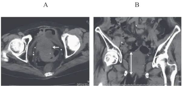
An 83 - year - old woman was admitted to a local clinic because of lower abdominal pain. As an abnormal rectal mass was observed on abdominal computed tomography (CT), she was referred to our hospital. A physical examination produced unremarkable results, and no superficial lymph node swelling was detected. Abdominal CT revealed an abnormal mass in the ventral rectum (Fig. 1A, B). Colon fiberscopy was performed, and the abdominal mass was found to be a rectal submucosal tumor (Fig. 2). An ultrasound -guided needle biopsy of the rectal submucosal tumor was per-formed via the perineum. A histological examination indicated that
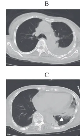
the lesion was predominantly composed of spindle - shaped cells, which were found to be diffusely positive for c- kit in a subsequent immunohistochemical analysis (Fig. 3A, B). In addition, a c-kit gene mutation (exon 11 deletion) was also detected in the speci-men. The patient’s laboratory findings on admission included a hemoglobin level of 10.7 g/dL, a carcinoembryonic antigen (CEA) level of 3.5 ng/mg, and a cancer antigen 19 - 9 (CA19 - 9) level of 5.0 U/ml. The patient did not exhibit severe anemia or elevated tumor marker levels. The possibility of rectal resection had been mentioned to the patient and her relatives, but they did not want to take this option. The patient and her relatives understood that if the tumor increased in size then the patient might develop ileus and that this would require a colostomy to be performed. The pa-tient was treated with imatinib (400 mg/day, orally), but the dose of imatinib was reduced after one week (300 mg/day, orally) as the patient suffered severe nausea and diarrhea. After that, the patient’s side effects improved. An abdominal CT scan performed at one month after the start of treatment showed that the patient’s abnormal findings had improved (a partial response (PR) accord-ing to the RECIST criteria), and chest and abdominal CT scans performed at three months after the start of treatment indicated that the patient’s abnormal findings continued to exhibit a PR, but left- sided pleural effusion was detected (Fig. 4A, B). As the patient complained of dyspnea, she underwent a thoracic cavity puncture, and a cytological examination revealed an adenocarcinoma, which was positive for an EGFR mutation (exon 19 deletion) (Fig. 5). A chest CT scan performed after the pleural effusion had been drained revealed a tumor (diameter : 22
!
20 mm) in the left lower lobe (Fig. 4C). The patient did not wish to undergo18F - fluorodeoxy glucose positron emission tomography. Brain magnetic resonance imaging did not detect any metastases. As a result, the patient was diagnosed with synchronous pulmonary adenocarcinoma (T1bNxM1a, Stage IV) and GIST (high risk).Although it was necessary to treat both the patient’s lung cancer and GIST, it was considered that administering both erlotinib (for the lung tumor) and imatinib (for the GIST) simultaneously would result in strong side effects. Thus, we decided to administer them
CASE REPORT
Efficacy of erlotinib and imatinib in a patient with a rectal
gastrointestinal stromal tumor and synchronous pulmonary
adenocarcinoma : A case report
Takanori Miyoshi1, Riho Mori2, Sachiko Amano2, Hiroyuki Sumitomo1, Mariko Aoyama1, Seiya Inoue1, Naoki Hino1, and Daisuke Wada3
1Department of Surgery, Tokushima Municipal Hospital, Tokushima, Japan,2Department of Pharmacy, Tokushima Municipal Hospital, Tokushima, Japan,3Department of Surgery, Takamatsu Municipal Hospital, Kagawa, Japan
Abstract : The synchronous existence of lung cancer and gastrointestinal stromal tumors (GIST) is considered to be extremely rare. To the best of our knowledge, this is the first report about the treatment of lung cancer and GIST with two kinds of molecular targeting drugs. An 83-year -old woman with a rectal GIST, which carried a c -kit mutation, and pulmonary adenocarcinoma, which exhibited an epidermal growth factor receptor (EGFR) mu-tation, was treated alternately with imatinib and erlotinib. Good control over both diseases was achieved for two years. The present case is not only of interest due to the rare co -occurrence of GIST and lung cancer, but also because it involved two tumors carrying different gene mutations, and both tumors were brought under control using different molecular targeting drugs. J. Med. Invest. 63 : 144-148, February, 2016
Keywords : Gastrointestinal stromal tumors, Synchronous lung cancer, Molecular targeting drugs
Received for publication October 2, 2015 ; accepted January 4, 2016. Address correspondence and reprint requests to Takanori Miyoshi, 2 -34, Kitajosanjima - cho, Tokushima city, Tokushima, 770 - 0812, Japan and Fax : +81 - 88 - 622 - 9379.
A
B
A
B
alternately. As we considered that the patient’s lung cancer was more serious than her GIST, we decided to administer the lung cancer treatment for longer periods than the GIST treatment. The patient was treated with imatinib for two weeks (300 mg/day, orally) and erlotinib (100 mg/day, orally) for four weeks in an al-ternating manner. Since side effects were expected, erlotinib was administered at a reduced dose from the beginning. The only side effect of this treatment was a mild rash (Grade 1). Good control of the lung cancer and the GIST was achieved (Fig. 6A, B), and the patient’s CEA level fell from 6.6 ng/ml to 3.3 ng/ml. The patient demonstrated a PR according to the RECIST criteria, but passed away two years after the start of therapy at another hospital. The patient’s lung cancer and rectal GIST did not worsen at the other hospital. The patient died of a urinary tract infection, but the above-mentioned therapy improved her quality of life (QOL) during the two - year treatment period.
Figure 1. An abdominal CT scan detected a tumor in the ventral rectum (arrows). A : axial view, B : coronal view
Figure 2. Colon fiberscopy showed a tumor in the rectal submucosa
Figure 3. A histological analysis demonstrated the dense proliferation of spindle - shaped cells (A : Hematoxylin and eosin (HE) staining
!
400) The positive immunohistochemical staining of c - kit (CD117) was consistent with a diagnosis of GIST (B :!
400).A
B
C
DISCUSSION
This case highlighted two important clinical issues : 1) There have only been a few reports about synchronous cases of GIST and lung cancer ; and 2) the administration of two kinds of molecular targeting drugs in an alternating manner brought both the lung cancer and GIST under control.
Regarding the first issue, GIST is a rare neoplasm that represents
about 0.1 - 1.0% of all malignant neoplasms of the gastrointestinal tract. Most GIST arise from the stomach (50 - 62%), small intestine (20 - 30%), colon (11%), or rectum (7%), while the esophagus is rarely affected (0.6 - 1%) (3, 11, 19, 20). GIST have been reported to occur synchronously, mostly with gastrointestinal carcinomas (21), but there are few reports about them occurring synchronously with lung cancer. Herein, we reported a case of synchronous pulmonary adenocarcinoma and GIST. The lung cancer was found incidentally during the treatment of the GIST with imatinib. The malignant cells in both tumors had specific gene mutations. Synchronous and asynchronous cases of GIST and other malignancies have been studied in several case series (5 - 12). Based on these reports, GIST associated with other primary malignancies occur at incidence rates ranging from 2.9% to 32.6%. The most common accompanying neoplasms are colorectal and gastric adenocarcinoma, and pan-creatic tumors (5, 9, 10). Therefore, cases in which GIST occur synchronously or asynchronously with other gastrointestinal ma-lignancies are relatively common. The high prevalence of other gastrointestinal malignancies in GIST patients is probably due to the frequent use of investigative or therapeutic surgery to treat/ investigate other malignancies. Concomitant GIST are thus usually discovered incidentally during surgery performed for other malig-nancies. Indeed, Kawanowaet al. (11) reported that microscopic GIST were found in 35% of gastric cancer patients that underwent stomach resections. However, synchronous GIST have rarely been detected in patients with lung cancer. Pandurenganet al. (6) sum-marized the clinical courses of 783 GIST patients and found only 10 cases in which lung cancer (1.3%) occurred before or after the GIST. Agaimyet al. (12) reported that lung cancer exhibited a fre-quency of 5% (26 cases) in GIST patients (486 cases). Furthermore, Figure 4. Patient’s CT findings at 3 months after the start of treatment
A : An abdominal CT scan revealed that the patient’s abnormal findings had improved slightly after 3 months’ imatinib therapy (arrow). B : A chest CT scan detected left - sided pleural effusion. C : Chest CT detected a tumor in the left lower lobe after pleural effusion drainage (arrowhead).
Figure 5. A cytological analysis resulted in a diagnosis of pulmonary adenocarcinoma (
!
100)B
A
Pontiet al. (7) and Ruka et al. (13) summarized 141 and 180 cases of GIST, respectively, and identified one and two patients, respec-tively, that developed non - small cell lung cancer after being diag-nosed with GIST. Therefore, the development of lung cancer in patients with GIST is not necessarily rare.
As for the second issue, in the present case two kinds of molecu-lar targeting drugs were administered in an alternating manner to treat the patient’s lung cancer and GIST, and these treatments were effective at bringing the tumors under control. The patient was treated with imatinib for two weeks and erlotinib for four weeks on an alternating basis. In the present case, we determined the optimal treatment strategy based on the expected side effects. The patient was initially treated with imatinib (400 mg/day, orally), but the dose was subsequently reduced to 300 mg/day (orally) since severe nausea and diarrhea appeared. After that, the patient’s side effects improved. Since side effects were expected, erlotinib was administered at a reduced dose from the beginning. Although it was necessary to treat both the patient’s lung cancer and GIST, it was considered that administering both drugs at the same time would result in strong side effects. Therefore, we decided to admin-ister them alternately. As we considered that the patient’s lung cancer was more serious than her GIST, we administered erlotinib for longer periods than imatinib. Good control of both the lung can-cer and the GIST was achieved (the patient exhibited a PR accord-ing to the RECIST criteria), but the patient died of a urinary tract infection at two years after the start of therapy. Despite this, no pleu-ral effusion or ileus occurred, which would have been indicative of the progression of the lung cancer and GIST, respectively, and the patient did not develop any treatment- related side effects. In addi-tion, the abovementioned therapy improved the patient’s QOL dur-ing the two - year treatment period.
We found an EGFR mutation in the pulmonary adenocarcinoma and c- kit mutation in the GIST in the present case. The detection of these gene mutations helped us to select treatment strategies involving molecular targeting agents that would have beneficial clinical effects on both tumors. The patient was initially treated with imatinib and erlotinib. Previous studies have attempted to detect EGFR and c- kit mutations in biphasic malignant lung tumors com-posed of primitive mesenchymal and epithelial components (17, 18). However, no coexisting mutations were found in these studies (17, 18). In the present case, the EGFR mutation status of the GIST and the c- kit mutation status of the lung tumor were not analyzed. It is therefore difficult to speculate on the interactions between the
EGFR and c- kit gene mutations and their etiological effects in the present case. Although the oncogenic interactions between the detected mutations remain to be determined, the coexistence of two different malignancies harboring specific EGFR and c- kit mu-tations in a single patient might represent useful information for future studies.
In summary, the present case is not only of interest due to the rare co - occurrence of a GIST and lung cancer, but also because it involved two tumors carrying different gene mutations, and both tumors were brought under control using different molecular tar-geting drugs.
CONFLICTS OF INTEREST STATEMENT
The authors’ state that they have no conflicts of interest.ACKNOWLEDGEMENTS
We would like to thank Dr. Kiyoku (Department of Pathology, Tokushima Municipal Hospital) for his pathological analysis and helpful discussion.
REFERENCES
1. Pidhorecky I, Cheney RT, Kraybill WG, Gibbs JF : Gastroin-testinal stromal tumors : current diagnosis biologic behavior and management. Ann Surg Oncol 7 : 70 5 - 712, 2000 2. Miettinen M, Lasota J : Gastrointestinal stromal tumors :
re-view on morphology, molecular pathology, prognosis, and dif-ferential diagnosis. Arch Pathol Lab Med 130 : 1466 - 1478, 2006
3. Tran T, Davila JA, El - Serag HB : The epidemiology of malig-nant gastrointestinal stromal tumors : an analysis of 1,458 cases from 1992 to 2000. Am J Gastroenterol 100 : 162 - 168, 2005 4. Hirota S, Isozaki K, Moriyama Y, Hashimoto K, Nishida T,
Ishiguro S, Kawano K, Hanada M, Kurata A, Takeda M, Muhammad Tunio G, Matsuzawa Y, Kanakura Y, Shinomura Y, Kitamura Y : Gain of function mutations of c- kit in human gastrointestinal stromal tumors. Science 279 : 577 - 580, 1998 5. Liu YJ, Yang Z, Hao LS, Xia L, Jia QB, Wu XT : Synchronous Figure 6. Patient’s CT findings at 6 months after the start of treatment
A : An abdominal CT scan revealed that the patient’s abnormal findings had improved slightly after 6 months’ imatinib therapy (arrow). B : A chest CT scan showed that the tumor in the left lower lobe had disappeared (arrowhead).
incidental gastrointestinal stromal and epithelial malignant tu-mors. World J Gastroenterol 15 : 2027 - 2031, 2009
6. Pandurengan RK, Dumont AG, Araujo DM, Ludwig JA, Ravi V, Patel S, Garber J, Benjamin RS, Strom SS, Trent JC : Survival of patients with multiple primary malignancies : a study of 783 patients with gastrointestinal stromal tumor. Ann Oncol 21 : 2107 - 2111, 2010
7. Ponti G, Luppi G, Martorana D, Rossi G, Losi L, Bertolini F, Sartori G, Pellacani G, Seidenari S, Boni E, Neri TM, Silini E, Tamburini E, Maiorana A, Conte PF : Gastrointestinal stromal tumor and other primary metachronous or synchronous neo-plasms as a suspicion criterion for syndromic setting. Oncol Rep 23 : 437 - 444, 2010
8. Wada Y, Koizumi T, Yokoyama T, Urushihata K, Yamamoto H, Hanaoka M, Kubo K : Synchronous gastrointestinal stromal tumor and primary lung adenocarcinoma. Intern Med 51 : 2407 - 10, 2012
9. Wronski M, Ziarkiewicz - Wroblewska B, Gornicka B, Cebulski W, Slodkowski M, Wasiutynski A, Krasnodebski IW : Synchro-nous occurrence of gastrointestinal stromal tumors and other primary gastrointestinal neoplasms. World J Gastroenterol 12 : 5360 - 5362, 2006
´
10. Liszka L, Zielinska- Pajak E, Pajak J, Golka D, Huszno J : Co-existence of gastrointestinal stromal tumors with other neo-plasms. J Gastroenterol 42 : 641 - 649, 2007
11. Kawanowa K, Sakuma Y, Sakurai S, Hishima T, Iwasaki Y, Saito K, Hosoya Y, Nakajima T, Funata N : High incidence of microscopic gastrointestinal stromal tumors in the stomach. Hum Pathol 37 : 1527 - 1535, 2006
12. Agaimy A, Wünsch PH, Sobin LH, Lasota J, Miettinen M : Oc-currence of other malignancies in patients with gastrointesti-nal stromal tumors. Semin Diagn Pathol 23 : 120 - 129, 2006 13. Ruka W, Rutkowski P, Nowecki Z, Nasierowska- Guttmejer A,
Debiec- Rychter M : Other malignant neoplasms in patients with gastrointestinal stromal tumors (GIST). Med Sci Monit 10 : LE13 - LE14, 2004
14. Antler AS, Ough Y, Pitchumoni CS, Davidian M, Thelmo W : Gastrointestinal metastases from malignant tumors of the lung. Cancer 49 : 170 - 172, 1982
15. Nilsson B, Bümming P, Meis - Kindblom JM, Odén A, Dortok A, Gustavsson B, Sablinska K, Kindblom LG : Gastrointestinal stromal tumors : the incidence, prevalence, clinical course, and prognostication in the preimatinib mesylate era : a population-based study in western Sweden. Cancer 103 : 821 - 829, 2005 16. Duchateau CS, Stokkel MP : Second primary tumors involving
non - small cell lung cancer : prevalence and its influence on survival. Chest 127(4) : 1152 - 1158, 2005
17. Macher - Goeppinger S, Penzel R, Roth W, Dienemann H, Thomas M, Schnabel PA, Schirmacher P, Bläker H : Expres-sion and mutation analysis of EGFR, c- KIT, andβ-catenin in pulmonary blastoma. J Clin Pathol 64 : 349 - 353, 2011 18. Bergmann F, Moldenhauer G, Herpel E, Gaida MM, Strobel
O, Werner J, Esposito I, Müerköster SS, Schirmacher P, Kern MA : Expression of L1 CAM, COX - 2, EGFR, c- KIT and Her2/ neu in anaplastic pancreatic cancer : putative therapeutic tar-gets? Histopathology 56 : 440 - 448, 2010
19. Gupta P, Tewari M, Shukla HS : Gastrointestinal stromal tu-mor. Surg Oncol 17 : 129 - 38, 2008
20. Miettinen M, El - Rifai W, Sobin HL, Lasota J : Evaluation of malignancy and prognosis of gastrointestinal stromal tumors : a review. Hum Pathol 33 : 478 - 83, 2002
21. Ferreira SS, Werutsky G, Toneto MG, Alves JM, Piantá CD, Breunig RC, Brondani da Rocha A, Grivicich I, Garicochea B : Synchronous gastrointestinal stromal tumors (GIST) and other primary cancers : case series of a single institution experience. Int J Surg 8 : 314 - 7, 2010