2) are the causative agents of acquired immune deficiency syndrome. Our interests have focused on HIV entry into target cells and HIV cell tropism. The cell tropism of HIV and the determinants of a cells susceptibility to HIV infection have been mainly explained by the combination of HIV surface proteins,Env,and expression of CD4 and coreceptors on the target cells. The coreceptors are molecules belong-ing to the G protein-coupled 7-transmembrane receptors, especially chemokine receptors. We propose that many researchers working in this field have noticed that our system of using a human glioma cell line,NP-2,has been quite useful for the identification of HIV corece-ptors,for the determination of coreceptor usage in HIV strains and for the isolation of primary HIV-1 strains, and for the titration of in-fectivity. The properties of the assay systems using NP-2 cells that we have developed are summarized in this review through an introduction of some of our work.(Kitakanto Med J 2012;62:1∼14)
Key words: HIV-1, CD4 receptor, coreceptor use, AIDS
Introduction
Human immunodeficiency virus type 1 (HIV-1) and type 2 (HIV-2) are retroviruses and are the causa-tive agents of acquired immune deficiency syndrome (AIDS). Retroviruses are grouped into three sub-families: oncovirinae, lentivirinae (lentivirus) and spumavirinae. The HIVs and simian im-munodeficiency viruses (SIVs) are members of the lentivirus subfamily. Lentiviruses are highly mutable, similar to influenza virus or hepatitis C virus,and thus the development of effective vaccines against lentivir-uses has not yet been achieved.
HIV-1 was isolated in 1983 by a French research group. As early as 1984, CD4 was identified as a factor necessary for HIV-1 entry,namely,as a receptor for HIV-1. It was soon realized that not all human cells positive for CD4 were susceptible to HIV-1 infec-tion, and another factor or secondary factor was
deemed to be necessary for HIV-1 entry into target cells in addition to CD4 (Fig.1A and 1B). After 12 years of intense competition to identify a second rece-ptor, it was reported in 1996 that a CXC chemokine receptor, CXCR4, could confer cells positive for CD4 but resistant to HIV-1 infection with the capacity to be infected by T-cell-line-tropic HIV-1 (HIV-1 strains preferentially grow in most T-cell lines). Then, a CC chemokine receptor, CCR5, was also found to be an indispensable factor for the entry of macrophage -tropic HIV-1 strains. Several papers that reported the chemokine receptors as HIV-1 coreceptors were ranked at or near the top of the list of the most cited papers of 1996. HIV-1, using CCR5 as a corece-ptor(i.e.,the CCR5-tropic virus),plays a critical role in the establishment of HIV-1 infection in humans as described below.
1 Department of Virology and Preventive Medicine,Gunma University Graduate School of Medicine,3-39-22 Showa-machi,Maebashi, Gunma 371-8511, Japan
Received : December 8, 2011
Address: HIROO HOSHINO Advanced Scientific Research-Leaders Development Unit, Gunma University Graduate School of Medicine, 3-39-22 Showa-machi, Maebashi, Gunma 371-8511, Japan
Viral and genomic structures of HIV-1
The size of HIV-1 is approximately 100 nm in diameter. There are three major structural protein genes, gag (p17, p24 and p15/p7), pol (RT : reverse transcriptase and protease), and env, encoded by its RNA genome. The envelope (Env) proteins gp120 and gp41 are localized on and throughout the viral membrane, which is mainly comprised of a lipid bilayer derived from host cells (Fig.2).Upon HIV-1 entry into cells, the CD4-binding region,C4 of gp120,binds to domain 1 of CD4,which leads to a conformational change of gp120 and permits the V3 region or loop of the Env protein to interact with its coreceptor, CCR5 or CXCR4. This binding results in dislocation of the viral transmembrane pro-tein gp41, and the viral particles (virions) will be connected to the cellular membrane through the fusion domain of gp41. This fusion will lead to further membrane fusion between virions and cells, and viral
Fig.1 NP-2, NP-2/CD4 and NP-2/coreceptor cells are resistant to HIV-1 infection while NP-2/CD4/ coreceptor cells are highly susceptible to infection with coreceptor-compatible HIV-1 strains(A,B, C and D). According to the trans-receptor mechanism,after infection of a mixture of NP-2/CD4 and NP-2/coreceptor cells with HIV-1, a few NP-2/coreceptor cells are expected to be infected with HIV-1 (E).
cores harboring HIV-1 genomic RNA enters into the cytoplasm of target cells, (Fig.3). Then, viral RNA is reverse transcribed, and the resultant viral DNA integrates into cellular genome, and thus, the early steps of HIV-1 infection are completed. Then, the late steps, namely, the transcription of viral RNA and synthesis of viral proteins, begin.
Importance of CCR5 in the establishment
of HIV-1 infection in vivo
CXCR4 and CCR5 belong to the G protein-cou-pled 7-transmembrane receptor (GPCR) family (Fig. 4A). In the asymptomatic phases of HIV-1 infection, CCR5-tropic (R5) viruses play a pivotal role in the establishment and persistence of HIV-1 infection in vivo. During the disease progression to symptomatic phases such as AIDS,the shift in the coreceptor use of HIV-1 frequently takes place in subjects infected with subtype B HIV-1. Subtype B is a major subtype of HIV-1 prevalent in developed countries like Japan or USA. CXCR4-tropic or dual-tropic (CCR5- and CXCR4-tropic) (R5X4) variants or HIV-1 strains using co-receptors other than CCR5 and CXCR4 are often detected in patients with early and late stages of immunodeficiency.
Some people have been found to be highly
resis-tant to HIV-1 infection,despite highly risky behaviors associated with contracting HIV-1, or to be relatively refractory to disease progression or to AIDS-related syndromes after infection. Molecular epide-miological studies on some of these people revealed that some individuals have a 32-base-pair deletion in the second extracellular loop of CCR5 gene. This deletion induces a premature termination in the second extracellular loop during translation of CCR5 protein (Fig.4B).
Those homozygous for this allele are known to be highly resistant to HIV-1 infection. Approximately 1% of Caucasians are heterozygous for this CCR5-delta-32 (CCR5Δ32) allele, and the peripheral blood lymphocytes (PBMCs) of subjects homozygous at this allele are highly resistant to infection by CCR5-tropic but not CXCR4-tropic HIV-1, even in tissue culture experiments. These findings indicate that CCR5 is required for the establishment of HIV-1 infection in humans.
The lack of CCR5 may not appreciably influence immunological functions of humans, and thus, CCR5 has been thought to be a good target for the develop-ment of new types of anti-HIV-1 agents that will block HIV-1 entry. Such an anti-CCR5 drug, Maraviroc, was approved in Japan in 2008. For the clinical use
Fig.2 Structure of HIV-1.
(A) Schematic presentation of HIV-1 virion. (B) Genetic structure of HIV-1 genome.
of this drug, an easy, rapid and accurate[TL2]assay system is necessary for determining the co-receptor use of HIV-1 strain, as the administration of this drug should be limited to subjects that are solely infected with CCR5-tropic HIV-1. The NP-2 cell system described below will be useful for this determination.
GUN strains of HIV-1
We cocultivated human T-cell leukemia virus type 1(HTLV-1)-positive MT-4,C8166 or ATL-3I T cell lines with PBMCs of HIV-1 infected subjects. HTLV-1-positive T cells are known to be highly sensitive to HIV-1 as they express abundant quantities
of CD4 and CXCR4 but not CCR5. From these cells, we isolated many HIV-1 strains and named them GUN viruses after Gunma, e.g.,GUN-1,GUN-2,GUN-3, GUN-4, GUN-5, GUN-6 and GUN-7.
GUN-6 was the first HIV-1 isolate from a foreign patient diagnosed in our University Hospital. Dr.H. Tanami,then a professor at the Central Laboratory in the Hospital,supplied us with this blood sample. We cocultivated the PBMCs from this sample with HTLV-1-positve MT-4 and ATL-3I cells and soon noticed that these T cells were positive for HIV-1 antigens. This viral strain was named GUN-6. The culture supernatant of these T cells was inoculated onto U937
Fig.3 Roles of CD4 and a coreceptor in HIV-1 infection.
(A) Interaction of the CD4-binding region and V3 loop of HIV-1 gp120 with CD4 and a coreceptor, respectively, of HIV-1-susceptible cells.
human monocytic cells to establish a cell line persist-ently infected with HIV-1,as HTLV-1-positive T cells could not survive once HIV-1 infection spread among them. A scanning electron microgram of HIV-1-posi-tive U937 cells was supplied from Dr.Tanami (Fig.5).
Selection of the NP-2 cell line as an
indicator cell line for HIV/SIV infection
We examined more than 20 human cell lines,
including brain-derived cell lines after transduction with a CD4-expression vector, to determine whether they became susceptible to T-cell-tropic, dual-tropic or macrophage-tropic HIV-1 strains. Most cell lines became susceptible to T-cell-tropic (CXCR4-tropic) viruses, such as the IIIB strain. A few cell lines were still resistant to typical HIV-1 strains that we had tested, and the NP-2 cell line was among them. No cell lines tested by us were susceptible to
CCR5-Fig.4 Schematic presentation of chemokine receptor CCR5. Structures of CCR5 (A) and CCR5Δ32 mutant with 32 base-pair deletion in the genome of the second extracel-lular loop (B). This deletion leads to premature termina-tion of CCR5 at the second extracellular loop.
Fig.5 Scanning electron micrograph of a human monocytic cell, U937, persistently infected with an HIV-1 strain, GUN-6.
Fig.6 Infection of NP-2/CD4/CCR5 cells with HIV-1.
(A) NP-2/CD4/CCR5 are cells highly susceptible to CCR5-tropic HIV-1 but completely resis tant to CXCR4-tropic HIV-1.
(B) NP-2/CD4/CCR5/iGFP cells were established after transfection of NP-2/CD4/CCR5 cells with the plasmid containing GFP gene fused with the nuclear localization signal of HIV-1 Rev. GFP is expressed under the control of HIV-1 long-terminal repeat (LTR) and tran sported into nuclei. Cell clones expressing GFP in the presence of Tat protein of HIV/SIV or after productive infection with HIV/SIV were selected and used for infection as described above.
-tropic HIV-1.
When NP-2/CD4 cells were further transduced with CCR5 or CXCR4,they became highly susceptible to CCR5-tropic or CXCR4-tropic HIV-1 strains,and large syncytia often formed a few days after inocula-tion of HIV-1,HIV-2 or SIV (Figs.1D,6 and 10B). To detect new HIV/SIV coreceptors, we transduced many GPCR genes and found that chemokine rece-ptors, such as CCR8, CXCR5, and XCR1,or the non -chemokine receptors FPRL1,GPR1,and RDC1 were new types of coreceptors for HIV-1,HIV-2 or SIV. Fig.7 shows the phylogenetic relationship between the HIV/SIV coreceptors and chemokine receptors. Most chemokine receptors can function as the coreceptors (Table 1).
Establishment of NP-2 cells with
inducible GFP (iGFP) cells
Several indicator cell systems in which indicator proteins, such as βgalactosidase or green fluorescent protein (GFP), are induced after the establishment of infection of HIV or SIV have been reported. How-ever, it has been difficult to accurately determine
CCR5 or CXCR4 usage in HIV/SIV strains using the indicator cell systems like the HeLa or HOS cell line reported previously because these cell lines are already expressing HIV/SIV co-receptors other than CCR5. Systems using T-cell lines, such as H9 and CEM can detect CXCR4-tropic HIV-1 but not CCR5-tropic HIV-1.
To work around these limitations,we have estab-lished an indicator cell system using the NP-2 cell line and GFP for direct detection of cells infected with HIV and SIV and for strict determination of their CCR5 and CXCR4 use. After transfection with a plasmid containing HIV-1 long terminal repeat and the green fluorescent protein (GFP) gene, the NP-2 indicator cells with inducible GFP (NP-2/iGFP cells) were established from cell clones that were mostly negative for GFP but that had become positive for GFP after HIV-1 infection,namely,after production of the HIV/ SIV transactivator protein Tat. One day after the infection of these cells with HIV-2 or SIV and HIV-1, GFP-positive cells could be identified (Fig.6B). The indicator cells formed GFP-positive syncytia 12 hours after cocultivation with HeLa cells expressing HIV-1
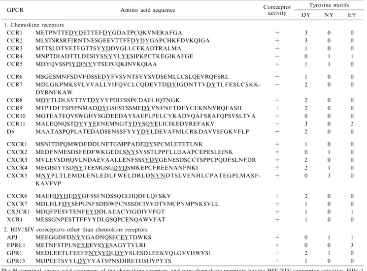
Table 1 Properties of the N-terminal regions of HIV/SIV coreceptors
GPCR Amino acid sequence Coreceptoractivity Tyrosine motifs DY NY EY 1. Chemokine receptors CCR1 METPNTTEDYDETTEFDYGDATPCQKVNERAFGA + 3 0 0 CCR2 MLSTSRSRFIRNTNESGEEVTTFFDYDYGAPCHKFDVKQIGA + 3 0 0 CCR3 MTTSLDTVETFGTTSYYDDVGLLCEKADTRALMA + 1 0 0 CCR4 MNPTDIADTTLDESIYSNYYLYESIPKPCTKEGIKAFGE − 0 1 1 CCR5 MDYQVSSPIYDINYYTSEPCQKINVKQIAA + 1 1 0 CCR6 MSGESMNFSDVFDSSEDYFVSVNTSYYSVDSEMLLCSLQEVRQFSRL − 1 0 0 CCR7 MDLGKPMKSVLVVALLVIFQVCLCQDEVTDDYIGDNTTVDYTLFESLCSKK DVRNFKAW − 2 0 0 -CCR8 MDYTLDLSVTTVTDYYYPDIFSSPCDAELIQTNGK + 2 0 0 CCR9 MTPTDFTSPIPNMADDYGSESTSSMEDYVNFNFTDFYCEKNNVRQFASH + 2 0 0 CCR10 MGTEATEQVSWGHYSGDEEDAYSAEPLPELCYKADVQAFSRAFQPSVSLTVA + 0 0 0 CCR11 MALEQNQSTDYYYEENEMNGTYDYSQYELICIKEDVREFAKV − 3 0 2 D6 MAATASPQPLATEDADSENSSFYYYDYLDEVAFMLCRKDAVVSFGKVFLP + 2 0 0 CXCR1 MSNITDPQMWDFDDLNFTGMPPADEDYSPCMLETETLNK + 1 0 0 CXCR2 MEDFNMESDSFEDFWKGEDLSNYSYSSTLPPFLLDAAPCEPESLEINK + 0 1 0 CXCR3 MVLEVSDHQVLNDAEVAALLENFSSSYDYGENESDSCCTSPPCPQDFSLNFDR + 2 0 0 CXCR4 MEGISIYTSDNYTEEMGSGDYDSMKEPCFREENANFNKI + 2 1 0 CXCR5 MNYPLTLEMDLENLEDLFWELDRLDNYNDTSLVENHLCPATEGPLMASF KAVFVP + 0 3 0 -CXCR6 MAEHDYHEDYGFSSFNDSSQEEHQDFLQFSKV + 2 0 0 CXCR7 MDLHLFDYSEPGNFSDISWPCNSSDCIVVDTVMCPNMPNKSVLL + 1 0 0 CX3CR1 MDQFPESVTENFEYDDLAEACYIGDIVVFGT + 1 0 1 XCR1 MESSGNPESTTFFYYDLQSQPCENQAWVFAT + 1 0 0
2. HIV/SIV coreceptors other than chemokine receptors
APJ MEEGGDFDNYYGADNQSECEYTDWKS + 0 1 1
FPRL1 METNFSTPLNEYEEVSYESAGYTVLRI + 0 0 3
GPR1 MEDLEETLFEEFENYSYDLDYYSLESDLEEKVQLGVVHWVSl + 2 1 0
GPR15 MDPEETSVYLDYYYATSPNSDIRETHSHVPYTS + 1 0 0
The N-terminal amino acid sequences of the chemokine receptors and non-chemokine receptors having HIV/SIV coreceptor activities,HIV-1 coreceptor activities and the presence of DY (DY or YD), NY (NY or YN) and EY (EY or YE) motifs are shown.
Env and Tat proteins. We have introduced this type of coculture experiments into the curriculum for medical students: it can be safely done by them and will help them to understand the infection mechanism of HIV-1. The NP-2/iGFP indicator cells enabled us to detect a small number of cells or a single focus of cells infected with HIV and SIV,to monitor the spread of their infection and to determine their CCR5/ CXCR4 usage without cell fixation.
Lack of the trans-receptor mechanism
in HIV-1 infection
We established NP-2 cells expressing CD4,CCR5 or CXCR4 alone to examine whether NP-2 cells ex-pressing CD4 or a coreceptor alone were more suscep-tible to cell-free virus infection or syncytium forma-tion induced by HIV-1 Env than NP-2 cells negative for both. For this purpose,we used cocultures of NP -2 cells expressing CD4 and those expressing CCR5 or CXCR4 and infected them with HIV-1 or cocultured these NP-2 cells with HIV-1 Env-expressing cells.
It has been previously reported that HIV-1 can infect cells using CD4 expressed on the surface of one cell and CCR5 or CXCR4 expressed on another cell. This type of infection has been referred to as the trans-receptor mechanism of virus infection (Fig.1E). According to the original report, up to 4% of
CD-4-negative, coreceptor-positive (CD4 coreceptor ) adherent cells can be infected with HIV-1 when CD4-positive human T cells had been cocultured with these adherent cells and infected with HIV-1.
We tried to confirm this mechanism using similar types of cells as those used in that report. That is, we used several assay systems to examine whether HIV-1 can infect CD4 coreceptor cells via CD4 expressed on neighboring cells. Firstly,C8166 human T cells, expressing CD4 but not CCR5, were mixed with NP-2/CCR5/iGFP(CD4 coreceptor cells)or NP-2/iGFP (CD4 coreceptor cells as control) indicator cells, and inoculated with a CCR5-tropic HIV-1 strain. No GFP-positive cells(<0.01%)were detected in these iGFP cells after 3 days post-inocula-tion (Fig.8A). In contrast,NP-2/CD4/CCR5/iGFP cells (CD4 coreceptor control cells) alone or mixed with C8166 cells yielded numerous GFP-posi-tive cells and syncytia after infection (Fig.8A). Thus, our data indicate that HIV-1 cannot infect CD4 CCR5 cells using the CD4 of adjacent cells.
We also confirmed this using DNA PCR. NP-2/ CCR5/iGFP, NP-2/iGFP, or NP-2/CD4/CCR5/ iGFP cells were mixed with C8166 cells,and inoculat-ed with a CCR5-tropic HIV-1 strain. After being in culture for 2 days, cellular DNAs were isolated and subjected to PCR. No HIV-1-specific PCR products
Fig.7 Phylogeny of the chemokine receptor genes and the HIV/SIV coreceptors not belonging to them. The chemokine receptors reported to function as HIV/SIV coreceptors are boxed or underlined. The major coreceptors CCR5 and CXCR4 and other so-called minor coreceptors relatively used by primary HIV-1 strains according to our analyses are boxed.
were detected using NP-2/CCR5/iGFP and NP-2/ iGFP cell samples (Fig.8B). HIV-1-specific PCR systems were sensitive enough to detect PCR bands when a mixture of NP-2/CD4/CCR5/iGFP and C8166 cells had been infected with 1/1,000-diluted virus. Thus,these results do not support that the trans -receptor mechanism of HIV-1 takes place frequently. We conclude that the expression of CD4 and a suitable coreceptor on the same cell is essential for the estab-lishment of HIV-1 infection under our culture condi-tions.
We also reported that some HIV-2 strains can efficiently infect CD4 coreceptor NP-2 cells in the presence of soluble CD4 (sCD4). Thus, NP-2/ CCR5/iGFP cells were infected with CCR5-tropic strain in the presence of sCD4. Approximately 2% of NP-2/CCR5/iGFP cells expressed GFP on day 3 post -infection (Fig.8A). These cells also gave discrete HIV-1-specific PCR bands (Fig.8B). Collectively, our data demonstrate that sCD4,but not cell-associat-ed CD4,can help HIV-1 to infect CD4 coreceptor cells.
Incorporation of HIV-1 resistant cells
into syncytia induced by HIV-1
HIV-1-resistant cells expressing either CD4 or a coreceptor are often found surrounding
HIV-1-suscep-tible cells, expressing both CD4 and a compaHIV-1-suscep-tible coreceptor, in vivo. When HIV-1-resistant NP-2 cells expressing CD4 or a coreceptor or lacking both were mixed with CD4 coreceptor NP-2 cells and inoculated with HIV-1, a small number of all these HIV-1-resistant cells(up to 2% under our assay condi-tions) were similarly incorporated into the syncytia induced by HIV-1, indicating a CD4-and coreceptor -independent incorporation of HIV-1-resistant cells into the syncytia. This incorporation was significant-ly impaired by the transfection of these cells with siRNAs for adhesion molecules,integrin β1 or cadher-in-11. Our study demonstrates that HIV-1-resistant cells can be incorporated into syncytia induced by HIV-1 and this incorporation may partially be mediat-ed through the adhesion molecules,although infection of HIV-1 resistant cells with cell-free HIV-1 will hardly take place even when these cells are co-cultured with HIV-1-susceptible cells. Syncytia have often been detected in the brain of HIV-1-infected subjects. The presence of syncytia is reported to be a hallmark of HIV-1 replication in the brain.
Promiscuous relationship between
chemokines and their receptors
As shown in Fig.9,many chemokines will bind to multiple chemokine receptors. MIP-1, MCP-2,
Fig.8 Lack of trans-receptor mechanism of HIV-1 infection.
(A) CD4-positive C8166 cells overlaid onto NP-2/CCR5/iGFP, NP-2/iGFP and NP-2/CD4/CCR5 cells and infected with CCR5-tropic HIV-1 strain. NP-2/CCR5 cells were infected with HIV-1 in the presence of sCD4 (10μg/ml). The cells were examine three days after HIV-1 inoculation.
(B) Mixtures of C8166 cells with NP-2/CCR5/iGFP,NP-2/iGFP and NP-2/CD4/CCR5 cells were infected with undiluted (1/1) or serially diluted HIV-1 as described above. HIV-1 DNA was detected 2 days after inocula tion by DNA PCR specific for HIV-1.
-MCP-4, RANTES, etc. will bind to CCR5. This relationship has been called promiscuous. I propose that this property is important for permitting the HIV -1 variability and its persistence in humans. HIV-1 is highly mutable, especially in its V3 region ; however, this region should also recognize a co-receptor,such as CCR5 (Figs. 1 and 2).
Generally speaking, a region necessary for an important function should not be mutable. Because CCR5 is promiscuous in its ligands, it may recognize HIV-1 strains with mutated V3 sequences. The V3 region is also known to be a major immunodominant domain, and human antibodies or mouse monoclonal antibodies that can neutralize HIV-1 can recognize this region. Thus, it is expected that HIV-1 mutants that can still recognize CCR5 can readily become immune escape mutants. The use of chemokine receptors,such as HIV/SIV coreceptors,may favor the survival and propagation of HIV/SIV mutants.
Use of the co-receptors by HIV-1
present in humans
We have made a panel of NP-2/CD4 cells express-ing various coreptors, to determine which coreceptors are used by HIV-1 present in humans. We have established NP-2/CD4 cells expressing CCR1, CCR2b, CCR3, CCR4, CCR5, CCR6, CCR7, CCR8, CCR9b, CCR10, CCR11, CXCR1, CXCR2, CXCR3,
CXCR4, CXCR5, CXCR6, XCR1, D6, APJ, FPRL1, GPR1, RDC1, etc.
Then, this panel of NP-2 cells was cocultivated with PBMC samples of 17 HIV-1-infected Japanese patients and examined for the establishment of HIV-1 infection by immunofluorescence assays to HIV-1 antigens. Twelve PBMC samples gave positive results. HIV-1 antigen-positive NP-2/CD4/CCR5 cells were obtained when cocultured with nine PBMC samples. Five and four samples gave positive results with the NP-2/CD4/CXCR4 and NP-2/CD4/GPR1 cells, respectively.
The HIV-1 virus present in the culture super-natants of the NP-2/CD4/CCR5 or NP-2/CD4/ CXCR4 cells that were positive for HIV-1 antigens were grown on NP-2/CD4 cells expressing CCR1, CCR3, CCR8, D6 or FPRL1. Thus, in addition to the chemokine receptors CCR5 and CXCR4, the chemokine receptors CCR8 and D6 and the non -chemokine receptors GPR1 and FPRL1 were used by HIV-1 infections in humans. NP-2 cells will be useful for isolating the primary HIV-1 strains from patients.
Characterization of HIV-1 variants
that can grow in U87/CD4 cells
I have been interested in HIV-1 replication in the human brain because neurological disorders are
fre-Fig.9 The chemokine receptors and their major ligands. CC chemokine(CCL)receptors,CXC chemo-kine (CXCL) receptors and CX3C and XC chemochemo-kine receptors are shown. Several CCRs recognize multiple ligands.
quently found in humans or animals infected with retroviruses. HTLV-1 causes HTLV-1-associated myelopathy(HAM)and adult T-cell leukemia(ATL). Nearly half of the HIV-1-infected subjects with or without immunodeficiency have been reported to develop some type of neurological disorder during the clinical course of infection with HIV-1.
Thus, we established several glioma cell lines transduced with CD4,including NP-2/CD4 and U87/ CD4, and examined whether these cells are infectable with HIV-1 as described above. The majority of glioma cell lines expressing CD4 were susceptible to CXCR4-tropic (X4) HIV-1 strains, although U87/ CD4 and NP-2/CD4 cells were highly resistant to HIV-1 strains. We had repeatedly cocultivated U87/ CD4 cells with human T cells infected with HIV-1 GUN primary isolates and in some cases, variant viruses were isolated. No viral mutants that could grow in NP-2/CD4 cells,however,have been isolated by us so far.
We isolated HIV-1 variants, such as GUN-1v, 4v and 7v, from the 1wt, GUN-4wt and GUN-7wt wild-type viruses,respectively,and then determined the variation of the genomic sequence
Fig.10 GPR1 as a new type of HIV-1 coreceptor.
(A ) Detection of GPR1 RNA by RT-PCR. The brain-derive fibroblast-like cells, BT20-N and BT3,glioblas toma cells transduced with CD4,U87/CD4,HTLV-1-positive and CD4-positeve T-cells,C8166,and human osteosarcoma cells,S+L-HOS,are positive for GPR1 RNA. These cells are susceptible to a variant of GUN-1wt strain, GUN-1v.
( B ) NP-2/CD4/GPR1 cells are susceptible to an HIV-1 strain,GUN-1v,and an HIV-2 strain,ROD,and formed many syncytia in a few days after infection. NP-2/CD4 cells are resistant to all HIV/SIV strains including GUN-1 and ROD we have tested.
-Fig.11 Isolation of GUN-1, GUN-4 and GUN-7 variants that can grow in U87/CD4 cells and production HIV-1 point-mutants at the tip of V3 region. The single point mutations affect coreceptor use from CCR5 to GPR1.
of the GUN viruses (Fig.11). We identified the genetic variation responsible for this phenotype,which turned out to be a single point mutation at the tip of the V3 region,from GPGR to GSGR (from proline to serine) of GUN-1wt and GUN-1v. Sequence ana-lyses revealed that in the cases of the GUN-4v and GUN-7v strains, GPGR to GAGR and GPGR to GTGR point mutations, respectively, were present.
Identification of GPR1 as a coreceptor
in infection of brain-derived cells
with HIV-1 variants
We established NP-2/CD4 cells expressing GPR1 and compared their susceptibility to GUN-1v variants and GUN-1 mutants,GUN-1/S,GUN-1/T,GUN-1/ A, GUN-1/L and GUN-1/R, harboring point muta-tions at the tip of the V3 region (Fig.11). NP-2/ CD4/GPR1 and U87/CD4 cells and brain-derived fibroblast-like cells (BT-20N cells) showed a similar HIV-1 susceptibility pattern to the HIV-1 variants and mutants(Fig.12). These results indicate that a single point mutation in the V3 region can determine the coreceptor usage or cell tropism of HIV-1.
We also examined GPR1 expression in various cells and their susceptibilities to GUN-1 variants and mutants. Figure 10A shows that cells expressing GPR1, BT-20N, BT-3, U87/CD4, C8166 and S+L-HOS, are susceptible to GUN-1v virus. These find-ings indicate that GPR1 is a coreceptor in the infection of brain-derived cells. NP-2/CD4/GPR1 cells were also susceptible to the ROD strain of HIV-2(Fig. 10B). Many HIV-2 and SIV strains use GPR1 as a coreceptor.
Inhibition of HIV-1 infection by the
N-terminal peptide of GPR1
As we noticed that the N-terminal sequences of GPCRs were important for the function of the HIV/ SIV coreceptors(Table 1),these sequences were expect-ed to bind to HIV/SIV virions. Therefore, we synthesized the N-terminal peptides of the coreceptors CCR5, CXCR4 and GPR1 and examined whether these peptides could affect HIV-1 infection. Un-expectedly, only the GPR1 peptide, consisting of N-terminal 27 amino acids, but not the others, inhibited infection of cells with not only GPR1-tropic viruses,
Fig.12 We isolated HIV-1 variants, GUN-1v, which could grow in U87/CD4 cells, from GUN-1wt strains. IIIB is CXCR4-tropic HIV-1 and SF162 and BaL are CXCR5-tropic HIV-1. GUN-1/P,GUN-1/S,GUN-1/T,GUN-1/A, GUN-1/L and GUN-1/R are point mutants at the tip of V3 region as shown in Fig.12. C8166, BT-20N, U87/CD4,NP-2/CD4/GPR1,NP-2/CD4 and NP-2 GPR1 cells were infected with HIV-1 strains and examined by indirect immunofluorescence using HIV-1-positive human sera as the first antibody. GUN-1/S, GUN-1/T, and GUN-1/A, plated onto BT-20N, U87/CD4 and NP-2/CD4/GPR1 cells as well as the human T-cell line C8166 expressing CXCR4 but not CCR5. IIIB,BaL and SF162 strains did not plated onto BT-20N,U87/CD4 and NP-2/CD4/GPR1 cells.
but also R5 and X4 viruses and R5-X4 dual-tropic viruses (Fig.13).
Trial to identify new HIV/SIV co-receptors
Approximately 20 GPCRs have been shown to function as HIV/SIV coreceptors (Fig.7). There may be another coreceptor belonging to GPCR family because it is one of the largest gene families present in the human genome. We have examined the N-termi-nal regions of the reported HIV/SIV coreceptors and noticed the frequent presence of tyrosine (Y) associat-ed with aspartic acid (D), asparagine (N) or glutamic acid (E). Namely, tyrosine motifs, DY, NY, or EY, are often found in the NTRs of the coreceptors(Table 1).After screening more than 900 different GPCRs, we selected 13 eligible candidates, including CCR6, CKR-L3, DRD, G2A, GPR12, GPR25, HCRTR2, LPAR2, LPAR3, NPY5R, OXGR1, PROKR1 and PROKR2. DNA microarray analyses of NP-2/CD4/ CCR5 cellular RNA showed that these cells abundant-ly express GPR12, NPY5R, OXGR1 and PROKR1. Because NP-2/CD4 cells were highly resistant to all of the HIV/SIV strains we tested,these GPCRs would not function as an HIV/SIV co-receptor under our assay conditions. The other candidate GPCRs, CCR6, CKR-L3, DRD2, G2A, GPR25, HCRTR2, LPAR2, LPAR3 and PROKR2 are now under investigation. Although so-called minor coreceptors may not play an
important role in HIV-1 infection, this type of infor-mation will give us insight into why HIV/SIV uses GPCRs as coreceptors for infection.
We have sent NP-2 cell sublines expressing one of the HIV/SIV coreceptors as well as CD4, especially NP-2/CD4/CCR5 and NP-2/CD4/CXCR4, to many researcher groups in Japan and several countries as shown by several references listed below. We are pleased to send them if requested.
Acknowledgements
I have been studying at the Department of Hygiene, Gunma University School of Medicine,later reorganized as the Department of Virology and Preventive Medicine, Gunma University Graduate School of Medicine since 1984. My thanks are offered for the understanding, assistance and collaboration to my works by the staff members of the Department,the Faculty and the University, graduate students, undergraduate students, researchers, technical assistants and secretaries. Most of the figures and the table used in this review were made by students and members of my lab. Especially, I have to thank Dr. Toshiro Kumanishi at Niigata University for providing me with the NP-2 cell line.
References
1. Barre-Sinoussi F, Chermann JC, Rey F, Nugeyre MT, Chamaret S, Gruest J, Dauguet C, Axler-Blin C, Vezinet-Fig.13 Effects of terminal peptides of the coreceptors on HIV-1 infection. The 27 amino-acid peptide of GPR1
N-terminal,but not the N-terminal sequence of CCR5 (R5),CXCR4 (X4)or CCR3 (R3)peptide,markedly inhibited infection of NP-2/CD4/GPR1,NP-2/CD4/CXCR4,NP-2/CD4/CCR3 and NP-2/CD4/CCR5 cells with various HIV-1 strains, such as IIIB, GUN-1wt, GUN-1Ser or BaL.
human immunodeficiency virus entry and infection in CD4-positive human brain and skin cells. J Virol 1990; 64: 215-221.
5. Feng Y,Broder CC,Kennedy PE,Berger EA : HIV-1 entry cofactor: functional cDNA cloning of a seven-transmem-brane, G protein-coupled receptor. Science 1996; 272: 872-877.
6. Deng H,Liu R,Ellmeier W,Choe S,Unutmaz D,Burkhart M,Di Marzio P,Marmon S,Sutton RE,Hill CM,Davis CB, Peiper SC,Schall TJ,Littman DR,Landau NR : Identifica-tion of a major co-receptor for primary isolates of HIV-1. Nature 1996; 381: 661-666.
7. Dragic T, Litwin V, Allaway GP, Martin SR, Huang Y, Nagashima A, Cayanan C, Maddon PJ, Koup RA, Moore JP,Paxton WA : HIV-1 entry into CD4+ cells is mediated by the chemokine receptor CC-CKR-5. Nature 1996; 381: 667-673.
8. Choe H, Farzan M, Sun Y, Sullivan N, Rollins B, Ponath PD,Wu L,Mackay CR,LaRosa G,Newman W,Gerard N, Gerard C, Sodroski J: The beta-chemokine receptors CCR3 and CCR5 facilitate infection by primary HIV-1 isolates. Cell 1996; 85: 1135-1148.
9. Wyatt R, Sodroski J, The HIV-1 envelope glycoproteins: fusogens, antigens, and immunogens, Science 1998; 280: 1884-1888.
10. Clapham PR, McKnight A : Cell surface receptors, virus entry and tropism of primate lentiviruses. J Gen Virol 2002; 83: 1809-1829.
11. Esser U, Speck RF, Deen KC, Atchison RE, Sweet R, Goldsmith MA : Molecular function of the CD4 D1 domain in coreceptor-mediated entry by HIV type 1. AIDS Res Hum Retroviruses 2000; 16: 1845-1854. 12. Hartley O, Klasse PJ,Sattentau QJ,Moore JP: V3: HIVs
switch-hitter. AIDS Res Hum Retroviruses 2005; 21: 171-189.
13. Cheng-Mayer C, Quiroga M,Tung JW,Dina D,Levy JA : Viral determinants of human immunodeficiency virus type 1 T-cell or macrophage tropism,cytopathogenicity,and CD4 antigen modulation. J Virol 1990; 64: 4390-4398. 14. Kiselyeva Y,Nedellec R,Ramos A,Pastore C,Margolis LB,
Mosier DE : Evolution of CXCR4-using human im-munodeficiency virus type 1 SF162 is associated with two unique envelope mutations. J Virol 2007; 81: 3657-3661. 15. Kuiken CL, de Jong JJ, Baan E, Keulen W, Tersmette M, Goudsmit J: Evolution of the V3 envelope domain in proviral sequences and isolates of human immunodeficiency virus type 1 during transition of the viral biological phenotype. J Virol 1992; 66: 4622-4627.
16. Koot M, Keet IP, Vos AH, de Goede RE, Roos MT, Coutinho RA, Miedema F, Schellekens PT, Tersmette M : Prognostic value of HIV-1 syncytium-inducing phenotype
Homozygous defect in HIV-1 coreceptor accounts for resis-tance of some multiply-exposed individuals to HIV-1 infec-tion. Cell 1996; 86: 367-377.
19. Paxton WA, Martin SR, Tse D, OBrien TR, Skurnick J, Van Devanter NL,Padian N,Braun JF,Kotler DP,Wolins-ky SM, Koup RA : Relative resistance to HIV-1 infection of CD4 lymphocytes from persons who remain uninfected despite multiple high-risk sexual exposure. Nature Med 1996; 2: 412-417.
20. Samson M, Libert F, Doranz BJ, Rucker J, Liesnard C, Farber CM, Saragosti S, Lapoumeroulie C, Cognaux J, Forceille C,Muyldermans G,Verhofstede C,Burtonboy G, Georges M,Imai T,Rana S,Yi Y,Smyth RJ,Collman RG, Doms RW,Vassart G,Parmentier M : Resistance to HIV-1 infection in caucasian individuals bearing mutant alleles of the CCR-5 chemokine receptor gene. Nature 1996; 382: 722-725.
21. Roche M,Jakobsen MR,Ellett A,Salimiseyedabad H,Jubb B, Westby M, Lee B, Lewin SR, Churchill MJ, Gorry PR : HIV-1 predisposed to acquiring resistance to maraviroc (MVC) and other CCR5 antagonists in vitro has an inher-ent, low-level ability to utilize MVC-bound CCR5 for entry. Retrovirology 2011; 8: 89.
22. Harada S, Koyanagi Y, Yamamoto N. Infection of HTLV-III/LAV in HTLV-I-carrying cells MT-2 and MT -4 and application in a plaque assay. Science 1985; 229 : 563-566.
23. Hoshino H, Esumi H, Miwa M, Shimoyama M,Minato K, Tobinai K, Hirose M, Watanabe S, Inada N, Kinoshita K, Kamihira S,Ichimaru,M,Sugimura T : Establishment and characterization of 10 cell lines derived from patients with adult T-cell leukemia. Proc Natl Acad Sci USA 1983; 80: 6061-6065.
24. Takeuchi Y, Inagaki M, Kobayashi N, Hoshino H : Isola-tion of human immunodeficiency virus from a Japanese hemophilia B patient with AIDS. Jpn J Cancer Res 1987; 78: 11-15.
25. Shimizu N,Takeuchi Y,Naruse T,Inagaki M,Moriyama E, Gojobori T, Hoshino H : Six strains of human im-munodeficiency virus type 1 isolated in Japan and their molecular phylogeny. J Mol Evol 1992; 35: 329-336. 26. Yamazaki, K.: Tumorigenicity of established human
glioma cell lines in lasat and nude mice ―In relation to differentiation and anaplasia of glioma cells―. Neur-opathology 1982; 3: 29-38 (in Japanese)
27. Soda Y,Shimizu N,Jinno A,Liu HY,Kanbe K,Kitamura T, Hoshino H : Establishment of a new system for determi-nation of coreceptor usages of HIV based on the human glioma NP-2 cell line. Biochem Biophys Res Commun 1999 ; 258: 313-321.
Hoshino H : Identification of the chemokine receptor TER1/CCR8 expressed in brain-derived cells and T cells as a new coreceptor for HIV-1 infection. Biochem Biophys Res Commun 1998; 243: 497-502.
29. Kanbe K,Shimizu N,Soda Y,Takagishi K,Hoshino H : A CXC chemokine receptor, CXCR5/BLR1, is a novel and specific coreceptor for human immunodeficiency virus type 2. Virology 1999 ; 265: 264-273.
30. Shimizu N,Soda Y,Kanbe K,Liu HY,Jinno A,Kitamura T, Hoshino H : An orphan G protein-coupled receptor, GPR1, acts as a coreceptor to allow replication of human immunodeficiency virus types 1 and 2 in brain-derived cells. J Virol 1999 ; 73: 5231-5239.
31. Tokizawa S,Shimizu N,Hui-Yu L,Deyu F,Haraguchi Y, Oite T, Hoshino H : Infection of mesangial cells with HIV and SIV: identification of GPR1 as a coreceptor. Kidney Int 2000; 58: 607-617.
32. Shimizu N,Soda Y,Kanbe K.,Liu HY,Mukai R,Kitamur-a T, Hoshino H : A putR,Kitamur-ative G protein-coupled receptor, RDC1, is a novel coreceptor for human and simian im-munodeficiency viruses. J Virol 2000; 74: 619-626. 33. Shimizu N,Tanaka A,Mori T,Ohtsuki T,Hoque A,Jinno
-Oue A, Apichartpiyakul C, Kusagawa S, Takebe Y, Hoshino H : A formylpeptide receptor, FPRL1, acts as an efficient coreceptor for primary isolates of human im-munodeficiency virus. Retrovirology 2008; 5: 52. 34. Shimizu N,Tanaka A,Oue A,Mori T,Ohtsuki
T,Apichar-tpiyakul C, Uchiumi H, Nojima Y, Hoshino H : Broad usage spectrum of G protein-coupled receptors as corece-ptors by primary isolates of HIV. AIDS 2009 ; 27: 761-769.
35. Hoque SA, Ohtsuki T, Tatsumi M, Shimizu N, Islam S, Jinno-Oue A, Hoshino H : Lack of the trans-receptor mechanism of HIV-1 infection : CD4- and coreceptor-in-dependent incorporation of HIV-1-resistant cells into syncytia induced by HIV-1. Microbes and Infection (in press)
36. Sakamoto T, Ushijima H, Okitsu S, Suzuki E, Sakai K, Morikawa S, Muller WE : Establishment of an HIV cell -cell fusion assay by using two genetically modified HeLa cell lines and reporter gene. J Virol Methods 2003; 114: 159-166.
37. Speck RF, Esser U, Penn ML, Eckstein DA, Pulliam L, Chan SY,Goldsmith MA : A trans-receptor mechanism for infection of CD4-negative cells by human immunodeficien-cy virus type 1. Curr Biol 1999 ; 9 : 547-550.
38. Liu HY, Soda Y, Shimizu N, Haraguchi Y, Jinno A, Takeuchi Y, Hoshino H : CD4-Dependent and CD4-in-dependent utilization of coreceptors by human im-munodeficiency viruses type 2 and simian imim-munodeficiency viruses. Virology 2000; 278: 276-288.
39. Jones MV, Bell JE, Nath A : Immunolocalization of HIV envelope gp120 in HIV encephalitis with dementia. AIDS 2000; 14: 2709-2713.
40. Bagasra O, Lavi E, Bobroski L, Khalili K, Pestaner JP, Tawadros R, Pomerantz RJ: Cellular reservoirs of HIV-1
in the central nervous system of infected individuals: identi-fication by the combination of in situ polymerase chain reaction and immunohistochemistry,AIDS 1996; 10: 573-585.
41. McKnight A, Weiss RA, Shotton C, Takeuchi Y, Hoshino H, Clapham PR : Change in tropism upon immune escape by human immunodeficiency virus. J Virol 1995; 69 : 3167-3170.
42. Lodowski DT, Palczewski K : Chemokine receptors and other G protein-coupled receptors. Curr Opin HIV AIDS 2009 ; 4: 88-95.
43. Takeuchi Y,Akutsu M,Murayama K,Shimizu N,Hoshino H : Host range mutant of human immunodeficiency virus type 1: modification of cell tropism by a single point mutation at the neutralization epitope in the env gene. J Virol 1991; 65: 1710-1718.
44. Shimizu NS,Shimizu NG,Takeuchi Y,Hoshino H : Isola-tion and characterizaIsola-tion of human immunodeficiency virus type 1 variants infectious to brain-derived cells: detection of common point mutations in the V3 region of the env gene of the variants. J Virol 1994; 68: 6130-6135.
45. Shimizu N, Haraguchi Y, Takeuchi Y, Soda Y, Kanbe K, Hoshino H : Changes in and discrepancies between cell tropisms and coreceptor uses of human immunodeficiency virus type 1 induced by single point mutations at the V3 tip of the env protein. Virology 1999 ; 259 : 324-333. 46. Jinno-Oue A, Shimizu N, Soda Y, Tanaka A, Ohtsuki T,
Kurosaki D,Suzuki Y,Hoshino,H : The synthetic peptide derived from the NH2-terminal extracellular region of an orphan G protein-coupled receptor, GPR1, preferentially inhibits infection of X4 HIV-1. J Biol Chem 2005; 280: 30924-30934.
47. Sabroe I, Peck MJ, Van Keulen BJ, Jorritsma A, Simmons G,Clapham PR,Williams TJ,Pease JE : A small molecule antagonist of chemokine receptors CCR1 and CCR3. Potent inhibition of eosinophil function and CCR3-mediat-ed HIV-1 entry. J Biol Chem 2000; 275: 25985-25992. 48. Lauren A, Vincic E, Hoshino H, Thorstensson R, Fenyo
EM : CD4-independent use of the CCR5 receptor by sequential primary SIVsm isolates. Retrovirology 2007; 4: 50.
49. Kajiwara K, Kodama E, Sakagami Y, Naito T, Matsuoka M : Dual-reporter phenotypic assay for human im-munodeficiency viruses. J Clin Microbiol 2008; 46: 792-5.
50. Tee KK, Kusagawa S, Li XJ, Onogi N, Isogai M, Hase S, Uenishi R,Liao H,Kamarulzaman A,Takebe Y : Isolation and characterization of a replication-competent molecular clone of an HIV-1 circulating recombinant form (CRF 33-01B). PLoS One. 2009 ; 4: e6666.
51. Nedellec R,Coetzer M,Shimizu N,Hoshino H,Polonis VR, Morris L, Martensson UE, Binley J, Overbaugh J, Mosier DE : Virus entry via the alternative coreceptors CCR3 and FPRL1 differs by human immunodeficiency virus type 1 subtype. J Virol 2009 ; 83: 8353-8363.