Contents lists available at ScienceDirect
MethodsX
j o u r n a l h o m e p a g e: www.elsevier.com/locate/mex
Method
Article
Technique
for
single-step
lymphocyte
isolation
from
an
endoscopic
biopsy
specimen
for
the
diagnosis
of
gastrointestinal
lymphoma
Masaya Iwamuro
a, ∗, Takahide Takahashi
b, Natsuki Watanabe
b,
Sizuma Omote
c, Katsunori Matsueda
d, Takehiro Tanaka
c,
Daisuke Ennishi
e, Fumio Otsuka
f, Tadashi Yoshino
c, Hiroyuki Okada
aa Department of Gastroenterology and Hepatology, Okayama University Graduate School of Medicine, Dentistry and
Pharmaceutical Sciences, Okayama 700-8558, Japan
b Division of Medical Support, Okayama University Hospital, Okayama 700-8558, Japan
c Department of Pathology, Okayama University Graduate School of Medicine, Dentistry and Pharmaceutical Sciences,
Okayama 700-8558, Japan
d Department of Gastrointestinal Oncology, Osaka International Cancer Institute, Osaka 541-8567, Japan e Department of Hematology and Oncology, Okayama University Graduate School of Medicine, Dentistry and
Pharmaceutical Sciences, Okayama 700-8558, Japan
f Department of General Medicine, Okayama University Graduate School of Medicine, Dentistry and Pharmaceutical
Sciences, Okayama 700-8558, Japan
abstract
In thispaper, weintroduceasimplified, one-step procedureforlymphocyteisolation froman endoscopically biopsied fragment.Forlymphocyteisolation,anendoscopicallyharvestedspecimenand5mL ofnormalsaline solutionwereplacedinawiremeshstrainersetinaporcelainbowl.Toobtainthelymphocytesuspension,the solidspecimenwascrushedusingtherubberportionofaplungerofa10mLinjectionsyringe.Flowcytometry wasperformedusingthelymphocytesuspension.Forvalidatingourmethods,theone-steplymphocyteisolation techniquewasusedtoperformflowcytometryonsamplesfrom23patientswith(n=12)orwithout(n=11) gastrointestinal lymphoma. Flow cytometry of light chain expression was performed in all patient samples (feasibility: 100%).Sensitivity was 83.3%(10/12) and specificity was 100%(11/11). Inconclusion, lymphocytes isolated froma singleendoscopicbiopsy specimen usingour simplifiedand quickprocedurearesuitable for flow cytometry.Considering that flowcytometryhas animportant advantageofprovidingthe results onthe examination day itself, the results of this study suggest that flow cytometric analysis using our single-step lymphocyteisolationtechniquecanbepotentiallyusedtodiagnoselymphomainthegastrointestinalmucosa.
• We introduce a simplified, one-step procedure for lymphocyte isolation from an endoscopically biopsied fragment.
∗ Corresponding author
E-mail address: [email protected] (M. Iwamuro). https://doi.org/10.1016/j.mex.2020.101095
2215-0161/© 2020 The Author(s). Published by Elsevier B.V. This is an open access article under the CC BY license ( http://creativecommons.org/licenses/by/4.0/ )
2 M. Iwamuro, T. Takahashi and N. Watanabe et al. / MethodsX 7 (2020) 101095
• Ourtechniqueisfeasibleforflowcytometric analysisinpatientswithgastrointestinallymphoma aswellas thosewithgastrointestinallesionsthataresuspectedtobelymphoma.
© 2020TheAuthor(s).PublishedbyElsevierB.V. ThisisanopenaccessarticleundertheCCBYlicense(http://creativecommons.org/licenses/by/4.0/) article info
Method name: Single-step lymphocyte isolation from an endoscopic biopsy specimen
Keywords: Flow cytometry, Light chain restriction, Gastrointestinal lymphoma, Lymphocyte isolation Article history: Received 20 August 2020; Accepted 5 October 2020; Available online 10 October 2020
Specifications table
Subject Area Medicine and Dentistry
More specific subject area Gastrointestinal lymphoma, flow cytometry
Method name Single-step lymphocyte isolation from an endoscopic biopsy specimen Name and reference of original
method
M. Iwamuro, K. Matsueda, T. Takahashi, S. Omote, T. Tanaka, D. Ennishi, F. Otsuka, T. Yoshino, H. Okada, An Endoscopic Biopsy Specimen Contains Adequate Lymphocytes for Flow Cytometric Analysis of Light Chain Expression in the Gastrointestinal Mucosa, Ann. Clin. Lab. Sci. 50 (2020) 348-353.
Resource availability N/A
Methoddetails Overview
Flow cytometric immunophenotyping is routinely used to aid the detection, classification, and monitoring of hematologic malignancies [1–3]. Because flow cytometry enables the rapid determination of surface antigens on the tested cells, the diagnosis of clonality and lymphoma subtypes can be completed on the day of examination [4, 5]. Therefore, this method has an advantage over the conventional pathological evaluations using paraffin-embedded specimens, in that it can provide results faster than the conventional methods.
Recently, we reported that flow cytometric analysis of light chain expression in endoscopic biopsy specimens was feasible for the diagnosis of gastrointestinal B-cell lymphoma using two endoscopic biopsy fragments or even a single endoscopic biopsy fragment [6, 7]. In our previous studies, we had used petri dishes, saline solution, a scalpel blade, tweezers, pipets, a pipet controller, a centrifugal separator, a tea strainer, and the plunger of a 10 mL syringe. Moreover, we performed the lymphocyte isolation procedure in a laminar flow cabinet. Because some of these apparatus are not available in most hospitals or clinics, we have modified the procedure so that it can be easily employed in a clinical setting. In this study, we introduce a simplified, one-step procedure for lymphocyte isolation from an endoscopically biopsied fragment. To the best of our knowledge, this is the simplest method reported thus far; lymphocyte isolation was completed within 2 min. In addition, laboratory wares and apparatus are not required for this procedure. Thus, lymphocyte isolation using our procedure can be performed in an endoscopy unit immediately after obtaining a biopsy specimen. This procedure would allow the widespread use of evaluating lymphocytes in the field of gastroenterology.
One-steplymphocyteisolation
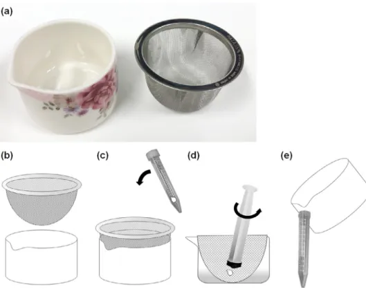
For lymphocyte isolation, a porcelain bowl with a spout (Narumi Co. Aichi, Japan) and a wire mesh tea strainer (Sun Co., Niigata, Japan) were used ( Fig.1A, B). The diameter (55 mm) and height (40 mm) of the mesh strainer were equal to the internal diameter and depth of the porcelain bowl. The porcelain bowl and wire mesh strainer were sterilized by autoclaving prior to use. During endoscopy, a single specimen was obtained using disposable biopsy forceps (EndoJaw TM, FB-230K, Olympus, Tokyo,
Japan). First, one endoscopically harvested specimen was placed in a 15 mL conical centrifuge tube containing 5 mL of normal saline solution. Next, a wire mesh strainer was set in a porcelain bowl and
Fig. 1. Scheme of the one-step lymphocyte isolation procedure. (a, b), Porcelain bowl with a spout and a wire mesh tea strainer were used; (c) One endoscopically harvested specimen was placed in a 15 mL conical centrifuge tube containing 5 mL of normal saline solution. A wire mesh strainer was set in a porcelain bowl, and saline solution containing specimen was decanted into it; (d) The tissue pieces were then crushed using the rubber portion of a plunger of a 10 mL injection syringe; (e) The saline solution with lymphocytes was then poured into the conical centrifuge tube.
saline solution containing the specimen was decanted into it ( Fig.1C). The conical centrifuge tube was mixed by inversion before decanting to ensure the removal of the specimen from the bottom of the tube. The solid specimens were then crushed using the rubber portion of a plunger of a 10 mL injection syringe ( Fig. 1D). To obtain the lymphocyte suspension, the sample was soaked in saline solution during grinding. Fibrous residues remained on the wire mesh tea strainer. Subsequently, the wire mesh tea strainer was removed and discarded. The saline solution containing lymphocytes was then poured into a conical centrifuge tube ( Fig.1E).
Flowcytometricanalysis
Flow cytometry was performed according to the guidelines for analyzing surface antigens on hematopoietic malignant cells, issued by the Japanese Committee for Clinical Laboratory Standards (JCCLS) [8]. Immediately before flow cytometry, samples were filtered using a 100
μ
m pore mesh strainer (Partec CellTrics, Sysmex, Kobe, Japan) and centrifuged at 400 × g for 5 min at room temperature. The cells were washed with phosphate-buffered saline solution and centrifuged again. The supernatant was removed, and the pellet was mixed with monoclonal antibodies, diluted at the optimal concentration according to the manufacturer’s instructions. The sample was kept in the dark for 15 min at room temperature. Monoclonal antibodies against CD45 (J33; Beckman Coulter, Pasadena, CA, USA), CD19 (J3-119; Beckman Coulter), CD20 (B-Ly1; Dako, Santa Clara, CA, USA), CD10 (ALB1; Beckman Coulter), CD3 (UCHT1; Beckman Coulter), and CD5 (BL1a; Beckman Coulter),4 M. Iwamuro, T. Takahashi and N. Watanabe et al. / MethodsX 7 (2020) 101095
were used and polyclonal antibodies were used for surface
κ
(Polyclonal F(ab’)2; Dako), andλ
(Polyclonal F(ab’)2; Dako) chains. Next, erythrocytes were lysed by adding VersaLyse Lysing Solution (Beckman Coulter). After 10 min, the sample was diluted with phosphate-buffered saline solution and centrifuged at 400 × g for 5 min at room temperature. The pellet was resuspended in 500μ
L phosphate-buffered saline solution and used for flow cytometry. The immunostained cells were analyzed by FACScan (Navios flow cytometer, Beckman Coulter) using Kaluza analysis software version 1.3 (Beckman Coulter).CD45, originally known as leukocyte common antigen, is used as a marker of hematopoietic cells except erythrocytes and platelets. CD20 is acquired during the late stages of B-cell lymphogenesis, and its expression is lost upon plasma cell differentiation. On the other hand, the surface expression of CD19 is highly regulated throughout B-cell development and maturation until the loss of its expression during differentiation into plasma cells. Therefore, CD45 was used as a leukocyte lineage-specific antigen and CD19 and CD20 are used as B-cell lineage-specific antigens. Because B-cell lymphomas typically occur after transformation and subsequent clonal expansion of specific lymphocytes, neoplastic cells express only one class of immunoglobulin containing either
κ
orλ
light chain. Therefore, light chain expression in a mature B-cell proliferation can be used as a surrogate marker to help diagnose B-cell lymphoma. Based on these principles, in this study, B-cell clonality was determined using theκ
:λ
ratio; theκ
andλ
light chain expression was quantified using a gated CD45 or CD20 population. Based on the previously published criteria for the flow cytometric analysis of restricted light chains, aκ
:λ
ratio within 0.5–3.0 was defined as negative for light chain restriction [6, 7, 9–11].The one-step lymphocyte isolation procedure is shown in Supplementary Video 1.
Methodvalidation Overview
For the conventional pathological analysis, a specimen was retrieved from the target lesion using disposable biopsy forceps. Based on the results of pathological diagnosis, patients were divided into lymphoma and benign groups. We analyzed two factors, i.e., the feasibility of the flow cytometric analysis of light chain expression ( Fig.2), and the sensitivity and specificity of the flow cytometric analysis of light chain expression, which were determined by comparing the results with those of pathological diagnosis. In addition, the percentage of CD10 + cells among CD19 +/CD20 + cells in the extranodal marginal zone of lymphoma in mucosa-associated lymphoid tissue (MALT lymphoma) patients—with light chain restriction—was compared with those of follicular lymphoma since CD10 is commonly expressed on the surface of follicular lymphoma cells, and MALT lymphoma cells are generally negative for CD10.
Patientsandethicsapproval
Flow cytometry was performed using the one-step lymphocyte isolation procedure with endoscopic biopsy specimens obtained from 23 patients between April 2019 and March 2020 at Okayama University Hospital (Okayama, Japan). In this prospective study, patients with previously diagnosed gastrointestinal lymphoma or those with gastrointestinal lesions suspected to be lymphoma were included.
Patients were prospectively registered and analyzed for this study. Flow cytometric analyses were performed as part of the standard clinical practice. Therefore, the need for written informed consent was waived. This study was approved by the ethics committee of Okayama University Hospital and adhered to the principles of the Declaration of Helsinki. The study protocol was registered in the UMIN Clinical Trials Registry (UMIN0 0 0 027730).
Fig. 2. A flow diagram summarizing the study. MALT lymphoma, extranodal marginal zone lymphoma of mucosa-associated lymphoid tissue.
Results
The characteristics of the patients (13 women and 10 men) are shown in Table 1. The median age was 65 years (range: 45–82 years). Biopsy sites included the stomach (n = 11), duodenum (n = 2), jejunum (n = 1), ileum (n = 2), cecum (n = 3), colon (n = 3), and rectum (n = 1). The final pathological diagnoses were lymphoma in 12 patients and benign lesions in 11 patients. The lymphoma subtype included gastric MALT lymphoma (n = 6), duodenal follicular lymphoma (grade 1; n = 2), cecal diffuse large B-cell lymphoma (DLBCL, n = 1), colonic DLBCL (n = 1), colonic MALT lymphoma (n = 1), and colonic follicular lymphoma (grade 1; n = 1). Benign lesions consisted of lymphoid hyperplasia (stomach, n = 1; rectum, n = 1), eosinophilic gastritis (n = 1), erosive gastritis (n = 1), gastric cancer (n = 1), non-specific ileitis (n = 1), ulcerative colitis (n = 1), colon cancer (n = 1), and the remission stage of lymphoma (ileal follicular lymphoma, n = 2; gastric MALT lymphoma, n = 1).
Flow cytometric analysis of light chain expression was performed in all patients. Therefore, the feasibility of flow cytometric analysis with the single-step lymphocyte isolation technique was 100%. The biopsy specimens of the 11 patients in the benign groups were light chain restriction negative ( Fig.3). The biopsy specimens from two patients with MALT lymphoma were light chain restriction negative, while those from the remaining 10 patients with lymphoma were light chain restriction positive ( Figs.4, 5). Overall, the sensitivity of light chain expression analysis for diagnosing lymphoma was 83.3% and specificity was 100% ( Table2).
In MALT lymphoma biopsy samples that were light chain restriction positive (n = 5), the percentages of CD10 + cells among CD19 +/CD20 + cells were 4.0%, 8.6%, 8.7%, 9.2%, and 16.5% ( Fig.4C), and 79.5% and 96.3% in follicular lymphoma (n = 2) ( Fig. 5C). Therefore, the follicular and MALT lymphoma cases could be correctly discriminated by flow cytometry, given that 20% is the threshold for CD10 + cells [10]. These results are in accordance with those of our previous study [5].
Conclusion
In conclusion, we showed that lymphocytes isolated from a single endoscopic biopsy specimen using our simplified and quick procedure are suitable for flow cytometry. The sensitivity of the flow cytometric analysis of light chain expression was 83.3%, and specificity was 100%. Therefore, although
6 M. Iwamuro, T. Takahashi and N. Watanabe et al. / MethodsX 7 (2020) 101095 Table 1
Characteristics of the enrolled patients
N Sex
Female 13
Male 10
Mean age (range, years) 65 (45–82)
Biopsy sites Stomach 11 Duodenum 2 Jejunum 1 Ileum 2 Cecum 3 Colon 3 Rectum 1 Pathological diagnosis MALT lymphoma 7 Follicular lymphoma 3 DLBCL 2
Benign lymphoid hyperplasia 2
Cancer 2
Eosinophilic gastritis 1
Erosive gastritis 1
Non-specific ileitis 1
Ulcerative colitis 1
Remission stage of lymphoma 3
MALT lymphoma, extranodal marginal zone lymphoma of mucosa-associated lymphoid tissue; DLBCL, diffuse large B-cell lymphoma
Fig. 3. Flow cytometric results for rectal lymphoid hyperplasia. (a) CD19 + /CD20 + cells were gated; (b) Light chain expression was analyzed and the κ/ λratio was found to be 1.69 (normal range), indicating that the sample was negative for light chain restriction.
Table 2
Cross-tabulation of the light chain expression analysis Lymphoma Benign Light chain restriction positive 10 0 Light chain restriction negative 2 11
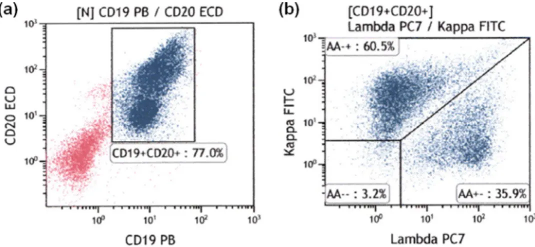
Fig. 4. Representative flow cytometric results for gastric MALT lymphoma. (a) CD19 + /CD20 + cells were gated; (b) There was predominant expression of the κlight chain, indicating that the sample was positive for light chain restriction; (c) The percentage of CD10 + cells was 7.4%.
Fig. 5. Representative flow cytometric results for duodenal follicular lymphoma. (a) CD19 + /CD20 + cells were gated; (b) the
κ/ λratio was 8.58, indicating positive results for light chain restriction; (c) The percentage of CD10 + cells was 79.5%. flow cytometric analysis and pathological diagnosis are complementary methods for the diagnosis of gastrointestinal lymphoma, attainment of results on the day of examination is a strong advantage of flow cytometric analysis. We hope that this single-step lymphocyte isolation technique will assist in popularizing the use of flow cytometry among gastroenterologists and endoscopists for the diagnosis of gastrointestinal lymphoma.
DeclarationofCompetingInterest
The authors declare that they have no known competing financial interests or personal relationships that could have appeared to influence the work reported in this paper.
Acknowledgement
The author thanks Ms. Hiromi Nakashima, Department of Hematology and Oncology, Okayama University Graduate School of Medicine, Dentistry and Pharmaceutical Sciences, for technical assistance.
Supplementarymaterials
Supplementary material associated with this article can be found, in the online version, at doi: 10. 1016/j.mex.2020.101095.
8 M. Iwamuro, T. Takahashi and N. Watanabe et al. / MethodsX 7 (2020) 101095
References
[1] D. Azoulay, H.I. Cohen, E. Dementiev, E. Eshel, L. Akria, E. Shaoul, N. Horowitz, Flow cytometry aneuploidy and cell cycle indexing as a possible tool for differentiating between CD10 + diffuse large B-cell lymphoma and follicular lymphoma, Cytom. B. Clin. Cytom. (2020) in press, doi: 10.1002/cyto.b.21861 .
[2] F.E. Craig , K.A. Foon , Flow cytometric immunophenotyping for hematologic neoplasms, Blood 111 (2008) 3941–3967 . [3] W.A. Geary , H.F. Frierson , D.J. Innes , D.E. Normansell , Quantitative criteria for clonality in the diagnosis of B-cell
non-Hodgkin’s lymphoma by flow cytometry, Mod. Pathol. 6 (1993) 155–161 .
[4] E. Gunduz , M. Celebioglu , O. Meltem Akay , H. Uskudar Teke , F. Sahin Mutlu , Z. Gulbas , The role of flow cytometry in the diagnosis of non-Hodgkin’s lymphoma, Hodgkin’s lymphoma, granulomatous inflammation and reactive lymph node specimens, J BUON 18 (2013) 739–745 .
[5] M. Iwamuro , K. Matsueda , T. Takahashi , S. Omote , T. Tanaka , D. Ennishi , F. Otsuka , T. Yoshino , H. Okada , An endoscopic biopsy specimen contains adequate lymphocytes for flow cytometric analysis of light chain expression in the gastrointestinal Mucosa, Ann. Clin. Lab. Sci. 50 (2020) 348–353 .
[6] C. Kawano-Yamamoto , K. Muroi , T. Izumi , K. Saito , K. Ozawa , Two color flow cytometry with a CD19 gate for the evaluation of bone marrow involvement of B-cell lymphoma, Leukem. Lymphoma 43 (2002) 2133–2137 .
[7] S.H. Kroft , A.M. Harrington , Flow cytometry of B-cell neoplasms, Clin. Lab. Med. 37 (2017) 697–723 .
[8] T. Ikemoto , K. Kitamura , N. Tatsumi , K. Nakahara , K. Higashi , K. Watanabe , Guidelines for performing surface antigen analysis by flow cytometry, Cytom. Res. 19 (2009) 39–44 .
[9] M.P. Leers , P.H. Theunissen , F.C. Ramaekers , B. Schutte , M. Nap , Clonality assessment of lymphoproliferative disorders by multiparameter flow cytometry of paraffin-embedded tissue: an additional diagnostic tool in surgical pathology, Hum. Pathol. 31 (20 0 0) 422–427 .
[10] K. Matsueda , M. Iwamuro , T. Takahashi , S. Omote , K. Nishida , T. Tanaka , D. Ennishi , F. Otsuka , T. Yoshino , H. Okada , Feasibility of flow cytometric analysis of restricted light chain in endoscopic biopsy specimens from patients with gastrointestinal tract B cell lymphoma: a pilot study, BMC Res. Notes 12 (2019) 571 .
[11] S. Oka , K. Muroi , K. Sato , S. Fujiwara , I. Oh , T. Matsuyama , K. Ohmine , T. Suzuki , K. Ozaki , M. Mori , T. Nagai , N. Fukushima , N. Fukushima , A. Tanaka , K. Ozawa , Flow cytometric analysis of kappa and lambda light chain expression in endoscopic biopsy specimens before the diagnosis of B-cell lymphoma, J. Clin. Exp. Hematop. 52 (2012) 127–131 .