Investigating the Underlying Intelligence
Mechanisms of the Biological Olfactory System
著者
Makino Yoshinari, Yano Masafumi
journal or
publication title
Advances in Artificial Intelligence, in press.
volume
2010
page range
478107
year
2010
URL
http://hdl.handle.net/10097/51783
Volume 2010, Article ID 478107,9pages doi:10.1155/2010/478107
Review Article
Investigating the Underlying Intelligence Mechanisms of the
Biological Olfactory System
Yoshinari Makino and Masafumi Yano
Research Institute of Electrical Communication, Tohoku University, 2-1-1 Katahira, Aoba-ku, Sendai 980-8577, Japan
Correspondence should be addressed to Yoshinari Makino,[email protected]
Received 8 September 2009; Revised 11 November 2009; Accepted 9 December 2009 Academic Editor: Naoyuki Sato
Copyright © 2010 Y. Makino and M. Yano. This is an open access article distributed under the Creative Commons Attribution License, which permits unrestricted use, distribution, and reproduction in any medium, provided the original work is properly cited.
The brain is the center of intelligence that biological systems have acquired during their evolutionary history. In unpredictably changing environments, animals use it to recognize the external world and to make appropriate behavioral decisions. Understanding the mechanisms underlying biological intelligence is important for the development of artificial intelligence. Olfaction is one of the sensory modalities that animals use to locate distant objects. Because of its relative simplicity compared with other sensory modalities and the wealth of knowledge at cellular, network, system, and psychophysical levels, it is possible that the biological olfactory system would be understood comprehensively. This paper reviews our biological and computational works with a focus on the temporal aspects of olfactory information processing. In addition, the paper highlights that the “time” dimension is essential for the functioning of the olfactory information processing system in the real world.
1. Introduction
Consider animals, such as honeybees, butterflies and so forth, collecting flower nectar in fields. For simplicity, all flowers have the same visual cues (e.g., shape, size, color, etc.), but each of them has a distinct odor. Some flowers can have the same odor but are spatially distributed randomly in the field. The quantity of flower nectar is fixed according to the type of odor; for example, flowers with odor A have 10 mL
nectar, odorB have 5 mL, and odor C have 0 mL. There might
be some dangerous flowers, such as insectivorous flowers, giving off specific odors. It is important to be aware of the cognitive abilities that an animal needs to possess in order to collect nectar efficiently and avoid danger.
An animal can sense an odor at various distances from its source, implying that an odor can be sensed at different concentrations. To approach or avoid flowers with a specific odor, the animal should be able to judge the odor at different concentrations as an odor with the same quality and should be able to determine its direction based on whether the odor signal becomes stronger or weaker. Thus, the animal must have the ability to identify odors in a concentration-invariant manner.
Suppose that the animal has visited many flowers in a certain field and has experienced the relationship of odors with nectar quantities or dangers and then moves to a different field. How will the animal behave in the new field where there are no flowers with odors identical to those experienced in the previous field? Based on the experience in the previous field, the animal should visit flowers with odors similar to those associated with a large amount of nectar and avoid flowers with odors similar to those of dangerous flowers. Thus, the animal must detect similari-ties and differences between odors and make appropriate behavioral decisions. These examples demonstrate that, at large, olfactory information processing is a typical example of pattern recognition. Problems of pattern recognition are further complicated in the real world where unpredictable noises or perturbations occur. Even in such situations, animals use the olfactory information to make appropriate behavior decisions in order to survive in the real world.
Olfaction is the sensory modality that many animals use to locate distant objects. Structures of the olfactory system in the brain, especially those from peripheral olfactory receptors to the primary olfactory network, have common
2 Advances in Artificial Intelligence characteristics among vertebrates, insects, terrestrial
gas-tropods (slugs/snails), and so forth [1]. Odorant molecules are received by olfactory receptor neurons (ORNs) that are distributed in the peripheral organs (e.g., the nasal cavity in vertebrates, the antennae in insects, and the tentacles in
slugs/snails) [1–4]. ORNs project axons to round-shaped
structures called glomeruli in the primary olfactory network
[1,5]. Axons of ORNs that express the same odor receptor
type converge in each glomerulus [6–8]. The primary
olfactory network (the olfactory bulb in vertebrates and the antennal lobe, AL, in insects) consists of output neurons and local inhibitory interneurons [2]. These neurons extend their dendrites into glomeruli and receive inputs from ORNs. The glomeruli are distributed as a glomerular layer along the surface of the olfactory bulb in vertebrates [2,3] and
botryoidally arranged in the insect antennal lobe [2, 4].
Physiological studies using an optical recording technique revealed that even monomolecular odorants evoke broadly distributed activation across glomeruli [9–12], so that an odor input is first represented as a spatial pattern of glomerular activation at the primary olfactory network.
The primary olfactory network transforms the input spa-tial pattern into the spatiotemporal activity pattern of output neurons [2] and sends it to various brain regions [5]. In general, brain functions emerge by cooperative information processing in various brain regions. To understand how the biological olfactory system solves pattern recognition problems in the real world, it is important to clarify the following two issues.
(i) How do odors spatiotemporally activate the olfactory regions in the brain, and how does the odor informa-tion flow within the brain as a whole?
(ii) To solve the problems of pattern recognition, what kind of computational algorithm is needed? How does the biological network implement it? To per-form the algorithm, how is the spatiotemporal odor representation in the primary olfactory network useful?
By understanding these issues comprehensively, the intel-ligence that the biological olfactory system has acquired evolutionarily to solve pattern recognition problems in the real world can be explained.
This paper reviews physiological and computational
studies on olfaction from these perspectives. Section 2
describes an experimental approach to explain the flow of olfactory information in the slug’s brain.Section 3describes the computational coding scheme of olfactory information
using the time dimension.Section 4summarizes the results
and provides directions for future work.
2. Information Flows in the Whole Brain
Understanding the flow of information within the brain as a whole is one of the goals that neuroscience research addresses, and for this purpose, several techniques have been developed to measure brain activities. However, it is still hard to observe the flow of information in the whole brain because
of the following difficulties: there is a tradeoff between spatial and temporal resolutions in any method, and the brain is too large and complex to be easily studied. So, it is important to select appropriate animals for measurement. In cases where the object to be measured is small, the optical imaging technique has a good spatiotemporal resolution. If we can use an animal that has a good learning ability, can discriminate objects, and has a small brain that is simple in structure, the flow of information can be visualized in such a brain, and it will be possible to explain the information processing principle in the brain as a whole.
From this perspective, a slug/snail is a useful animal model. In these animals, olfaction is a dominant sensory modality for recognizing external objects (visual or auditory systems have not been developed in their brain) [1,13,14].
The slug/snail has a good odor learning ability [15–20].
It has a small and simple brain; so whole brain activity
can be measured with good spatiotemporal resolutions [21–
26]. Furthermore, there are several experimental
advan-tages of their olfactory system. Noses of these animals are located on the tips of two pairs of tentacles (superior and inferior tentacles, STs and ITs). Several behavioral and physiological studies have revealed that there are functional
differences between STs and ITs [27–31]; ST is involved
in olfactory orientation [28], whereas IT is involved in
learning [29] or retrieving odors [27,31]. Their brain can be dissected and isolated as a whole without a lose in the function of these olfactory organs [23, 24, 27, 32, 33]. These features are useful for explaining the relationship between the flow of information and the emergence of brain functions.
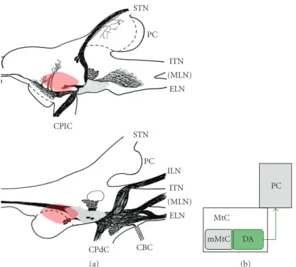
Figure 1(a) shows the slug we used, the Japanese slug
Incilaria fruhstorferi, andFigure 1(b)schematically illustrates the slug’s brain, its cerebral ganglia (CG). The STs and ITs are connected to CG via superior and inferior tentacle nerves (STNs and ITNs), respectively. CG comprises three lobes: the procerebrum (PC), mesocerebrum (MsC), and metacerebrum (MtC). Anatomical studies indicate that after entering the body of CG, afferent fibers of the tentacle nerves segregate into several bundles and terminate in PC and MtC [1,14,35]. The PC is regarded as the olfactory center, and many studies have investigated its role in the processing of olfactory information [22–24,27,30,36–40]. It is suggested that PC is involved in acquiring and retrieving odor memory [27,30,38].
In contrast, few studies have investigated olfactory
infor-mation processing in MtC. Anatomically, afferent fibers from
STN and ITN converge into the medial region of MtC
(mMtC) (Figures1(c)and1(d)) [26]. The MtC is thought
to collect olfactory, taste, and other sensory information and to command motor actions when appropriate signals are
received [14]. The relationships between PC and MtC in the
slug’s brain might correspond to those between cortical and subcortical structures in the vertebrate’s brain. Hence, the slug’s brain is suitable for our research purpose to observe the flow of information within the whole brain.
We optically recorded the brain activity evoked by electrical stimulations of STN and ITN and obtained the following results [26].
STs l IT (a) (MLN) ITN ILN STN ELN CPdC CPIC PC MtC MsC CC MtC IM TM CM PC (MLN) ITN ILN STN ELN CPdC CPIC (b) (c) ST mMtC MtC PC IT (d)
Figure 1: Slug’s brain. (a) The Japanese giant terrestrial slug Incilaria fruhstorferi. A pair of superior tentacles (STs) can be seen at the head end. A part of the left inferior tentacle (l IT) can be seen close to the base of the left ST. Scale bar: 5 cm. (b) Gross anatomy of the slug brain, cerebral ganglia. Left and right sides show dorsal and ventral views of the cerebral ganglia, respectively. The cerebral ganglion is divided into three lobes: the procerebrum (PC), mesocerebrum (MsC), and metacerebrum (MtC). Olfactory information received at the tips of STs and ITs reaches the brain through superior and inferior tentacle nerves (STNs and ITNs, resp.). See [26] for details. (c) Anatomical projections of fibers from STN (blue arrows) and ITN (red arrows). Both ST and IT send olfactory information into PC (left panel) and to the medial region of MtC (right panel). (d) Schematic illustration of anatomical olfactory projections from ST and IT in the brain. Figures1(b)and
1(c)are adapted and modified, with permission, from [34] 2006 IEEE.
(1) STN and ITN stimulations activate both PC and mMtC.
(2) Regardless of STN or ITN stimulations, the mMtC response is about 50 milliseconds earlier than the PC response.
(3) STN and ITN stimulations evoked different activa-tion patterns of mMtC: the ITN stimulaactiva-tion activated the lateral half of mMtC more strongly than its medial half, whereas the STN stimulation activated both halves evenly. In contrast, there seems to be no difference between the activation patterns in PC evoked by STN and ITN stimulations.
It is interesting that PC responses to STN and ITN stim-ulations are the same, in spite of memory functions of
PC [27, 30, 38, 40] and functional differences between
STs (orientation) and ITs (memory) [27–31]. This implies
that direct olfactory inputs to PC would not explain the differences in memory functions between STs and ITs.
Since mMtC responds faster than PC to the nerve stimulation, there is a possibility that mMtC activations
evoked by STN and ITN stimulations affect PC differently,
and as a result, a functional difference between ST and IT
for memory functions might emerge. We hypothesized that monoamine- (such as serotonin, dopamine, etc.) containing neurons might mediate the transfer of information from mMtC to PC because, as neuromodulators, monoamines are known to have important roles in memory functions
[41–43] and in changing the functional modes of neural
networks [44–46]. Therefore, we stained the
dopamine-containing neurons in the slug’s brain [34]. Main results
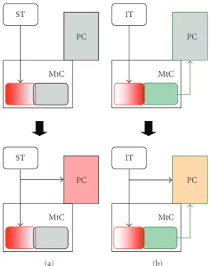
are summarized inFigure 2. The dopamine-containing
neu-ropils are intensively distributed in the central and lateral regions of MtC (Figure 2(a), dotted area), and this central MtC region seems to spatially overlap with lateral area of the mMtC region that was strongly activated by the ITN
stimulation (Figure 2(a)). The dopamine containing output
fibers from the central MtC region seem to project into PC. Gelperin and his colleagues physiologically suggested that dopamine might modulate the network function of the PC
[47, 48]. So, the revealed morphological features suggest
that the dopamine-containing neurons might act as a bridge between mMtC and PC, especially when the mMtC region
is activated through the ITs [Note1]. Dopamine-containing
fibers project densely into the STN and ITN, and these come at the tentacle ganglion of ST and IT, suggesting another possibility that dopaminergic modulation of olfactory
infor-4 Advances in Artificial Intelligence CPdC ELN (MLN) ITN ILN PC STN CBC CPIC ELN (MLN) ITN PC STN (a) MtC mMtC DA PC (b)
Figure 2: A major dopamine system in the slug brain. (a) Schematic illustrations of the major dopamine system. These figures are made on the basis of brain sections treated using the histofluorescent method for staining catecholamine (dopamine). Top and bottom panels are based on sections from dorsal and ventral parts, respectively. Filled ovals indicate dopamine-containing cell bodies. Dotted area indicates the area that the dopamine-containing fibers are densely distributed. The estimated mMtC region that would be activated by the STN or ITN stimulation is indicated by red shade. A large open-oval in the bottom indicates the metacerebral giant cell. (b) Function of the major dopamine system (DA) suggested by its morphology. The major dopamine system might function as a bridge between the medial region of MtC (mMtC) and the procerebrum (PC).Figure 2(a)is adapted and modified, with permission, from [34] 2006 IEEE.
mation processing might occur more peripherally through the tentacle ganglia.
Olfactory functions in the slug brain can be explained on the basis of the flow of olfactory information (Figure 3). Based on the mMtC activity, ST and IT would have different roles in olfactory functions. The medial half of mMtC (that is strongly activated by the STN stimulation) might be related to olfactory orientation. Indeed, several motor neurons related to olfactory orientation movements are
known in regions close to mMtC [49–51]. The lateral half
of mMtC (that is strongly activated by the ITN stimulation) might activate the monoamine system, and this system would modulate functional modes of PC to acquire or retrieve odor memory.
It is suggested that the brain (cerebral ganglia) can evaluate important odors and can command motor actions without PC, even though the odor discriminating ability of the brain without PC is not good compared to that
with PC [49]. Based on this observation and data from
our study [26], it can be hypothesized that mMtC might
contribute to the rough evaluation of important odors. The quick response of mMtC to STN and ITN stimulations might result in quick responses of the slug/snail to important odors and limit the odors that are processed by PC for fine
discrimination [26]. Such a temporal process would be
eco-logically advantageous, since various odors exist in the real world.
3. Information Representation Using Time
Dimension Solves Pattern-Recognition
Problems
There is growing evidence that biological systems use time
as a dimension for sensory information coding [52–57].
Computationally, a central aspect of pattern recognition is achieving a “good” representation in which the main features of an object are simply and naturally revealed [58,
59]. Neurobiological information representation is crucial in facilitating such a computation [58,60].
Regardless of biological or artificial systems, recognition systems must extract invariant features of objects from varying input signals depending on situations and must detect and judge similarities and differences between objects appropriately. These requirements are for a system that can identify objects and make appropriate behavioral decisions in the real world. Thus, the temporal representation of objects should satisfy certain computational criteria for it to be useful for recognition. Criterion 1 is invariance. For instance, the size or strength of sensory signals varies when the same object is sensed from different distances. Even in this situation, object representation should be invariant. Criterion 2 is similarity. The degree of similarity of objects must be reflected in their representation while also expressing subtle differences. Regardless of sensory modalities, biological recognition shows a speed/accuracy
MtC PC ST MtC PC ST (a) MtC PC IT MtC PC IT (b)
Figure 3: The flow of olfactory information in the slug brain. Brain activities evoked by superior tentacle (ST) (a) and inferior tentacle (IT) (b) stimulations in early phase (top) and late phase (bottom). Based on the activation patterns evoked by ST and IT stimulations, it was suggested that only the IT stimulation would activate the dopamine system in the early phase (a), (b) top. It may be possible to modulate the functional mode of the procerebrum (PC) activation by the activated dopamine system, so that the processing of olfactory information at PC differs between ST and IT stimulations (a), (b) bottom.
tradeoff, which is the relationship between sampling time
and accuracy in object discrimination [61–63]. This
psy-chophysical principle constrains the temporal representation format of the biological system by imposing Criterion 3, which is coarse-to-fine nature. The coarse similarity or
difference of objects should be encoded in the early epoch
of the temporal representation, and the information encoded in the late epoch should help in detecting subtle differences among objects.
The question arises as to what kind of information representation format would satisfy the three representation criteria. Suppose that there is a set ofn feature dimensions
each of which describes a specific object feature. By prepro-cessing of sensory signals from an object, the intensity of each feature is obtained, and consequently, the input repre-sentation of the object is an analog vector of n-dimensional feature space. Changing the signal intensity or size from the same object would only cause changes in the coefficients or bias of input analog vectors. In this case, the order of feature dimensions (in other words, the ranking of feature dimensions) determined by elemental analog values of the input vector would be scale invariant. So, if a computational algorithm transforms the order of analog values of the input vector into a temporal sequence of spike activity of neurons
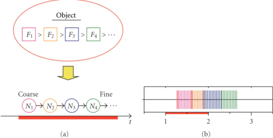
(or neural assemblies), each of which controls the respective feature dimension (Figure 4(a)), then the emergent temporal representation has scale invariance, satisfying Criterion 1. A temporal sequence of neural activities represents the hierarchical relationship of feature dimensions: as time advances, an object is coarsely classified on the basis of a feature represented by the first activated neuron, further subclassified by subsequent ones, and finally identified
by taking all activated neurons into account (Figure 4(a),
bottom), implying that the representation satisfies Criteria 2 and 3.
Using then-dimensional feature space, a system adopting
such a coding scheme can theoretically represent and discriminate n! objects (“!” is the factorial operator). If the system can set a temporal range for object representation, it can control the clustering levels of objects and make appropriate behavioral decisions that are flexible according to situations.
The proposed coding scheme is highly consistent with information transformation in the primary olfactory net-work of biological systems, especially in that of insects and vertebrate zebra fish [52–55]. As mentioned in Section 1, odor input representation is the spatial pattern of glomerular
activation [9–12]. Even monomolecular odorants evoke a
broadly distributed activation across glomeruli, such that each glomerulus is regarded as a functional module detecting a particular feature of odorant molecular structure [3, 9–
12, 64]. Odors at very low concentrations activate a few
glomeruli above the background level. As the concentration increases, several additional glomeruli are recruited, and the glomerular activity is a logarithmic function of concentra-tion [9,11,65–67]. Furthermore, similarity in the molecular structures of different odor molecules is correlated with sim-ilarity in spatial patterns of glomerular activation [10,11,68,
69] and with perceptual similarity [69–71]. Spatiotemporal activity patterns of the insect primary olfactory network are
robust to odor concentration changes [54], whereas those
of the zebra fish primary olfactory network are suggested to reflect odor similarity in a temporally coarse-to-fine manner
[53]. These imply that the input spatial pattern of
odor-evoked glomerular activation can be represented as an analog vector with a concentration bias [67]; the glomerular ranking that can be determined by the odor-evoked glomerular
activation would be concentration invariant [72–74]; if
the glomerular ranking can be transformed into temporal sequence of neural activities according to the proposed coding algorithm, the emergent spatiotemporal patterns would reflect the odor similarity as physiologically observed [72–74].
By means of biologically plausible neural components and their interactions, we constructed a primary olfactory network model that can implement the coding algorithm
shown inFigure 4(a)for odor information. The model
con-sists of glomerular modules, each consisting of one output neuron and two types (intraglomrular and interglomeru-lar types) of inhibitory local interneurons. In the single glomerular module model, the intraglomerular inhibition helps to cause a phasic activity of the output neuron in response to a constant glomerular input, and onset latency
6 Advances in Artificial Intelligence F1 > F2 > F3 > F4 > . . . N1 N2 N3 N4 . . . Object Coarse Fine Object t (a) 1 2 3 (b)
Figure 4: Object representation using temporal sequence of neural activities. (a) The order of feature intensities of an object (e.g.,F1 >
F2 > F3> F4) is transformed into a temporal sequence of neural activities in control of respective features (e.g.,N1 → N2 → N3 → N4).
The emergent temporal sequence of neural activities represents a hierarchical relationship of object features in coarse-to-fine manner on the temporal axis. (b) A typical simulated result of spike activities of output neurons of the four-glomerular module model. Spikes of each output neuron are indicated in different colors.
of the phasic response of the output neuron decreases as the input strength increases. In the network model, the glomeru-lar modules are connected by the interglomeruglomeru-lar inhibitory interneurons. Through these interglomerular connections, the activated output neuron inhibits other glomerular output neurons and delays their phasic activation, and consequently phasic activities of the output neurons are ordered according to the strength order of the glomerular inputs (Figure 4(b)). The network model successfully implements this spatiotem-poral transformation regardless of absolute input analog values or concentration biases [72–74].
In the insect higher center network, that is, the mush-room body (MB), odors are represented by spikes sparsely distributed in space and time [53]. In our primary olfactory network model, the odor information is represented as the temporal sequence of the output neurons’ activities, in which the information about the activity sequence is intensively-coded around transient overlaps of the neural activities (see
Figure 4(b)). So, we further constructed a higher center model that evaluates the transient overlaps of the neural activities of the output neurons of the primal olfactory network model. The higher center model can extract odor information as a temporal sequence of sparse spikes with a coarse-to-fine nature and can store the odor information as network connectivity with the help of simple Hebbian connections [72–74].
These indicate that the essential problems of olfactory recognition can be solved by information representation using the time dimension. In the proposed coding scheme, significant features characterizing objects are temporally placed first, followed by more subtle features. This strategy is crucial for the system to survive in an unpredictably changing environment because it allows the system to respond quickly to vitally important object features. Even though it may be just one dimension mathematically, “time” is an important dimension for systems that live and survive in the real world. Thus, the information representation using the time
dimension is a basis for pattern recognition that warrants further investigation in biological systems.
4. Towards an Understanding of Biological
System Intelligence
In Section 2, we have shown that the olfactory system
in the slug’s brain consists of two pathways: one is the mMtC-pathway that probably has a role for quickly and roughly discriminating important odors; the other is the PC-pathway that is rather slow but important for finely discriminating odors. With respect to the computational
model shown inSection 3, the physiological data about the
spatiotemporal neural activities are mainly based on those
obtained from the insect AL-MB pathway [52, 54, 55].
The insect MB is suggested to have important roles in fine
discrimination of odors [75, 76] and odor learning and
memory [77–79], as the slug/snail PC is suggested to have
such roles [33, 38, 40]. So, the computational model in
Section 3 is thought to correspond to the PC-pathway in
Section 2, and we can suggest that the olfactory system of the brain uses “time” dimension doubly (i.e., the anatomically
differentiated two pathways that have different temporal
responses to odors, and the coarse-to-fine odor representa-tion using the temporal sequence of neural activity in the fine discrimination pathway) to quickly and dominantly process important odors or odor-features. These temporal aspects of olfactory information processing might be essential for systems working in the real world.
To comprehensively understand the mechanisms
involved in olfactory information processing, we must explain how global information flow and computational information processing are related and integrated on the systems level. In the information representation proposed inSection 3, odor is described as the permutation of odor features, each of which works like letters of an alphabet.
So, information represented by the temporal sequence of neuronal activities is regarded as symbolic information. Symbolic information is useful for identifying objects and detecting their similarities and differences. However, symbolic information of objects itself cannot help the system make a behavioral decision because it does not represent information on the values of objects. We call this “value information.” Value information cannot be defined by attributes of objects alone. Value information of an object should be evaluated and determined by the system in real time on the basis of experience with the object and current conditions of the environment and the system. From invertebrates (including the slugs and snails mentioned inSection 2) to vertebrates (including primates), monoamine systems in the brain are known to be strongly related to internal and motivational states of biological systems and to play important roles in learning and memory [32,41–43,80–86], so that monoamine systems might be involved in determining the value information of objects. As neuromodulators, monoamines would affect the brain activity in a wide spatial and temporal range, and their spatiotemporal dynamics in the whole brain would be
critical for neural network functions [84]. The challenge
is to explain biological and computational mechanisms relating temporally represented symbolic information with value information that might be determined by global information flows within the brain. This approach would clarify the design principles of an intelligent system that works well in the unpredictably changing environment of the real world.
Note 1. This hypothesis predicts that the PC should respond
differently between the STN and ITN stimulation by the
indirect input through the mMtC. However, we could not observe such difference. In the experiment, both STN and ITN stimulations evoked transient depolarization followed by the strong and long lasting (about 10 seconds)
hyper-polarization in the PC [26]. The transient depolarization
of the PC is caused by the direct input from the STN and ITN, whereas the following PC hyperpolarization is probably caused by inhibitory effects of the intrinsic PC neurons [87]. In the experiment of [26], this intrinsic hyperporlaization in the PC might mask the PC response caused by the mMtC pathway.
Acknowledgments
This study was supported by a Grant no. 21500294 for Y.Makino from the Japan Society for the Promotion of Science, and by a Grant no. 17075003 for Y.Makino and M.Yano from the Japanese Ministry of Education, Culture, Sports, Science, and Technology.
References
[1] R. Chase and B. Tolloczko, “Tracing neural pathways in snail olfaction: from the tip of the tentacles to the brain and beyond,” Microscopy Research and Technique, vol. 24, no. 3, pp. 214–230, 1993.
[2] G. Laurent, “Olfactory network dynamics and the coding of multidimensional signals,” Nature Reviews Neuroscience, vol. 3, no. 11, pp. 884–895, 2002.
[3] K. Mori, Y. K. Takahashi, K. M. Igarashi, and M. Yamaguchi, “Maps of odorant molecular features in the mammalian olfactory bulb,” Physiological Reviews, vol. 86, no. 2, pp. 409– 433, 2006.
[4] L. B. Vosshall and R. F. Stocker, “Molecular architecture of smell and taste in Drosophila,” Annual Review of Neuroscience, vol. 30, pp. 505–533, 2007.
[5] G. M. Shepherd, Neurobiology, Oxford University Press, New York, NY, USA, 2nd edition, 1988.
[6] L. Buck and R. Axel, “A novel multigene family may encode odorant receptors: a molecular basis for odor recognition,”
Cell, vol. 65, no. 1, pp. 175–187, 1991.
[7] R. Vassar, S. K. Chao, R. Sitcheran, J. M. Nunez, L. B. Vosshall, and R. Axel, “Topographic organization of sensory projections to the olfactory bulb,” Cell, vol. 79, no. 6, pp. 981–991, 1994. [8] L. B. Vosshall, A. M. Wong, and R. Axel, “An olfactory sensory
map in the fly brain,” Cell, vol. 102, no. 2, pp. 147–159, 2000. [9] R. W. Friedrich and S. I. Korsching, “Combinatorial and
chemotopic odorant coding in the zebrafish olfactory bulb visualized by optical imaging,” Neuron, vol. 18, no. 5, pp. 737– 752, 1997.
[10] C. G. Galizia, S. Sachse, A. Rappert, and R. Menzel, “The glomerular code for odor representation is species specific in the honeybee Apis mellifera,” Nature Neuroscience, vol. 2, no. 5, pp. 473–478, 1999.
[11] B. D. Rubin and L. C. Katz, “Optical imaging of odorant representations in the mammalian olfactory bulb,” Neuron, vol. 23, no. 3, pp. 499–511, 1999.
[12] N. Uchida, Y. K. Takahashi, M. Tanifuji, and K. Mori, “Odor maps in the mammalian olfactory bulb: domain organization and odorant structural features,” Nature Neuroscience, vol. 3, no. 10, pp. 1035–1043, 2000.
[13] R. Chase, “The olfactory sensitivity of snails, Achatina fulica,”
Journal of Comparative Physiology A, vol. 148, no. 2, pp. 225–
235, 1982.
[14] R. Chase, “Structure and function in the cerebral ganglion,”
Microscopy Research and Technique, vol. 49, no. 6, pp. 511–520,
2000.
[15] A. Gelperin, “Rapid food aversion learning by a terrestrial mollusk,” Science, vol. 189, no. 4202, pp. 567–570, 1975. [16] C. Sahley, J. W. Rudy, and A. Gelperin, “An analysis of
associative learning in a terrestrial mollusc—I. Higher-order conditioning, blocking and a transient US pre-exposure effect,” Journal of Comparative Physiology A, vol. 144, no. 1, pp. 1–8, 1981.
[17] C. L. Sahley, K. A. Martin, and A. Gelperin, “Analysis of asso-ciative learning in the terrestrial mollusc Limax maximus—II. Appetitive learning,” Journal of Comparative Physiology A, vol. 167, no. 3, pp. 339–345, 1990.
[18] A. Yamada, T. Sekiguchi, H. Suzuki, and A. Mizukami, “Behavioral analysis of internal memory states using cooling-induced retrograde amnesia in Limax flavus,” Journal of
Neuroscience, vol. 12, no. 3, pp. 729–735, 1992.
[19] H. Suzuki, T. Sekiguchi, A. Yamada, and A. Mizukami, “Sensory preconditioning in the terrestrial mollusk, Limax
flavus,” Zoological Science, vol. 11, pp. 121–125, 1994.
[20] T. Teyke, “Food-attraction conditioning in the snail, Helix
pomatia,” Journal of Comparative Physiology A, vol. 177, no.
4, pp. 409–414, 1995.
[21] D. Kleinfeld, K. R. Delaney, M. S. Fee, J. A. Flores, D. W. Tank, and A. Gelperin, “Dynamics of propagating waves in
8 Advances in Artificial Intelligence
the olfactory network of a terrestrial mollusk: an electrical and optical study,” Journal of Neurophysiology, vol. 72, no. 3, pp. 1402–1419, 1994.
[22] S. Kawahara, S. Toda, Y. Suzuki, S. Watanabe, and Y. Kirino, “Comparative study on neural oscillation in the procerebrum of the terrestrial slugs Incilaria bilineata and Limax
margina-tus,” Journal of Experimental Biology, vol. 200, no. 13, pp.
1851–1861, 1997.
[23] S. Toda, S. Kawahara, and Y. Kirino, “Image analysis of olfactory responses in the procerebrum of the terrestrial slug
Limax marginatus,” Journal of Experimental Biology, vol. 203,
no. 19, pp. 2895–2905, 2000.
[24] E. S. Nikitin and P. M. Balaban, “Optical recording of odor-evoked responses in the olfactory brain of the naive and aversively trained terrestrial snails,” Learning and Memory, vol. 7, no. 6, pp. 422–432, 2000.
[25] S. Watanabe, S. Shimozono, and Y. Kirino, “Optical recording of oscillatory neural activities in the molluscan brain,”
Neuro-science Letters, vol. 359, no. 3, pp. 147–150, 2004.
[26] H. Makinae, Y. Makino, T. Obara, and M. Yano, “Specific spatio-temporal activities in the cerebral ganglion of Incilaria
fruhstorferi in response to superior and inferior tentacle nerve
stimulation,” Brain Research, vol. 1231, pp. 47–62, 2008. [27] T. Kimura, S. Toda, T. Sekiguchi, S. Kawahara, and Y. Kirino,
“Optical recording analysis of olfactory response of the procerebral lobe in the slug brain,” Learning and Memory, vol. 4, no. 5, pp. 389–400, 1998.
[28] R. Chase and R. P. Croll, “Tentacular function in snail olfactory orientation,” Journal of Comparative Physiology A, vol. 143, no. 3, pp. 357–362, 1981.
[29] A. Friedrich and T. Teyke, “Identification of stimuli and input pathways mediating food-attraction conditioning in the snail, Helix,” Journal of Comparative Physiology A, vol. 183, no. 2, pp. 247–254, 1998.
[30] T. Kimura, S. Toda, T. Sekiguchi, and Y. Kirino, “Behavioral modulation induced by food odor aversive conditioning and its influence on the olfactory responses of an oscillatory brain network in the slug Limax marginatus,” Learning and Memory, vol. 4, no. 5, pp. 365–375, 1998.
[31] T. Kimura, A. Iwama, and T. Sekiguchi, “Contributions of superior and inferior tentacles to learned food-avoidance behavior in Limax marginatus,” Zoological Science, vol. 16, no. 4, pp. 595–602, 1999.
[32] M. E. Egan and A. Gelperin, “Olfactory inputs to a bursting serotonergic interneuron in a terrestrial mollusk,” Journal of
Molluscan Studies, vol. 47, pp. 80–88, 1981.
[33] T. Teyke and A. Gelperin, “Olfactory oscillations augment odor discrimination not odor identification by Limax CNS,”
NeuroReport, vol. 10, no. 5, pp. 1061–1068, 1999.
[34] Y. Makino, H. Makinae, T. Obara, H. Miura, and M. Yano, “Observations of olfactory information flows within brain of the terrestrial slug, Inciralia fruhstorferi,” in Proceedings
of IEEE International Conference on Neural Networks (IJCNN ’06), pp. 3874–3881, Vancouver, Canada, July 2006.
[35] R. Chase and B. Tolloczko, “Interganglionic dendrites consti-tute an output pathway from the procerebrum of the snail
Achatina fulica,” Journal of Comparative Neurology, vol. 283,
no. 1, pp. 143–152, 1989.
[36] R. Chase, “Responses to odors mapped in snail tentacle and brain by [14C]-2-deoxyglucose autoradiography,” Journal of
Neuroscience, vol. 5, no. 11, pp. 2930–2939, 1985.
[37] A. Galperin and D. W. Tank, “Odour-modulated collective network oscillations of olfactory interneurons in a terrestrial mollusc,” Nature, vol. 345, no. 6274, pp. 437–440, 1990.
[38] T. Kimura, H. Suzuki, E. Kono, and T. Sekiguchi, “Mapping of interneurons that contribute to food aversive conditioning in the slug brain,” Learning and Memory, vol. 4, no. 5, pp. 376– 388, 1998.
[39] A. Sch¨utt, E. Bas¸ar, and T. H. Bullock, “Power spectra of ongoing activity of the snail brain can discriminate odorants,”
Comparative Biochemistry and Physiology A, vol. 123, no. 1, pp.
95–110, 1999.
[40] Y. Kasai, S. Watanabe, Y. Kirino, and R. Matsuo, “The procerebrum is necessary for odor-aversion learning in the terrestrial slug Limax valentianus,” Learning and Memory, vol. 13, no. 4, pp. 482–488, 2006.
[41] S. A. Siegelbaum, J. S. Camardo, and E. R. Kandel, “Serotonin and cyclic AMP close single K+channels in Aplysia sensory neurones,” Nature, vol. 299, no. 5882, pp. 413–417, 1982. [42] M. Hammer, “An identified neuron mediates the
uncondi-tioned stimulus in associative olfactory learning in honey-bees,” Nature, vol. 366, no. 6450, pp. 59–63, 1993.
[43] W. Schultz, P. Apicella, and T. Ljungberg, “Responses of monkey dopamine neurons to reward and conditioned stimuli during successive steps of learning a delayed response task,”
Journal of Neuroscience, vol. 13, no. 3, pp. 900–913, 1993.
[44] E. Marder and S. Hooper, “Neurotransmitter modulation of the stomatogastric ganglion of decapod crustaceans,” in Model
Neural Networks and Behavior, A. I. Selverston, Ed., pp. 319–
337, Plenum Press, New York, NY, USA, 1985.
[45] R. E. Flamm and R. M. Harris-Warrick, “Aminergic modula-tion in lobster stomatogastric ganglion—I. Effects on motor pattern and activity of neurons within the pyloric circuit,”
Journal of Neurophysiology, vol. 55, no. 5, pp. 847–865, 1986.
[46] Y. Makino, M. Akiyama, and M. Yano, “Emergent mechanisms in multiple pattern generation of the lobster pyloric network,”
Biological Cybernetics, vol. 82, no. 6, pp. 443–454, 2000.
[47] A. Gelperin, L. D. Rhines, J. Flores, and D. W. Tank, “Coherent network oscillations by olfactory interneurons: modulation by endogenous amines,” Journal of Neurophysiology, vol. 69, no. 6, pp. 1930–1939, 1993.
[48] L. D. Rhines, P. G. Sokolove, J. Flores, D. W. Tank, and A. Gelperin, “Cultured olfactory interneurons from Limax max-imus: optical and electrophysiological studies of transmitter-evoked responses,” Journal of Neurophysiology, vol. 69, no. 6, pp. 1940–1947, 1993.
[49] M. Peschel, V. Straub, and T. Teyke, “Consequences of food-attraction conditioning in Helix: a behavioral and electrophys-iological study,” Journal of Comparative Physiology A, vol. 178, no. 3, pp. 317–327, 1996.
[50] S. A. Prescott, N. Gill, and R. Chase, “Neural circuit mediating tentacle withdrawal in Helix aspersa, with specific reference to the competence of the motor neuron C3,” Journal of
Neurophysiology, vol. 78, no. 6, pp. 2951–2965, 1997.
[51] E. S. Nikitin, I. S. Zakharov, E. I. Samarova, G. Kemenes, and P. M. Balaban, “Fine tuning of olfactory orientation behaviour by the interaction of oscillatory and single neuronal activity,”
European Journal of Neuroscience, vol. 22, no. 11, pp. 2833–
2844, 2005.
[52] G. Laurent, M. Wehr, and H. Davidowitz, “Temporal rep-resentations of odors in an olfactory network,” Journal of
Neuroscience, vol. 16, no. 12, pp. 3837–3847, 1996.
[53] R. W. Friedrich and G. Laurent, “Dynamic optimization of odor representations by slow temporal patterning of mitral cell activity,” Science, vol. 291, no. 5505, pp. 889–894, 2001. [54] M. Stopfer, V. Jayaraman, and G. Laurent, “Intensity versus
identity coding in an olfactory system,” Neuron, vol. 39, no. 6, pp. 991–1004, 2003.
[55] J. Perez-Orive, O. Mazor, G. C. Turner, S. Cassenaer, R. I. Wilson, and G. Laurent, “Oscillations and sparsening of odor representations in the mushroom body,” Science, vol. 297, no. 5580, pp. 359–365, 2002.
[56] J. Hegd´e and D. C. Van Essen, “Temporal dynamics of shape analysis in macaque visual area V2,” Journal of
Neurophysiol-ogy, vol. 92, no. 5, pp. 3030–3042, 2004.
[57] M. D. Menz and R. D. Freeman, “Stereoscopic depth process-ing in the visual cortex: a coarse-to-fine mechanism,” Nature
Neuroscience, vol. 6, no. 1, pp. 59–65, 2003.
[58] R. D. Luce, Vision, Freeman, New York, NY, USA, 1982. [59] R. O. Duda, P. E. Hart, and D. G. Stork, Pattern Classification,
John Wiley & Sons, New York, NY, USA, 2nd edition, 2001. [60] J. J. Hopfield, “Pattern recognition computation using action
potential timing for stimulus representation,” Nature, vol. 376, no. 6535, pp. 33–36, 1995.
[61] R. D. Luce, Response Times, Oxford University Press, Oxford, UK, 1986.
[62] N. M. Abraham, H. Spors, A. Carleton, T. W. Margrie, T. Kuner, and A. T. Schaefer, “Maintaining accuracy at the expense of speed: stimulus similarity defines odor discrimina-tion time in mice,” Neuron, vol. 44, no. 5, pp. 865–876, 2004. [63] D. Rinberg, A. Koulakov, and A. Gelperin, “Speed-accuracy
tradeoff in olfaction,” Neuron, vol. 51, no. 3, pp. 351–358, 2006.
[64] K. Mori and G. M. Shepherd, “Emerging principles of molec-ular signal processing by mitral/tufted cells in the olfactory bulb,” Seminars in Cell and Developmental Biology, vol. 5, no. 1, pp. 65–74, 1994.
[65] M. Wachowiak, L. B. Cohen, and M. R. Zochowski, “Dis-tributed and concentration-invariant spatial representations of odorants by receptor neuron input to the turtle olfactory bulb,” Journal of Neurophysiology, vol. 87, no. 2, pp. 1035– 1045, 2002.
[66] S. Sachse and C. G. Galizia, “The coding of odour-intensity in the honeybee antennal lobe: local computation optimizes odour representation,” European Journal of Neuroscience, vol. 18, no. 8, pp. 2119–2132, 2003.
[67] C. D. Brody and J. J. Hopfield, “Simple networks for spike-timing-based computation, with application to olfactory processing,” Neuron, vol. 37, no. 5, pp. 843–852, 2003. [68] S. Sachse, A. Rappert, and C. G. Galizia, “The spatial
representation of chemical structures in the antennal lobe of honeybees: steps towards the olfactory code,” European Journal
of Neuroscience, vol. 11, no. 11, pp. 3970–3982, 1999.
[69] F. Guerrieri, M. Schubert, J. C. Sandoz, and M. Giurfa, “Perceptual and neural olfactory similarity in honeybees,”
PLoS Biology, vol. 3, no. 4, article e60, 2005.
[70] M. Laska, C. G. Galizia, M. Giurfa, and R. Menzel, “Olfactory discrimination ability and odor structure-activity relation-ships in honeybees,” Chemical Senses, vol. 24, no. 4, pp. 429– 438, 1999.
[71] C. Linster and M. E. Hasselmo, “Behavioral responses to aliphatic aldehydes can be predicted from known electro-physiological responses of mitral cells in the olfactory bulb,”
Physiology & Behavior, vol. 66, no. 3, pp. 497–502, 1999.
[72] Y. Makino, M. Yasuike, Y. Naka, H. Miura, and M. Yano, “Olfactory computation using spatiotemporal pattern of net-work activity for odor representation,” Neuroscience Research, vol. 58, supplement, p. S103, 2007.
[73] Y. Makino, M. Yasuike, Y. Naka, H. Miura, and M. Yano, “Principal characteristics in odor recognition naturally emerge from spatiotemporal coding,” Neuroscience Research, vol. 61, supplement, p. S249, 2008.
[74] Y. Makino, M. Yasuike, Y. Naka, H. Miura, and M. Yano, “A computational algorithm for odor representation using a spatiotemporal sequence,” Society for Neuroscience Abstract, vol. 362, p. 15, 2008.
[75] K. MacLeod and G. Laurent, “Distinct mechanisms for synchronization and temporal patterning of odor-encoding neural assemblies,” Science, vol. 274, no. 5289, pp. 976–979, 1996.
[76] M. Stopfer, S. Bhagavan, B. H. Smith, and G. Laurent, “Impaired odour discrimination on desynchronization of odour-encoding neural assemblies,” Nature, vol. 390, no. 6655, pp. 70–74, 1997.
[77] J. S. de Belle and M. Heisenberg, “Associative odor learning in Drosophila abolished by chemical ablation of mushroom bodies,” Science, vol. 263, no. 5147, pp. 692–695, 1994. [78] J. B. Connolly, I. J. H. Roberts, J. D. Armstrong, et al.,
“Associative learning disrupted by impaired Gs signaling in
Drosophila mushroom bodies,” Science, vol. 274, no. 5295, pp.
2104–2107, 1996.
[79] J. Dubnau, L. Grady, T. Kitamoto, and T. Tully, “Disruption of neurotransmission in Drosophila mushroom body blocks retrieval but not acquisition of memory,” Nature, vol. 411, no. 6836, pp. 476–480, 2001.
[80] R. Gillette and W. J. Davis, “The role of the metacerebral giant neuron in the feeding behavior of Pleurobranchaea,” Journal of
Comparative Physiology A, vol. 116, no. 2, pp. 129–159, 1977.
[81] K. R. Weiss and I. Kupfermann, “Homology of the giant serotonergic neurons (metacerebral cells) in Aplysia and pulmonate molluscs,” Brain Research, vol. 117, no. 1, pp. 33– 49, 1976.
[82] M. S. Livingstone, R. M. Harris-Warrick, and E. A. Kravitz, “Serotonin and octopamine produce opposite postures in lobsters,” Science, vol. 208, no. 4439, pp. 76–79, 1980. [83] C. M. Lent and M. H. Dickinson, “Serotonin integrates
the feeding behavior of the medicinal leech,” Journal of
Comparative Physiology A, vol. 154, no. 4, pp. 457–471, 1984.
[84] G. Bicker and R. Menzel, “Chemical codes for the control of behaviour in arthropods,” Nature, vol. 337, no. 6202, pp. 33– 39, 1989.
[85] E. A. Kravitz, “Serotonin and aggression: insights gained from a lobster model system and speculations on the role of amine neurons in a complex behavior,” Journal of Comparative
Physiology A, vol. 186, no. 3, pp. 221–238, 2000.
[86] K. M. Crisp and K. A. Mesce, “To swim or not to swim: regional effects of serotonin, octopamine and amine mixtures in the medicinal leech,” Journal of Comparative Physiology A, vol. 189, no. 6, pp. 461–470, 2003.
[87] S. Watanabe, S. Kawahara, and Y. Kirino, “Glutamate induces CI− and K+ currents in the olfactory interneurons of a
terrestrial slug,” Journal of Comparative Physiology A, vol. 184, no. 6, pp. 553–562, 1999.