Anatomy of the Mid-gut Gland of the Prawn,
Penaeus japonicus BATE
著者
NAKAMURA Kaworu, YONEKURA Ken-ichirou
journal or
publication title
鹿児島大学水産学部紀要=Memoirs of Faculty of
Fisheries Kagoshima University
volume
29
page range
259-266
別言語のタイトル
クルマエビの中腸腺に関する解剖学的研究
Vol.29 pp. 259-266 (1980)
Anatomy of the Mid-gut Gland of the Prawn,
Penaeus japonicus Bate
Kaworu Nakamura* and Ken-ichirou Yonekura*
Abstract
Morphological investigations of the mid-gut glands were performed in the prawns,
macro-anatomically. Of particular interest was the elucidation of the system of proximal branching
ducts assembling in the collecting cavity, and an opening of the latter connecting with the
mid-gut. Tissues were extirpated from specimens fixed previously with 10% formalin; and
following dissection, their cut-ends were observed under the dissecting microscope. Copy-drawings of the tissues as observed horizontally, sagittally, and transversally were made. Subse quently, three dimensional restorations were constructed for a better understanding. As a result, it was recognized that there were individual differences in: volumes of the cavities,
degrees of developments of the ducts, and conditions of the diverticula. It seems therefore that
the above differences of inner structure may be due to nutritional habits of individual prawns.
In the Crustacea and Mollusca a mid-gut bears one or more pairs of exocrine
glandular tissues composed of compound branched tubular or acinar arrangements of
cuboidal cells; and functions as an important organ, concerned with: secretion of the
digestive fluid, absorption of the digested diet, and accumulation of the nutriment.
As for the decapod, Crustacea, it has been considered by van Weel1), Vonk2)3) and
other workers that simple blind-ending butules or diverticula are the composing units
of the gland; and that their secreted fluid is collected in the primary duct, a main
cavity of the gland, then is poured into the mid-gut which is situated close behind
a pylorus.
Considering its usefulness and few reports of the glandular structure,
morphological investigations of the mid-gut glands were performed here in the prawns macro-anatomically, especially from a stand point of elucidation of the system of
proximal branching ducts assembling in the cavity and an opening of the latter
connecting with a pyloric area.
Materials and Methods
Mid-gut glands used in this study were extirpated from thoracic regions of the prawn, Penaeus japonicus Bate, which had been fixed with 10% formalin for a long
period previously at laboratory.
For intact extirpation, specimens were provided
with neighbouring tissues such as the latter half of a pylorus and short anterior part of an intestine. Before observation they were re-fixed with Bouin's solution to harden
260 Mem. Fac. Fish., Kagoshima Univ. Vol. 29 (1980)
tissues, enabling easier dissection.
They were then separated into two pieces by
dissecting horizontally, sagittally or transversally.
Observations and copy-drawings
of each cut-end were dealt with setting them in the watered petri dish under a dis
secting microscope, X 10 or 20 in magnification. After experiments, restorations of the three dimensional view were undertaken for a better understanding.
Results and Discussion
The mid-gut gland is situated on a posterior region of the pylorus holding in the latter. Behind the pylorus an intestine passes through the upper part of the mid-gut gland at the midline. Its anterior part shows some blue-green coloration. On the other hand, the posterior part shows whitish, and through its surface membrane the crowded diverticula appear pale brown in some cases. A few grooves run distinctly on the surface of the mid-gut gland, especially in the larger one. They separate a surface layer of the latter into three or four parts, maintaining some regularity, and have many connections with shallower ones. It seems that the grooves are produced by an unequal protuberance of the inner tissue. As for a superficial vascular system, its distribution seems to run along above the grooves and many small pores arrange themselves along the latter on the surface membrane of the mid-gut gland. The gland is also provided on its dorsal surface with a paired .artery, arteria hepatica, which is directly.derived from the heart. After passing through each side of the intestine, they reach to an upper wall of the cavity of the gland and diverge there into
many capillaries. On the other hand, another paired artery is recognized at the
anterior surface of the gland. Each of them branches from a paired aorta, aorta
anterior. They run along the front of the mid-gut gland, passing by both of the
lateral sides of the pylorus, and arrive upon a paired lymphoid organ. True func
tions of the latter are still unknown.
The mid-gut gland has a large paired cavity surrounded ventro-laterally by main
mass of diverticula along its midline. The paired cavity is provided with a
heart-shaped opening, which has been completed by fusion of another pore. This opening
is a passage for the digestive fluid, and probably also for digested nutrients, connecting
with the ventro-posterior area of the pylorus or the mid-gut. The opening has a
thick border and further at its superior, there exists the valve of the pylorus on which
surface many setae are observed. It is considered that the valve may function as a
regulator against the inflow of undigested materials. Both sides of inner wall of the
posterior pylorus become gradually a. paired plate or rod. which occupy the limited
posterior lumen of the digestive tract, from the point of opening of the mid-gut gland.
The inner wall of the posterior pylorus shows sagittal runnings, of shallow grooves, and
has a chitinous. layer provided with setae on its surface. Setae are also observed on
the above plates. The previous valve is situated under this paired plate, .which has
been named as a funnel-shaped organ. It appears that the shivering movement, the
Fig. 1. Sagittal cut-end of anterior region of the mid-gut gland. The cavity wall of the mid-gut
gland shows many openings of the 1st ducts, the latter possessing pores of the 2nd ducts, and at the ventro-anterior an invagination is observed. The entrance of the mid-gut gland, that is a fused opening of the paired cavity, is provided with a broad thick border and also the pylorus valve at the superior. Posterior of the pylorus plate, that is a projection of each side of the pylorus wall, is cut off since at the point of the opening of the cavity. At the periphery of the mid-gut gland, diverticula show a decreasing tendency of their calibers.
Abbrev., div.: diverticula, d.por.: pore of the 1st duct, 1st d.: 1st duct, int.: intestine,
inv.: invagination of the cavity, m.gl.cav.: cavity of the mid-gut gland, m.gl.op.: opening of the cavity of the mid-gut gland, pyl.pl.: pylorus plate, pyl.val.: pylorus valve.
262 Mem. Fac. Fish., Kagoshima Univ. Vol. 29 (1980)
Sagittal
Fig. 2. Diagrammatic representation of the pyloric region and the opening area of the mid-gut gland. At the posterior region of the pylorus, each side of inner wall projects posteriorly becoming the pylorus plate, further its posterior lengthens and
inserting deeply into the intestine its rod-shaped tissue. The pylorus valve, derived
from the lower part of the pyloric wall, is situated under these plates, which covers
superiorly the opening of the cavity of the mid-gut gland. The latter shows a
heart-shape at the point of horizontal view. Abbrev., int.: intestin, m.gl.op.: opening
of the mid-gut gland, pyl.: pylorus, pyl.pl.: pylorus plate, pyl.val.: pylorus valve.
chyme along the alimentary canal. Further, the seate observed on the surfaces of
the pylorus and the paired plate may possibly function as a filter during this procedure. The paired cavity is spindle-shaped; and on each inner surface wall, a certain
arrangement of small openings of ducts is recognized. These ducts are named here
as the 1st ducts, which diverge distally to smaller 2nd ducts. The latters diverge
Ventral Lateral/left ictd ''nv. 2ndd. 1st d. Dorsal 2nd d. Frontal 2nd d.-m.gl.op. 3rd d. 2ndd m.gl.op. Fig. 3. Diagrams of outer structures on the surface of the cavity wall, restored from inner observations of the mid-gut gland. Specimen: 11.5 mm in length of the mid-gut gland. Outer membranous sac, diverticula and peripheral ducts arc neglected in these drawings. The cavity consists fundamentally of a paired spindle-shaped sac, possessing its entrance at the front which is fused openings of both cavities. Each of the ventral and the dorsal surface is provided with a paired low protu berance at the anterior. The arrangement of the 1st ducts shows a certain regularity. The cavity volume of this specimen is more expanded but its duct system on the surface is not so developed, compared with the larger specimen such as in Fig. 4.1-4.2. Abbrev., 1st d.: 1st duct, inv.: invagination, m.gl.op.: opening of the mid-gut gland. 2nd d.: 2nd duct, 3rd d.: 3rd duct.
Ventral Lateral/left Istd. Dorsal Frontal 2ndd ^2ndd 2ndd Istd m.gLop. 2ndd rrtgl op. Fig. 4. Diagrams of outer structures on the surface of the cavity wall, restored from inner obsci'vations of the cut-ends of the mid-gut gland. Specimen: 22.0 mm in length ofthe mid-gut gland. Outer membranous sac, diverticula and peripheral ducts are neglected for simplifi cation in these drawings. The cavity lengthens possessing well developed duct system, of which main members are the 1st and the 2nd ducts. Abbrev., Istd.: 1st duct, inv.: invagination, m.gl.op.: opening of the mid-gut gland, 2nd d.: 2nd duct. 2 sr d 3 < O CO 00 O
the primary secretion or collecting duct. Such ducts as the 1st, 2nd, etc. have been
all united to the name of secondary ducts. The paired cavity possesses a thin mem
branous wall for its border, and at each of its ventro-anterior areas there exists a large
hollow provided with about three of the 1st ducts. There exists similar dome-like
protuberance at each dorso-anterior of the cavity wall. It arranges several of the
1st ducts in file on its surface. In smaller samples, undeveloped 1st ducts are observed
allowing a direct connection of diverticula with the cavity and the 1st ducts number
about 63, some accompanying the 2nd ducts. As for larger ones, a ratio of caliber
of the 1st duct to the cavity has a tendency for enlargement. Further in the latter,
the 1st ducts number more than 80, and up to the 6th, their branches are recognized. According to age, the distance of each 1st duct seems to become shorter, and sends
off the 2nd duct more proximally. At the ventral of the cavity, the 1st and the 2nd
ducts are recognized more than at the dorsal, but such ducts as the 5th or the 6th are
seen more in the latter area. It seems that the development of the duct system may
submit to some regularity though there exists a certain fluctuation.
As for diverticula, a mass of simple blind-ending tubules, whose tubule calibers
become smaller distally and also at the peripheral region of the mid-gut gland. In
one case, such a decrease showed about 1/5 to 1/6 at the peripheral compared to the
proximal. A compartment of each diverticulum has a hexagonal boundary based
on the basement membrane. In some cases disappearance of diverticular cells was
observed, leaving behind only their basement membranes. In such samples, cavities
are more extended compared to compact ones in same size and their high divergence
is recognized. Further, in such cases, the openings of the 1st ducts on the ventral of
the cavity show a increasing tendency in their number. It is well known that the
cells of diverticula secret their digestive fluid by holocrine secretion. It is probable
that if an individual suffered a severe condition for a long period such as a starvation, a physiological burden of the frequent molting, or maturation, etc., the cells might
be exhausted as principal energy reserves for compensation of these states. Con
sidering together the above results, it may be suggested that different appearances of
diverticula are due to nutritional differences of the individuals.
Finally, it may be concluded that morphological investigations of mid-gut glands
allow indirect understanding of the nutritional conditions of the prawns. That is,
such examinations of the mid-gut glands as: the sizes of the cavities, the numbers of the ducts and their branchings, and appearances of the diverticula may be useful for a diagnosis of culture conditions; provided the exact relations are clarified between the structure of the mid-gut gland and each physiological factor.
Acknowledgement
Thanks are due to Masakazu Namita, the chief of the Yaku-Suisan Co., for obliging supply of the materials.
266 Mem. Fac. Fish., Kagoshima Univ. Vol. 29 (1980)
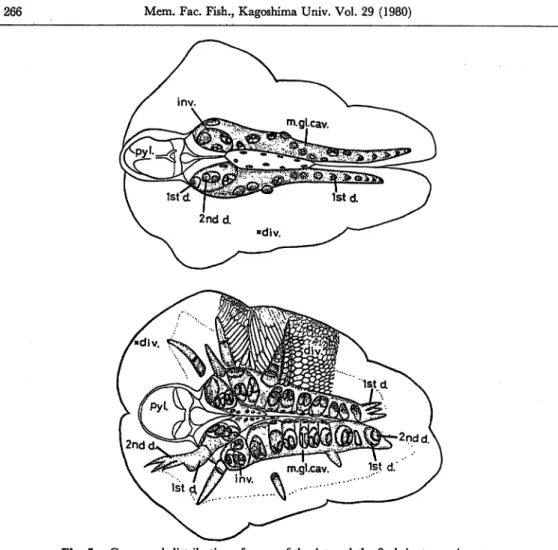
Fig. 5. Compared distribution of pores of the 1st and the 2nd duct openings to
the ventral of the cavity ofthe mid-gut gland. Specimens: upper, 11.0 mm
in length; lower, 12.0 mm in length. Differences are recognized in cavity
volumes, developmental degrees of the ducts, and diverticular appearances between these two specimens of almost similar size. The upper shows in distinguishable compartments of diverticula because of compact states of
them filled with reserves. On the other hand, the lower shows a disap
pearance of proximal diverticula, and only membranous remnants such as the basement membranes or the compartments of diverticula. Abbrev.,
div.: diverticula, Istd.: 1st duct, inv.: invagination, m.gl.cav.: cavity
of the mid-gut gland, pyl.: pylorus, 2nd d.: 2nd duct.
References
1) van Weel, P. B. (1955): Processes of secretion, restitution and resorption in gland of midgut of
Atya spinipes Newport. Physiol. Zool., 28, 40-54.
2) Vonk, H. J. (1955): Comparative physiology: nutrition, feeding, and digestion. Ann. Rev.
Physiol, 17, 483-498.
3) Vonk, H.J. (1960): Digestion and Metabolism, in "The Physiology of Crustacea" (ed. by T. H. Waterman), vol. 1, Academic Press, New York and London, 291-316.