Generation of retinal pigment epithelium from
human induced pluripotent stem cells showed
polarized secretion of VEGF and PEDF
著者
鎌尾 浩行
著者(英)
Kamao Hiroyuki
学位名
博士(医学)
学位授与機関
川崎医科大学
学位授与年度
平成25年度
学位授与年月日
2014-03-13
学位授与番号
35303甲第609号
URL
http://doi.org/10.15111/00000016
155 Kawasaki Medical Journal 39(4):155−162,2013
Generation of retinal pigment epithelium from human induced
pluripotent stem cells showed polarized secretion of VEGF and PEDF.
Hiroyuki KAMAO
1), Junko ISHIDA
1), Taiji SUDA
2), Junichi KIRYU
1)1) Department of Ophthalmology, 2) Tissue Biology & Electron Microscopy Research Center, Kawasaki Medical School, 577 Matsushima, Kurashiki 701-0192, Japan
ABSTRACT Age-related macular degeneration (AMD) is one of the major diseases that cause severe visual impairment in developed countries and is related to dysfunction of the retinal pigment epithelium (RPE). Human induced pluripotent stem cells (hiPSCs) have been drawing attention as a valuable source of RPE for basic research on or treatment of AMD. Here we investigated whether RPE could be generated from hiPSCs in Kawasaki Medical School. We maintained hiPSCs retaining pluripotent markers after a series of regular culture steps involving passaging, freezing, and thawing. hiPSCs were then cultured in RPE differentiation medium, and pigmented colonies were manually isolated for further differentiation into RPE. Differentiated cells formed pigmented cells with a typical RPE cobblestone appearance, abundant apical microvilli, adherens junctions, and tight junctions. Furthermore, generated pigmented cells expressed typical RPE marker genes, exhibited a barrier function, and secreted growth factors in a polarity-dependent manner similar to native RPE. These results indicate that RPE derived from hiPSCs in our facility can be used for in vitro and in vivo research.
(Accepted on August 6, 2013)
Key words: induced pluripotent stem cell, Retinal pigment epithelium, Regeneration therapy
Corresponding author Hiroyuki Kamao
Department of Ophthalmology, Kawasaki Medical School, 577 Matsushima, Kurashiki 701-0192, Japan
Phone : 81 86 462 1111 Fax : 81 86 463 0923
E-mail: hironeri@med.kawasaki-m.ac.jp
INTRODUCTION
Age-related macular degeneration (AMD)1)
is caused by dysfunction of the retinal pigment epithelium (RPE), which plays an essential role in the maintenance of photoreceptors2) and a healthy
ocular environment. The recent introduction of treatments such as laser therapy3), photodynamic
therapy4), and anti-vascular endothelial growth
factor therapy5) has resulted in significant overall
maintenance or improvement of vision in AMD patients; however, a large number of AMD patients
still have severe visual impairment with the limited effects of these therapies, such as non-responders, dry AMD, or advanced AMD. RPE transplantation with the aim of replacing pathological RPE with healthy RPE was introduced more than two decades ago, and RPE transplantation for AMD has been reported using allologous fetal RPE6) or autologous
peripheral RPE7). In summary, the former exhibited
complications associated with immune rejection, while the latter required invasive surgery to harvest peripheral RPE and place them in the submacular
156 Kawasaki Medical Journal
area. Therefore, RPE derived from autologous human induced pluripotent stem cells (hiPSCs)8)
emerged as an ideal alternative cell source to overcome these disadvantages.
hiPSCs are typically reprogramed by transfecting stem cell-associated genes into adult somatic cells. Since hiPSCs share major characteristics with embryonic stem cells such as pluripotency, self-renewal capacity, gene/protein expression, and epigenetic status, hiPSCs have been generated from various resources for research on and treatment of various diseases including retinitis pigmentosa9),
Parkinson's disease10), and cardiovascular disease11).
The hiPSC culture is completely different from other types of cell lines and requires specialized methods. Here we investigated whether a widely used hiPSC line could be cultured that retained pluripotency markers after a series of culture steps involving passaging, freezing, and thawing, and also tested if we could differentiate hiPSC-derived RPE (hiPSC-RPE) from them in our facility.
MATERIALS AND METHODS
Culture of hiPSCs and hiPSC-RPE
The utilization of hiPSCs was approved by the Research committee of Kawasaki Medical School. The hiPS cell line, 253G1 produced from healthy human dermal fibroblast cells using 3 transcription factors (Oct3/4, Sox2, and Klf4) was purchased from RIKEN BioResource Center. These hiPSCs were seeded on Mitomycin C-treated mouse feeder cells (SNL cells) after frozen hiPSCs were immediately thawed by Primate ES Cell Medium (ReproCELL). hiPSCs were maintained in Primate ES Cell Medium supplemented with 5 ng/ml bFGF (Wako) and passaged by pipetting into small cell aggregates when hiPSC colonies became large. hiPSCs were put into freeze stock after three months culture using the vitrification method. To differentiate into RPE, hiPSCs were cultured on gelatin-coated dishes in the differentiation medium
(GMEM; GMEM supplemented with 1 mM sodium pyruvate, 0.1 mM non-essential amino acids and 0.1 mM 2-mercaptoethanol) and 20% KnockOut Serum Replacement (KSR) for 4 days, GMEM and 15% KSR for 6 days, and GMEM and 10% KSR for 20 days. The Rho-associated kinase inhibitor Y-27632 (10 µM, Wako), the ALK4 inhibitor SB431542 (5 µM, Sigma), and the casein kinase I inhibitor CKI7 (3 µM, Sigma) was added to the differentiation medium for 18 days. After the pigmented cells focally appeared, the differentiation medium was switched to SFRM [DMEM/F12 (7:3) supplemented with B27 (Invitrogen), 2 mM L-glutamine (Sigma)] for 7 days. Pigmented colonies were then transferred to laminin-coated dishes in SFRM supplemented with 10 ng/ml bFGF (Wako) and grown until confluence was reached.
Immunocytochemistry
hiPSCs and hiPSC-RPE were fixed with 4% paraformaldehyde (PFA) and, after permeabilization with 0.3% TritonTM X-100 and blocking with
blocking one (NACALAI), samples were incubated with the primary antibody overnight at 4℃ followed by secondary antibody reactions for 1 hr at room temperature. Antigens detected with primary antibodies (spices, company, and dilution) were; Oct3/4 (mouse, Santa Cruz, 1/500), Nanog (rabbit, ReproCELL, 1/500), SSEA-4 (mouse, Millipore, 1/300), Tra-1-60 (mouse, Millipore, 1/300), Pax6 (rabbit, Covance, 1/600), MITF (mouse, Abcam, 1/1000), Bestrophin (mouse, Abcam, 1/500), RPE65 (rabbit, 1/1000), and ZO-1 (rabbit, Zymed, 1/100). The bound primary antibodies were detected with secondary antibodies labeled with Alexa Fluor 546; goat anti-rabbit IgG (1/1000; Molecular Probes), or goat anti-mouse IgG (1/1000; Molecular Probes), and nuclei were stained with 4'6-diamindino-2-phenylindole (DAPI) (Molecular Probes, 1 µg/ml). Specimens were imaged using a laser-scanning confocal microscope (FV1000-D, OLYMPUS).
157 Kamao H, et al. : Generation of hiPSC-RPE for basic research
Transmission electron microscopy (TEM)
hiPSC-RPE were fixed in 2.5% glutaraldehyde, postfixed with 1% osmium tetroxide, dehydrated with ascending concentrations of ethanol, and infiltrated by epoxy resin. Two days after drying under the hood, ultrathin sections, about 60 nm thick, were obtained with a microtome. hiPSC-RPE were visualized with a transmission electron microscope (JEM-1400, JEOL).
Reverse transcription-polymerase chain reaction
Total RNA was extracted from confluent hiPSCs (253G1) and hiPSC-RPE (253G1) using RNeasy Plus Mini Kit® (QIAGEN) according to the
manufacturer's instructions. The RNA concentration and quality were assessed with a NanoDrop 1000 spectrophotometer (NanoDrop Technoligies). The RNA was reversetranscribed using 20 µl reaction consisted of: 11 µl RNA (1,000 ng) in DNase RNase Free water (QIAGEN), 1 µl 50 µM Oligo(dT)20
(Invitrogen), 1 µl 10 mM dNTP Mix (Invitrogen), 1 µl Rnase OUT (20 U/µl; Invitrogen), SuperScriptTM
III Reverse Transcriptase (4 µl 5×First-Standard Buffer, 1 µl 0.1M DTT, 1 µl SuperScript III 200 U/µl; Invitrogen) and the cDNA synthesis was performed as follows: 15 min at 25℃, 60 min at 50℃, and 15 min at 70℃. PCR reactions were performed using Ex Taq® DNA polymerase (Takara
Bio). Thermal cycling conditions were performed as follows: one cycle at 94℃ for 120 sec; 30 cycles at 94℃ for 30 sec, 57℃ for 30 sec, and 72℃ for 60 s; one cycle at 72℃ for 60 sec. Primer sequences used to amplify the three RPE-specific genes are listed in Table 1.
Measurement of transepithelial electrical resistance (TER)
T E R w a s p e r f o r m e d a c c o r d i n g t o t h e manufacturer's instructions of the electrical resistance system (Millicell®, Millipore) at 37℃. The
TER value (Ωcm2) was calculated by subtracting
the value of medium alone in the insert as a blank from the experimental value and multiplying it by the area of the insert membrane.
Enzyme linked immunosorbent assay (ELISA) for vascular endothelial growth factor (VEGF) and pigment epithelium-derived factor (PEDF)
The culture medium from confluent hiPSC-RPE was collected 24 hr after the medium was changed. Secretion levels were measured by the human VEGF-ELISA Kit (eBioscience) for VEGF and human PEDF-ELISA Kit (BioVender) for PEDF, according to the manufacturers' instructions.
RESULTS
Maintenance culture and characterization of hiPSCs
To determine that we could stably culture hiPSCs in Kawasaki Medical School, we investigated if we could maintain hiPSCs that retained pluripotency markers after performing a series of culture steps involving passaging, freezing, and thawing. Firstly, provided hiPSCs (253G1) were thawed and seeded on Mitomycin C-treated SNL feeder cells (SNL). A medium change was made every day and hiPSC colonies are transferred onto new dishes with SNL feeder cells every 4 to 5 days. Three months after initiating the culture, hiPSCs were frozen and stocked in liquid nitrogen (the passage number was 20). One week after being kept as a frozen stock, thawed hiPSCs were fed again for three months (the passage number was 38). The performed procedure
Table 1. Primer sequences used to amplify the four RPE-specific genes
primers Sequences Bestrophin 1 Forward TAGAACCATCAGCGCCGTC
Reverse TGAGTGTAGTGTGTATGTTGG RPE65 Forward TCCCCAATACAACTGCCACT
Reverse CCTTGGCATTCAGAATCAGG MERTK Forward TCCTTGGCCATCAGAAAAAG
Reverse CATTTGGGTGGCTGAAGTCT GAPDH Forward ACCACAGTCCATGCCATCAC Reverse TCCACCACCCTGTTGCTGTA
158 Kawasaki Medical Journal
Fig. 1. Maintenance culture and characterization of hiPSCs
(a) Culture steps involved in thawing, feeding, and freezing of hiPSCs (b) Phase image of a hiPSC colony with differentiated cells (arrow). Scale bar, 100 µm. (c) Phase image of hiPSCs. Scale bar, 100 µm (d-e) hiPSCs expressed the pluripotency markers OCT3/4, Nanog, SSEA4, and Tra-1-60. Scale bar, 200 µm.
was summarized in Figure 1a. When differentiated cells exhibiting a polygonal morphology appeared, we manually picked up the colonies including these differentiated cells (Fig. 1b). hiPSCs that went through the series of culture steps (Fig. 1c) retained the expression of pluripotency markers (OCT3/4, Nanog, SSEA-4, and Tra-1-60) in immunocytochemistry (Fig. 1d-g).
Generation of RPE from hiPSCs
hiPSCs were then differentiated to RPE using a modified protocol established previously12). Three
weeks after initiating differentiation, pigmented colonies presented a typical RPE cobblestone appearance (Fig. 2a). To purify these cells, we manually picked up, plated them onto a laminin coat dish, and cultured them until confluency was reached (Fig. 2b). To become fully mature cells, purified pigmented cells were continuously
cultured at a confluent state (Fig. 2c, d). These pigmented cells had the structural characteristics of RPE including abundant apical microvilli, adherens junctions, and tight junctions under transmission electron microscopy (TEM) (Fig. 2e-g). Furthermore, generated pigmented cells expressed typical RPE markers related to chloride channels (BEST1; Bestrophin 1)13), the retinoid
cycle (RPE65)14,15), and phagocytosis (MERTK)16) in
reverse transcriptase-polymerase chain reaction (RT-PCR) (Fig. 3a) and eye field specification markers (PAX6 and MITF), BEST1, RPE65, and tight junction marker (ZO-1) in immunocytochemistry (Fig. 3b-f).
Functional assessment of hiPSC-RPE in vitro
We investigated whether generated pigmented cells exhibited RPE functions typically observed in native RPE. Since RPE are known to contribute
b DAPI e Nanog DAPI g DAPI f Tra-1-60 DAPI a culture for 3 m Plate on SNLs Day 0 Thawing of iPSCs Day 97 Freezing of iPSCs Day 90 stock for 1 w culture for 3 m Test the pluripotency of iPSCs Day 187 OCT 3/4 SSEA-4 d Figure 1 c
159 Kamao H, et al. : Generation of hiPSC-RPE for basic research
Fig. 2. Generation of RPE from hiPSCs
(a) Approximately 3 weeks after the initiation of differentiation, some cells formed pigmented colonies with a typical RPE cobblestone appearance (arrow). (b) Phase image of transferred pigmented cells cultured until confluency. Scale bar, 100 µm. (c) Purified pigmented cells became confluent. (d) Phase image of purified pigmented cells forming a typical RPE cobblestone appearance. Scale bar, 100 µm. (e) Transmission electron microscopy image of pigmented cells, (f) apical microvilli, and (g) adherens junctions and tight junctions.
Fig. 3. Gene expression of RPE from hiPSCs
(a) Purified pigmented cells expressed typical RPE markers (BEST1, RPE65, and MERTK) in RT-PCR. (b-f) Purified pigmented cells derived from hiPSC expressed early (MITF, PAX6) and mature (BEST1, RPE65, ZO-1) RPE markers in immunocytochemistry. Scale bar, 50 µm.
b e f g a d Figure 2 c b d BEST1 a c Figure 3 e f Mitf PAX6 RPE65 ZO-1 ① BEST1 ② RPE65 ③ MERTK ④ sterile water ⑤ GAPDH ① ② ③ ④ ⑤ BEST1 RPE65 MERTK GAPDH hiPSC s hiP S-RPE hiPS-RPE
160 Kawasaki Medical Journal
to the barrier function and secrete growth factors including vascular endothelial growth factor (VEGF)17) and pigment epithelium-derived
factor (PEDF)18), we evaluated the functional
tight junction, or a barrier function by measuring transepithelial electrical resistance (TER)19), and
measured the polarity-dependent concentration of VEGF and PEDF in the culture medium using ELISA. We seeded pigmented cells on the TranswellTM for these experiments. Four weeks after
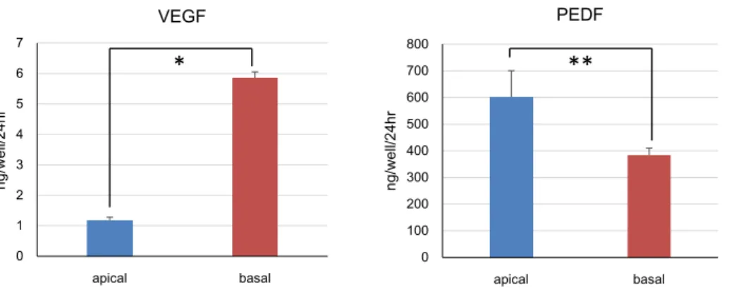
they became confluent, we measured the TER and concentration of PEDF and VEGF in the apical and basal media of the TranswellTM. The mean TER was
410 Ω cm2 (n = 6) and concentration of VEGF was
higher in the basal medium (5.9 ± 0.2 ng/well/24hr
n = 3) than in the apical medium (1.2 ± 0.1 ng/
well/24hr n = 3), whereas that of PEDF was higher in the apical medium (600 ± 99 ng/well/24hr n = 3) than in the basal medium (380 ± 27 ng/well/24hr
n = 3) (Fig. 4). All data are expressed as mean ±
SEM (standard error of the mean), with P < 0.05 being considered statistically significant. Statistical analyses were performed using StatMate4 (ATMS). Secretion of PEDF and VEGF was evaluated using paired t-tests.
DISCUSSION
In this study, we demonstrated that we could successfully maintain hiPSCs and induce hiPSC-RPE to be used for research in Kawasaki Medical School.
hiPSCs are currently a useful cell source for regenerative medicine, disease modeling, and drug testing, even though the hiPSC culture requires specialized methods. We had to demonstrate the pluripotency of hiPSCs and the function of generated differentiation cells. Therefore, we firstly confirmed that hiPSCs retained pluripotency after the series of regular culture steps involving passaging, freezing, and thawing. As shown Figure 1, our hiPSCs retained the expression of OCT3/4, Nanog, SSEA-4, and Tra-1-60, which indicated that we could maintain these cells without losing their pluripotency.
The limitation of existing clinical treatments for AMD has resulted in attention being given to replacing pathological RPE with healthy RPE. Research using hiPSC-RPE as an ideal cell source for transplantation is currently underway and RPE derived from embryonic stem cells is being examined in a clinical trial20). The differentiation
methods of RPE from hiPSCs and generated
hiPSC-Figure 4 each n = 3 0 1 2 3 4 5 6 7 apical basal ng/ w el l/24hr VEGF 0 100 200 300 400 500 600 700 800 apical basal ng /w el l/24hr PEDF
*
**
Fig. 4. Functional assessment of hiPSC-RPE in vitro
The concentration of VEGF was higher in the basal medium (5.9 ± 0.2 ng/well/24hr n = 3) than in the apical medium (1.2 ± 0.1 ng/well/24hr n = 3), whereas that of PEDF was higher in the apical medium (600 ± 99 ng/well/24hr n = 3) than in the basal medium (380 ± 27 ng/well/24hr n = 3). All data are expressed as means ± SEM. *P < 0.01, **P < 0.05.
161 Kamao H, et al. : Generation of hiPSC-RPE for basic research
RPE have displayed appropriate physiological properties in vitro and in vivo in previous reports21,22). As was shown in the results of the
present study, our differentiated RPE also exhibited morphological and genetic properties consistent with typical RPE characteristics, which indicates that they can be used as tools for further RPE research.
Another important observation in this study was that our hiPSC-RPE secreted VEGF and PEDF, as seen in Figure 4. Considering the roles of these factors in physiology and pathology, these results indicate that hiPSC-RPE transplantation could also be applied to various clinical purposes such as the protection of degenerating photoreceptors or regeneration of choroid vasculature. Therefore, we can utilize our hiPSC-RPE for clinically oriented researchin vitro and in vivo in Kawasaki Medical School.
ACKNOWLEDGEMENTS
We thank M. Takahashi and M Mandai (Laboratory for Retinal Regeneration, Center for Developmental Biology, RIKEN) for their valuable comments and discussions on this work.
This study was supported by a Research Project Grant from Kawasaki Medical School.
REFERENCES
1) Bressler NM, Bressler SB, Fine SL: Age-related macular degeneration. Surv. Ophthalmol. 32: 375-413, 1988 2) Young RW, Bok D: Participation of the retinal pigment
epithelium in the rod outer segment renewal process. J. Cell Biol. 42: 392-403, 1969
3) Macular Photocoagulation Study Group: Argon laser photocoagulation for neovascular maculopathy. Three-year results from randomized clinical trials. Arch Ophthalmol 104: 694-701, 1986
4) Treatment of age-related macular degeneration with photodynamic therapy (TAP) Study Group.: Photodynamic therapy of subfoveal choroidal neovascularization in age-related macular degeneration
with verteporfin: one-year results of 2 randomized clinical trials—TAP report. Arch Ophthalmol 117: 1329-1345, 1999
5) Heier JS, Antoszyk AN, Pavan PR, et al.: Ranibizumab for treatment of neovascular age-related macular degeneration: a phase I/II multicenter, controlled, multidose study. Ophthalmology 113: 633-642, 2006 6) Algvere PV, Berglin L, Gouras P, Sheng Y:
Transplantation of fetal retinal pigment epithelium in age-related macular degeneration with subfoveal neovascularization. Graefes Arch Clin Exp Ophthalmol. 232: 707-716, 1994
7) Binder S, Stolba U, Krebs I, et al.: Transplantation of autologous retinal pigment epithelium in eyes with foveal neovascularization resulting from age-related macular degeneration: a pilot study. Am. J. Ophthalmol. 133: 215-225, 2002
8) Takahashi K, Tanabe K, Ohnuki M, Narita M, Ichisaka T, Tomoda K, Yamanaka S: Induction of pluripotent stem cells from adult human fibroblasts by defined factors. Cell 131: 861-872, 2007
9) Jin ZB, Okamoto S, Xiang P, Takahashi M: Integration-free induced pluripotent stem cells derived from retinitis pigmentosa patient for disease modeling. Stem Cells Trans Med. 1: 503-509, 2012
10) Wernig M, Zhao JP, Pruszak J, Hedlund E, Fu D, Soldner F, Broccoli V, Constantine-Paton M, Isacson O, Jaenisch R: Neurons derived from reprogrammed fibroblasts functionally integrate into the fetal brain and improve symptoms of rats with Parkinson's disease. Proc Natl Acad Sci U S A 105: 5856-5861, 2008
11) Zhang J, Wilson GF, Soerens AG, Koonce CH, Yu J, Palecek SP, Thomson JA, Kamp TJ: Functional cardiomyocytes derived from human induced pluripotent stem cells. Circ Res 104: e30-41, 2009
12) Osakada F, Ikeda H, Mandai M, Wataya T, Watanabe K, Yoshimura N, Akaike A, Sasai Y, Takahashi M: Toward the generation of rod and cone photoreceptors from mouse, monkey and human embryonic stem cells. Nat. Biotechnol. 26: 215-224, 2008
13) Stone EM, Nichols BE, Streb LM, Kimura AE, Sheffield VC: Genetic linkage of vitelliform macular degeneration (Best's disease) to chromosome 11q13. Nat Genet. 1: 246-250, 1992
14) Gu SM, Thompson DA, Srikumari CR, et al.: Mutations in RPE65 cause autosomal recessive childhood-onset
162 Kawasaki Medical Journal severe retinal dystrophy. Nat Genet. 17: 194-197, 1997
15) Marlhens F, Bareil C, Griffoin JM, et al.: Mutations in RPE65 cause Leber's congenital amaurosis. Nat Genet. 17: 139-141, 1997
16) Gal A, Li Y, Thompson DA, Weir J, Orth U, Jacobson SG, Apfelstedt-Sylla E, Vollrath D: Mutations in MERTK, the human orthologue of the RCS rat retinal dystrophy gene, cause retinitis pigmentosa. Nat Genet. 26: 270-271, 2000
17) Adamis AP, Sima DT, Yeo KT, Yeo TK, Brown LF, Berse B, D'Amore PA, Folkman J: Synthesis and secretion of vascular permeability factor/vascular endothelial growth factor by human retinal pigment epithelial cells. Biochem Biophys Res Commun. 193: 631-638, 1993 18) Steele FR, Chader GJ, Johnson LV, Tombran-Tink J.
Pigment epithelium-derived factor: neurotrophic activity and identification as a member of the serine protease inhibitor gene family. Proc Natl Acad Sci USA 90: 1526-1530, 1993
19) Stevenson BR, Siliciano JD, Mooseker MS, Goodenough DA: Identification of ZO-1: a high molecular weight polypeptide associated with the tight junction (zonula occludens) in a variety of epithelia. J. Cell Biol. 103: 755-766, 1986
20) Schwarts SD, Hubschman JP, Heilwell G, Franco-Gardenas V, Pan CK, Ostrick RM, Mickunas E, Gay R, Klimanskaya I, Lanza R: Embryonic stem cell trials for macular degeneration: a preliminary report. Lancet. 379: 713-720, 2012
21) Hirami Y, Osakada F, Takahashi K, Okita K, Yamanaka S, Ikeda H, Yoshimura N, Takahashi M: Generation of retinal cells from mouse and human induced pluripotent stem cells. Neurosci Lett. 458: 126-131, 2009
22) Carr AJ, Vugler AA, Hikita ST, et al.: Protective effects of human iPS-derived retinal pigment epithelium cell transplantation in the retinal dystrophic rat. PLoS One 4: e8152, 2009