INTRODUCTION
The provision of quality information on specific pathogens associated with Japanese Infectious Diseases (SPJID) through the activities of the National Bioresource Project of the Japanese Ministry of Education, Culture, Sports, Science and Technology, is especially important for both the col-lection of cultures and their use. The pathogenic factors of Biosafety Level 3 (BSL3) pathogens and SPJID are often encoded on plasmids or insertion sequences on the bacterial chromosomes. These fac-tors are, therefore, unstable and are often “cured” (i.e. eliminated) from the host. The important patho-genic factor invE (virB) of Shigella spp. and entero-invasive Escherichia coli is required for the inva-siveness of these bacteria (Mitobe et al., 2009). This factor is encoded on a plasmid and is easily cured from the host when stock bacteria are cultured. Once the factor has been eliminated from the host, it is difficult to differentiate these pathogens from non-pathogenic E. coli. The Vi antigen of Salmonella enterica serovar Typhi is the most important viru-lence factor used for differentiating this serovar from the 2000 other serovars of S. enterica, but the
Vi synthesis gene clusters are naturally deleted from the host chromosome (Hashimoto et al., 1996, Hashimoto & Khan, 1997).
Capsular antigen fraction 1 (caf1) (Galyov, 1990) and plasminogen activator surface protease (pla) are located on plasmids of Yersinia pestis (Guinet & Carniel, 2012). Once the plasmids are cured from the host, it becomes very difficult to differentiate Y. pestis from the closely related Yersinia pseudotuber-culosis.
Type strains and strains used for serotyping of SPJID are especially important for maintaining qual-ity information on the pathogenic factors in these bacteria. Simple and rapid confirmation methods are required so that these strains can be checked before they are distributed. To fulfill this need, we developed a tagged-cocktail PCR method (Hayashi, 2013) and established a protocol to differentiate the PCR amplicons by using a DNA strip containing six printed anti-tags and three position markers. We applied this technology to confirm the presence of pathogenic factors of BSL3 and SPJID strains. The PCR amplification method was also optimized for this with the use of a low-profile PCR tube and a quick amplification PCR machine. We established a protocol to confirm the presence of pathogenic fac-tors of BSL3 and SPJID strains within 40 min from the time bacteria were collected from a single colo-ny on a culture plate.
A cocktail PCR and DNA strip method for quick confirmation
of multiple pathogenic factors in BSL3 stock cultures
Takayuki Ezaki
1)*, Izumi Kanazawa
1), Sayoko Hayashi
3), Masahiro Hayashi
2),
Ibrahim Eldesoky
1)and Hajime Fukunaga
1)1)Regeneration and Advanced Medical Science, Gifu University Graduate School of Medicine,
2)Anaerobic Laboratory, Life Science Research Center, Gifu University
1-1 Yanagido, Gifu 501-1194, Japan
3)Gifu University of Medical Science, 795-1 Aza-Nagamine, Ichihiraga, Seki, Gifu 501-3892, Japan
Repositories of culture collections of Biosafety Level 3 (BSL3) pathogens are expected to provide information on the pathogenic factors present in stock bacteria. However, it is difficult to maintain valid pathogenic information on each of the different BLS3 pathogens because quality control of the stock strains generally has to be performed by a few staff members.
We developed a common platform to differentiate six different PCR amplicons on a DNA strip. The six amplicons can be differentiated with the help of three position markers printed on the strip. With this method, the pathogenic factors in BSL3 pathogens can be determined within 40 min after cultured bacteria are picked up from a single colony.
*Corresponding author
E-mail: ezaki2016@gmail.com Accepted: February 26, 2016
MATERIALS AND METHODS
Cell cultures
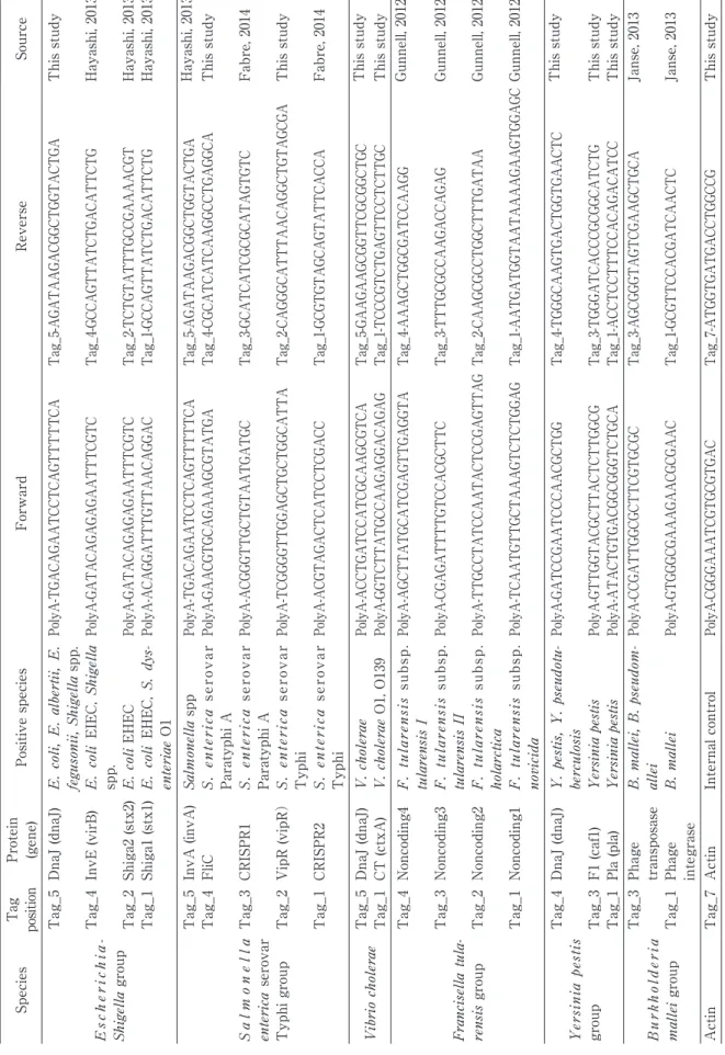
Strains from the Gifu Type Culture Collection (GTC), and the Japan National Collection of Bacterial Pathogens (JNBP), along with wild Y. pestis strains stocked at the Mongolian National Center for Zoonotic Diseases (Ulaanbaatar, Mongolia) were used. The tagged primers used are listed in Table 1. Upstream primers were labeled with a common polyA sequence and downstream primers were labeled with six different tags. These primers were synthesized by Sigma-Aldrich Japan (Tokyo, Japan) and Tohoku Bio-Array Corporation (Sendai, Japan). The DNA strips printed with six different anti-tags and three position markers were kindly provided by Tohoku Bio-Array Corporation (Fig. 1).
Examination methods
Bacteria from a single colony plated the day before and incubated overnight were suspended in 500 ml of distilled water, and the solution was boiled to disrupt the cells. A volume of 2.5 ml of the solu-tion was used for cocktail PCR analysis. The con-centrations of the single-colony suspensions ranged
from 107 to 108 cfu/ml. To determine the detection
limit, cell suspensions of 101 to 108 cfu/ml were also
prepared. For rapid PCR, a low-profile PCR tube was selected and a total of 10 ml of the reaction mix-tures was used for analysis. The reaction mixmix-tures contained 2.5 ml of 4× primer mixture (0.8 mM), 2.5 ml of crude DNA suspension, and 5 ml of 2× premixed Taq polymerase (TakaraBio, Kyoto, Japan). After denaturation at 97℃ for 2 min, samples were ampli-fied under the following two-step PCR conditions: 60℃ for 10 s and 97℃ for 5 s. Thirty PCR cycles were performed. The total amplification time was within 30 min.
After PCR amplification, 10 ml of a 0.5% blue latex suspension labeled with polyT was added to the PCR tube, and a DNA strip was inserted into the PCR tube for chromatography. Ten minutes later, the blue latex had been completely absorbed by the DNA strip and the target line that captured the PCR amplicon on the DNA strip had turned blue.
Sensitivity of the DNA strip
Two-fold serial dilutions of the PCR products of E. coli shiga2-positive strain 157:H7 (GTC 14509) were prepared. PCR products in the diluted solutions were detected with a standard latex solution to
determine the endpoint of detection. Ten-fold serial dilutions of a Salmonella enterica GTC 3P0637 cell
suspension from 108 to 101 cfu/ml were prepared,
and the invA amplicon was detected by using SYBR green (Takara Bio, Kyoto, Japan) and a Step One Plus PCR machine (Applied Bio, Tokyo, Japan). After amplification, the amplicon was also detected by using the DNA-strip method, and the sensitivities of the two methods were compared.
Screening for pathogenic factors
Bacteria from a single colony plated the day before and incubated overnight were suspended in
500 ml of distilled water to prepare a 108 cfu/ml cell
suspension. The solution was boiled at 100℃ for 3 min. Next, 2.5 ml of the boiled cell suspension was mixed with 2.5 ml of 4× cocktail primers (0.8 mM) and 5 ml of 2× SYBR Premix EX Taq polymerase (Takara Bio). All cocktail primers were denatured at 97℃ for 2 min. The following two-step PCR cycle was applied 30 times for all primers: 60℃ for 10 s and 97℃ for 5 s.
All JNBP strains of genus Shigella listed in Table 2 were analyzed for invE, stx1 (Shiga 1) and stx2 (Shiga 2). Reference serotyping strains, including 13 Shigella dysenteriae, 28 Shigella flexneri, 26 Shigella boydii and 10 Shigella sonnei, were analyzed. dnaJ primer to detect both Shigella species and E. coli was also included in the cocktail primers.
Salmonella enterica serovar Typhi and serovar Paratyphi A are SPJID pathogens. The invA gene is shared among all 2000 Salmonella serovars. To differentiate these two serovars from other Salmonella serovars, Vi antigen regulator (vipR) and flagellar antigen gene (fliC:a) were prepared. Vi antigen is specific for serovar Typhi, but it is often deleted from the chromosome (Hashimoto et al., 1996, Hashimoto & Khan, 1997). We therefore select-ed another differential factor, CRISPR2, which is a recently reported serovar Typhi-specific factor that was found by complete genome analysis (Fabre et al., 2014). CRISPR1 and FliC:a were selected for dif-ferentiating serovar Paratyphi A. FliC:a is a stable gene, but several other salmonella serovars belong-ing to group A flagellar groups cross-react with FliC:a primers. CRISPR-1 has thus far been detect-ed only in serovar Paratyphi A (Fabre et al., 2014).
CT toxin of
Table 1 Pathogenic factors and cocktail primers Species Ta g pos iti on Protein (gene) Positive species Forward Reverse Source E sc h er ic h ia -Shigella group Tag_5 DnaJ (dnaJ) E. coli, E. albertii, E. fegusonii, Shigella spp. Po ly A -TG A CA GA ATC CT CA GT TT TT CA Ta g_ 5-A GATA A GA CG GC TG GTA CTG A This study Tag_4 InvE (virB) E. coli EIEC, Shigella spp. Po ly A-GA TA CA GA GA GA A TT TC GT C Ta g_ 4-G CCA GT TAT CTG A CAT TC TG Hayashi, 2013 Tag_2 Shiga2 (stx2) E. coli EHEC Po ly A-GA TA CA GA GA GA A TT TC GT C Ta g_ 2-T CTG TAT TTG CCG A A A A CG T Hayashi, 2013 Tag_1 Shiga1 (stx1) E. coli EHEC, S. dys -enteriae O1 Po ly A-A CA GGA TT TG TT A A CA GGA C Ta g_ 1-G CCA GT TAT CTG A CAT TC TG Hayashi, 2013 S a lm o n e ll a enterica serovar Typhi group Tag_5 InvA (invA) Salmonella spp Po ly A -TG A CA GA ATC CT CA GT TT TT CA Ta g_ 5-A GATA A GA CG GC TG GTA CTG A Hayashi, 2013 Tag_4 FliC S. enterica serovar Paratyphi A Po ly A -G A A CG TG CA GA A A GCG TATG A Ta g_ 4-CG CAT CAT CA A GG CC TG A GG CA This study Tag_3 CRISPR1 S. enterica serovar Paratyphi A Po ly A -A CG GG TTG CTG TA ATG ATG C Ta g_ 3-G CAT CAT CG CG CATA GTG TC Fabre, 2014 Tag_2 VipR (vipR) S. enterica serovar Typhi Po ly A -T CG GG GT TG GA GC TG CTG GCAT TA Ta g_ 2-CA GG GCAT TTA A CA GG CTG TA GCG A This study Tag_1 CRISPR2 S. enterica serovar Typhi Po ly A -A CG TA GA CT CATC CT CG A CC Ta g_ 1-G CG TG TA GCA GTAT TCA CCA Fabre, 2014 Vibrio cholerae Tag_5 DnaJ (dnaJ) V. cholerae Po ly A -A CC TG AT CCAT CG CA A GCG TCA Ta g_ 5-GA A GA A GC GG TT CG CGG CT GC This study Tag_1 CT (ctxA) V. cholerae O1, O139 Po ly A-GG TC TT A TG CC A A GA GGA CA GA G Ta g_ 1-TCCC GTC TG A GT TCC TC TT GC This study Francisella tula -rensis group Tag_4 Noncoding4 F. tularensis subsp. tularensis I Po ly A -A GC TTATG CAT CG A GT TG A GG TA Ta g_ 4-AAAG CT GG CG A TC CAAG G Gunnell, 2012 Tag_3 Noncoding3 F. tularensis subsp. tularensis II Po ly A -CG A GAT TT TG TC CA CG CT TC Ta g_ 3-T TT GC GC CAA GAC CAG AG Gunnell, 2012 Tag_2 Noncoding2 F. tularensis subsp. holarctica Po ly A -T TG CC TATC CA ATA CT CCG A GT TA G Ta g_ 2-CA A GCG CC TG GC TT TG ATA A Gunnell, 2012 Tag_1 Noncoding1 F. tularensis subsp. novicida Po ly A -T CA ATG TTG CTA A A GT CT CTG GA G Ta g_ 1-AA TG A TG GT AA TAAAAG AAG TG GA GC Gunnell, 2012 Yersinia pestis group Tag_4 DnaJ (dnaJ) Y. pestis, Y. pseudotu -berculosis Po ly A -G AT CCG A AT CC CA A CG CTG G Ta g_ 4-TG GG CA A GTG A CTG GTG A A CT C This study Tag_3 F1 (caf1) Yersinia pestis Po ly A -G TTG GTA CG CT TA CT CT TG GCG Ta g_ 3-TG GG AT CA CC CG CG GCAT CTG This study Tag_1 Pla (pla) Yersinia pestis Po ly A -ATA CTG TG A CG GCG GG TC TG CA Ta g_ 1-A CC TCC TT TCC A CA GA CA TCC This study Burkholderia mallei group Tag_3 Phage transposase B. mallei, B. pseudom -allei Po ly A -C CG AT TG GCG CT TCG TG CG C Ta g_ 3-A GCG GG TA GT CG A A GC TG CA Janse, 2013 Tag_1 Phage integrase B. mallei Po ly A-G TGGG CGA A A GA A CG CGA A C Ta g_ 1-G CG TT CCA CG AT CA A CT C Janse, 2013 Actin Tag_7 Actin Internal control Po ly A -CG GG A A AT CG TG CG TG A C Ta g_ 7-ATG GTG ATG A CC TG GC CG This study
cocktail was prepared to detect both CT-positive and -negative strains of V. cholerae. Strains belong-ing to the O1 and O139 serotypes are generally both ctx- and dnaJ-positive. The other 140 serotypes were expected to be CT-negative and dnaJ-positive.
and of
Both the plasmid-encoded plasminogen activator gene pla (Guinet & Carniel, 2012) and the chromo-somal chaperone gene dnaJ of Y. pestis were pre-pared. pla was recently reported in a wild strain of Escherichia coli and in Citrobacter koseri (Hänsch et al., 2015). Pathogenic wild-type strains of Y. pestis were expected to be both pla- and dnaJ-positive, but plasmid-cured strains were expected to be only dnaJ-positive. Because another plasmid-encoded specific F1 antigen gene, caf1 (Galyov et al., 1990; Tsui et al., 2015), produced nonspecific amplicons, calf1 was not mixed with pla, but was instead ampli-fied independently from the pla and dnaJ cocktail. After PCR, the two amplicons were combined and detected with a single DNA strip. The closely relat-ed Y. pseudotuberculosis has an identical dnaJ sequence, and the dnaJ PCR was expected to be positive. Once the plasmids are cured from Y. pes-tis, it cannot be differentiated from Y. pseudotuber-culosis by using this cocktail primer (Table 3).
Subspecies and pathovar differentiation of
Genome analyses have revealed sequences for dif-ferentiating subspecies of Francisella tularensis (Gunnell et al., 2012). We added tags to these four primers and made a cocktail to differentiate two subspecies, namely F. tularensis subsp. holarctica and F. tularensis subsp. novicida, from F. tularensis
Fig. 1 A DNA strip for differentiating six cocktail PCR amplicons. Six anti-tags and three marker
lines are printed on a nitrocellulose membrane to differentiate six PCR amplicons. Anti-Tag 7 is internal control (I.C.)
Anti-tag 1 Anti-tag 2 Anti-tag 4 Anti-tag 3 Anti-tag 5 Anti-tag 7 (I.C.)
Table 2 Pathogenic factors and percentage of positive results
Species Virulent factor % of positive results among stock strains
Shigella dysenteriae stx1 8% (1/13)
Shigella dysenteriae InvE 15% (2/13)
Shigella flexneri InvE 25% (7/28)
Shigella boydii InvE 31% (8/26)
Shigella sonnei InvE 20% (2/10)
Salmonella enterica serovar Typhi vipR 85% (17/20) Salmonella enterica serovar Typhi CRISPR2* 100% (3/3) Salmonella enterica serovar Paratyphi CRISPR2* 0% (0/3) Salmonella enterica serovar Typhi CRISPR1* 0% (0/5) Salmonella enterica serovar Paratyphi CRISPR1* 100% (3/3)
Vibrio cholerae O1/O139 ctxa 87% (13/15)
Vibrio cholerae other serotypes ctxa 0% (0/140)
Yersinia pestis caf1 60% (3/5)
Yersinia pestis pla 60% (3/5)
Yersinia pseudotuberculosis pla 0% (0/5) *CRISPAR1, CRISPAR2, are not pathogenic factors but cited in this table.
subsp. tularensis.
Differentiation and identification of and
Burkholderia mallei and Burkholderia pseudomal-lei are genetically similar organisms (Yabuuchi et al., 1992). Housekeeping genes that are useful for differ-entiating two such closely related species are rare. Several multiplex PCR methods have been reported (Rattanathongkom et al., 1997; Wongratanacheewin et al., 2000), but closely related species, such as Burkholderia thailandensis and environmental iso-lates of B. pseudomallei are not clearly differentia-ble. However, a recent whole-genome analysis has revealed several sequences that are specific to each species (Janse, 2013). Using information on these noncoding sequences, we prepared tagged primers to make cocktail PCR mixtures to differentiate the two species. Phage transposase, which is common to both species, and phage integrase, which is found
only in B. mallei (Table 1), were prepared for differ-entiating the two closely related species.
RESULTS AND DISCUSSION
The standard PCR method employs a high-profile PCR tube to run 20 to 50 ml of PCR reaction mix-tures, and 96 specimens can usually be amplified with a standard PCR machine.
To reduce the amplification time, we designed a four-well PCR machine (Quick Mobile), and employed low-profile PCR tubes to run experiments with a 10 ml reaction volume. The DNA extraction protocol was also simplified. Bacteria from a single colony were suspended in 500 ml of distilled water
(to a concentration of around 107 to 108 cfu/ml) and
boiled for 3 min. With this simple sample prepara-tion, the entire experiment could be completed with-in 40 mwith-in from the time bacteria were collected from a single colony. Gram-positive Mycobacterium tuber-culosis and Clostridium botulinum, which have
Table 3 Differentiation of closely related subspecies and serovars
Francisella tularensis group Noncoding1 Noncoding2 Noncoding3 Noncoding4Tag 1 Tag 2 Tag 3 Tag 4 Tag 5dnaJ
subsp. tularensis GTC 3P0421 − − − + +
subsp. holarctica GTC 3P0422 − + − − +
subsp. novicida GTC 3P0425T + − − − +
Salmonella enterica subsp. enterica group Tag 1 Tag 2 Tag 3 Tag 4 Tag 5
CRISPR2 vipR CRISPR1 FliC:a invA
serovar Typhi GTC 3P0637 Vi positive + + − − +
serovar Typhi GTC 3P0612 Vi negative + − − − −
serovar Paratyphi A GTC 3P0433 − − + + +
serovar Dublin GTC 02558 Vi positive − − − − +
Citrobacter freundii GTC 14916 Vi positive − − − − −
Bukholderia mallei group
Tag 1 Tag 2 Tag 3 Tag 4 Tag 5
Phage
integrase transposasePhage
B. pseudomallei GTC 3P0028 − +
B. pseudomallei 10 wild-type strains (0/10) (10/10)
B. mallei GTC 3P0003T + +
B. mallei 4 wild-type strains (4/4) (4/4)
B. thailandensis GTC 3P0407T − −
B. vietnamiensis GTC 3P0436T − −
Yersinia pestis group Tag 1 Tag 2 Tag 3 Tag 4 Tag 5
pla caf1 dnaJ
P. pestis GTC 3P0417T + − +
P. pestis Russian vaccine strain + not tested +
P. pestis 3 Mongolian wild-type strains (2/3) not tested (3/3)
tough cell walls, were successfully analyzed by using this simple protocol, (Data not shown).
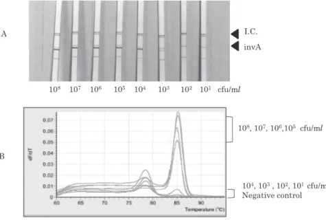
Sensitivity of the DNA strip
The PCR amplicon of the E. coli Shiga2-positive strain O157:H7 (GTC 14509) was diluted by two-fold serial dilution. Up to 64-fold diluted amplicon was visualized (Fig. 2). PCR amplicon in the same dilut-ed solutions was also detectdilut-ed by electrophoresis
and ethidium bromide staining; the amplicon could be detected at dilutions up to eight-fold (Data not shown.)
The PCR product from cell suspensions of S.
enterica serovar Typhi GTC 3P0637 at 108 to 103 cfu/
ml could be clearly visualized by using the DNA-strip method (Fig. 3A). However, the real-time PCR
results for the solutions at 104 to 102 cfu/ml could
not be differentiated from that for the negative
con-Fig. 2 Sensitivity of the DNA strip
Serially diluted PCR amplicons of Escherichia coli Shiga2-positive strain O157:H7 (GTC 14509) were detected with a stan-dard latex solution. The DNA-strip method could detect the amplicon in all dilutions up to the 64-fold dilution.
I.C. Stx2
0 2 4 8 16 32 64 N.C.
Fig. 3 Real-time PCR and cocktail PCR DNA strips
A. The PCR products from the cell suspensions of Salmonella enterica GTC 3P0637 at 108 cfu/ml to 103 cfu/ml could be clearly visualized using the DNA-strip method.
B. Real-time PCR using the cell suspensions at 108 cfu/ml to 105 cfu/ml could detect amplicon, but the method could not differentiate the cell suspensions at 104 cfu/ml to 102 cfu/ml. The Tm of the invA amplicon is 85.1℃.
104, 103, 102, 101 cfu/ml Negative control 108, 107, 106,105 cfu/ml 108 107 106 105 104 103 102 101 cfu/ml invA I.C. A B
trol (Fig. 3B).
Screening for pathogenic factors
Freshly isolated Shigella species usually carry the invE (virB) plasmid. Among 76 stock strains of
Shigella species, only 25% were invE-positive, and only the Shigella dysenteriae O1 GTC 00786 strain was stx1-positive (Table 1). Data for S. dysenteriae O1 GTC 00786 and other representative stock strains are shown in Figure 4.
The Vi antigen is generally believed to be specific to S. enterica serovar Typhi. However, some strains of S. enterica serovar Dublin and Citrobacter freun-dii also carry Vi genes. The vipR primer that we prepared did not amplify the DNA from Vi gene-positive S. enterica serovar Dublin GTC 02558 or C. freundii GTC 14916 (Fig. 5). Salmonella enterica serovar Typhi GTC 10007-1 was originally Vi-positive, but the strain has lost its Vi gene (GTC 10007-3) (Hashimoto et al., 1996). Among the 20 stock strains analyzed, three had lost the Vi antigen (Table 2). The recently described Crispr-2 and Crispr-1 were specific to each serovar (Table 3).
The CT toxin of V. cholerae is reportedly present only in serotypes O1 and 139 (Shi et al., 1998). The CT gene is reported to be stable, but two strains among 15 stock cultures of O1 and O139 were CT-negative (Table 2). The non O1-O139 V. choler-ae were all CT-negative among the 140 strains ana-lyzed (Table 2).
The Fl antigen (caf1) and pla genes of Y. pestis are located on plasmids, and these genes are often cured from the host. The type strain GTC 3P0417 of Y. pestis had already lost the caf1. Among four
Fig. 4 Cocktail to amplify and
Freshly isolated Shigella species usually carry the invE plasmid. Among the 76 stock strains of Shigella species analyzed, only 25% of the strains were invE-positive. Shigella dysenteriae O1 GTC 00786 was the only strain that was stx1-positive. Data for S. dysenteriae O1 GTC 00786 and the following representative stock strains are shown.
1. Escherichia coli (O157:H-) GTC 14508 (Shiga1+Shiga2+
dnaJ),
2. Shigella dysenteriae (O1) GTC 00786 (Shiga1+invE+
dnaJ)
3. Shigella flexneri GTC 01924 (invE+dnaJ)
4. Shigella flexneri GTC 01927 (invE negative+dnaJ) 5. Shigella boydii GTC 14835 (invE negative+dnaJ)
Fig. 5 Cocktail to differentiate closely related
serovars and
1. S. enterica serovar Typhi GTC 10007-1 (Vi+ invA) 2. S. enterica serovar Typhi GTC 10007-3 (del Vi, invA) 3. S. enterica serovar Dublin GTC 02558 (Vi positive) 4. C. freundii GTC 14916 (Vi positive)
strains isolated from animals in Mongolia, three were pla-positive, but one strain had lost pla (Table 3). The dnaJ primer amplified sequences in both Y. pestis and the closely related Y. pseudotuberculosis. Both F1 antigen-gene and pla-deleted strains were positive for dnaJ, and it was difficult to differentiate Y. pestis from Y. pseudotuberculosis by using PCR-based methods.
The three subspecies of F. tularensis need to be differentiated from each other, because only F. tular-ensis subspecies tulartular-ensis and subspecies holarctica are classified as SPJID pathogens. Housekeeping genes such as the 16S rRNA gene and dnaJ gene are almost identical among species (Siddaramappa et al., 2012). Sequences specific to subspecies have been found by complete genome analysis (Gunnell et al., 2012). Four cocktail primers designed from these genomic sequences could be used to differentiate each of the three subspecies and the wild-type strains (Table 3).
Burkholderia pseudomallei and B. mallei are genetically similar species (Yabuuchi et al., 1992) and the 16S rRNA gene and housekeeping genes cannot be used to differentiate them. Burkholderia pseudo-mallei is a motile species, whereas B. pseudo-mallei is high-ly adapted to its host animals and has lost its motili-ty. Complete genome analysis, has revealed many phage-derived genes in their chromosomes (Janse et al., 2013). Phage-derived sequences and insertion sequences were selected to differentiate the two species. Primers for amplifying the phage integrase in B. mallei did not amplify any sequences in B. pseudomallei (Table 2). However, primers for the integrase in B. pseudomallei could amplify sequenc-es in all clinical strains. Thsequenc-ese two integrase primer sets did not amplify any sequences in the closely related B. thailandensis.
CONCLUSIONS
The cocktail PCR and DNA-strip method for dif-ferentiating six different amplicons is simple to per-form. The same device can be used to detect patho-genic factors in BLS3 and SPJID pathogens. Culture collection involves the handling of a broad range of different pathogens. Such collections are generally maintained by only a few staff members, who are expected to offer pathogenic information on all items in collections. This quick and simple method of detecting pathogenic factors in BLS3 and SPJID strains in less than an hour is expected to be very
useful in the handling of culture collections. REFERENCES
Fabre, L., Le Hello, S., Roux, C., Issenhuth-Jeanjean, S. & Weill, F.-X. 2014. CRISPR is an optimal target for the design of specific PCR assays for Salmonella enterica serotypes Typhi and Paratyphi A. PLoS Negl. Trop. Dis. 8: e2671. Galyov, E.E., Smirnov, O.Y., Karlishev, A.V.,
Volkovoy, K.I., Denesyuk, A.I., Nazimov, I.V., Rubtsov, K.S., Abramov, V.M., Dalvadyanz, S.M. &
Zav’yalov, V.P. 1990. Nucleotide sequence of the
Yersinia pestis gene encoding F1 antigen and the primary structure of the protein. Putative T and B cell epitopes. FEBS Lett. 277: 230-232.
Guinet, F. & Carniel, E. 2012. Impact on the host of the Yersinia pestis-specific virulence set and the contribution of the pla surface protease. Adv. Exp. Med. Biol. 954: 211-216.
Gunnell, M.K., Lovelace, C.D., Satterfield, B.A., Moore,
E.A., O’Neill, K.L. & Robison, R.A. 2012. A
multiplex real-time PCR assay for the detection and differentiation of Francisella tularensis subspecies. J. Med. Microbiol. 61: 1525-1531. Hänsch, S., Cilli, E., Catalano, G., Gruppioni, G.,
Bianucci, R., Stenseth, N.C., Bramanti, B. & Pallen, M.J. 2015. The pla gene, encoding plasminogen activator, is not specific to Yersinia pestis. BMC Res. Notes 8: 535. doi: 10.1186/s13104-015-1525-x. Hashimoto, Y. & Khan, A.Q. 1997. Comparison of
ViaB regions of Vi-positive organisms. FEMS Microbiol. Lett. 157: 55-57.
Hashimoto, Y., Khan, A.Q. & Ezaki, T. 1996. Positive autoregulation of vipR expression in ViaB region-encoded Vi antigen of Salmonella typhi. J. Bacteriol. 178: 1430-1436.
Hayashi, M., Natori, T., Hayashi, S.K., Miyata, M., Ohkusu, K., Kawamoto, K., Kurazono, H., Makino, S. & Ezaki, T. 2013. A new protocol to detect multiple foodborne pathogens with PCR dipstick DNA chromatography after six-hour enrichment c u l t u r e i n a b r o a d - r a n g e f o o d p a t h o g e n enrichment broth. Bio. Med. Research Int. 2013: 295050. doi: 10.1155/2013/295050.
Janse, I., Hamidjaja, R.A., Hendriks, A.C.A. & Rotterdam, B.J. 2013. Multiplex qPCR for reliable detection and differentiation of Burkholderia mallei and Burkholderia pseudomallei. BMC Infect. Dis. 13: 86-93.
Watanabe, H. 2009. Involvement of RNA-binding protein Hfq in the osmotic-response regulation of invE gene expression in Shigella sonnei. BMC Microbiol. 9: 110. doi: 10.1186/1471-2180-9-110. R a t t a n a t h o n g k o m , A . , S e r m s w a n , R . W . &
Wongratanacheewin, S. 1997. Detection of Burkholderia pseudomallei in blood samples using polymerase chain reaction. Mol. Cell. Probe. 11: 25-31.
Shi, L., Miyoshi, S., Hiura, M., Tomochika, K., Shimada, T. & Shinoda, S. 1998. Detection of genes encoding cholera toxin (CT), zonula occludens toxin (ZOT), accessory cholera enterotoxin (ACE) and heat-stable enterotoxin (ST) in Vibrio mimicus clinical strains. Microbiol. Immunol. 42: 823-828.
Siddaramappa, S., Challacombe, J.F., Petersen, J.M., Pillai, S. & Kuske, C.R. 2012. Genetic diversity within the genus Francisella as revealed by comparative analyses of the genomes of two
North American isolates from environmental s o u r c e s . B M C G e n o m i c s 13: 422. d o i : 10.1186/1471-2164-13-422.
Tsui, P.Y., Tsai, H.P., Chiao, D.J., Liu, C.C. & Shyu, R.H. 2015. Rapid detection of Yersinia pestis recombinant fraction 1 capsular antigen. Appl. Microbiol. Biotechnol. 99: 7781-7789.
Yabuuchi, E., Kosako, Y., Oyaizu, H., Yano, I., Hotta, H., Hashimoto, Y., Ezaki, T. & Arakawa, M. 1992. Proposal of Burkholderia gen. nov. and transfer of seven species of the genus Pseudomonas homology group II to the new genus, with the type species Burkholderia cepacia (Palleroni and Holmes 1981) comb. nov. Microbiol. Immunol. 36: 1251-1275.
Wongratanacheewin, S., Komutrin, K. & Sermswan, R.W. 2000. Use of multiplex PCR patterns as genetic markers for Burkholderia pseudomallei. Acta Trop. 74: 193-199. BSL 3の保存細菌株の病原因子を迅速に確認するカクテル PCR-DNA クロマト法 江崎孝行1),金澤 泉1),林佐代子3),林 将大2),エルデソキー・イブラヒム1),福永 肇1) 1)岐阜大学大学院医学研究科再生分子統御学講座, 2)岐阜大学生命科学センター嫌気性菌実験施設,3)岐阜医療科学大学 BSL3 の細菌の分譲にあたって分譲株が不安定な病原性因子を保有しているかどうかの情報が要求される.多種類にわた る菌種の病原因子を確認し,分譲することは少数のスタッフで運営されている分譲業務には大きな負担になる.我々は最大 6 種類のプライマーに Tag をつけ,一本の PCR チューブ内で増幅するカクテルプライマー増幅法を作成し,増幅産物を DNA-Strip 上にプリントした Anti-Tag に結合させ,3 本の位置マーカー情報と対比させ,増幅産物を目視判定する方法を 作成した.この方法で単一集落から PCR 産物の目視判定まで 40 分以内で BSL3 の病原体の病原因子の有無を判定する方法 を確立した.