INTRODUCTION
Phagocytes migrate across the endothelium along a chemoattractant gradient to the site of infection where they phagocytize invading microorganisms. Typical chemoattractants are bacterial derived formyl-peptides (e.g. formyl-methionyl-leucyl-phenylalanine (fMLP)), activated complements (C5a, C3a) and CXC chemokines (e.g. IL-8). Receptors against the chemoattractants are heptahelical ones coupled to pertussis toxin (Bordetella pertussis islet-activating protein (IAP)) sensitive heterotrimeric G-protein (Gi) (1, 2). Platelet-activating factor (PAF) also activates phagocytes via its receptor which is coupled to Gq-and Gi-protein. Binding of the chemoattractant to its receptor in phagocytes induces rapid activation of chemotaxis, respiratory burst, phagocytosis, and degranulation, the result being host defense.
There are two known major types of phagocytes ; polymorphonuclear leukocytes (PMNs) and cells of the monocyte-macrophage series. Phagocytes require extracellular glucose as an energy source for defense-related activities (3, 4).
Glucose uptake across the plasma membrane of mammalian cells is mediated by a family of integral membrane glycoproteins, namely glucose trans-porters. Glucose uptake is mainly regulated by the number of glucose transporters present on the cell surface (5). There are at least seven types of glucose transporters with different intracellular distributions despite similar structures (5). In insulin-responsive tissues such as adipose tissues and muscle, glucose transporter type 4 (GLUT4) is mainly expressed in the intracellular region under basal conditions. In response to various agents such as insulin, GLUT4 is rapidly translocated to the plasma membrane
Activation of G-protein coupled fMLP or PAF receptor
directly triggers glucose transporter type 1 (GLUT1)
translocation in Chinese hamster ovary (CHO) cells
stably expressing fMLP or PAF receptor
Akifumi Hagi, Hideki Hayashi, Kazuhiro Kishi, Lihong Wang, and Yousuke Ebina
Division of Molecular Genetics, Institute for Enzyme Research, The University of Tokushima, Japan Abstract : The chemoattractants, fMLP and PAF, stimulate glucose uptake in phagocytes to obtain an energy source for host defense. Glucose uptake in phagocytes is mainly regulated via glucose transporter type 1 (GLUT1). To examine molecular mechanisms of facilitated glucose uptake in response to fMLP or PAF, we established CHO cells stably expressing fMLP or PAF receptor with c-myc epitope tagged GLUT1 which could immunologically detect GLUT1 on the cell surface. In the CHO cells, both fMLP and PAF directly triggered GLUT1 translocation from the intracellular pool to the cell surface, and stimulated glucose uptake. Therefore, in phagocytes, we propose that fMLP and PAF also trigger GLUT1 translocation to stimulate glucose uptake as an energy source for host defense. J. Med. Invest. 47 : 19-28, 2000Key words : fMLP ; PAF ; glucose transport ; GLUT1 ; G-protein coupled receptor
Abbreviations : fMLP, formyl-methionyl-leucyl-phenylalanine ; IAP,
Bordetella pertussisislet-activating protein ; PAF, platelet-activating factor ; PMN, polymorphonuclear leukocyte ; GLUT1, glucose transporter type 1 ; CHO cell, Chinese hamster ovary cell ; GLUT1myc, c-myc epitope-tagged GLUT1 ; FBS, fetal bovine serum ; 2-DG, 2-deoxy-D-glucose ; TGB, thioglycolate broth ; PMA, phorbol 12-myristate 13-acetate ; PI 3-kinase, phosphatidylinositol 3-kinase ; PDK, 3-phosphoinositide-dependent protein kinase ; MAPK, mitogen-activated protein ; ERK, extracellular signal-regulated
kinase ; PLC, phospholipase C ; IP3, inositol 1,4,5-triphosphate ; DAG, diacylglycerol ; PKC, protein kinase C.
Received for publication November 25, 1999 ; accepted December 13, 1999.
Address correspondence and reprint requests to Yousuke Ebina, M.D., Ph.D., Division of Molecular Genetics, Institute for Enzyme Research, The University of Tokushima, Kuramoto-cho, Tokushima 770-8503, Japan and Fax : +81-88-633-7437.
The Journal of Medical Investigation Vol. 47 2000
19 19
resulting in stimulation of glucose uptake (5). In contrast, GLUT1 is ubiquitously expressed in many tissues and resides mainly on the cell surface to contribute to basal glucose uptake (5). A significant amount of GLUT1 remains in the intracellular pool and is translocated to the cell surface on the stimu-lation of insulin, but the degree of translocation is less than that for GLUT4 (6, 7). It is technically difficult to estimate the amount of endogenous GLUT1 translocated to the cell surface of phagocytes.
We developed a highly sensitive and quantitative method to detect directly and immunologically the translocation of glucose transporters on the cell surface of intact cells, using a c-myc epitope-tagged glucose transporter (8). Using this method, the translocation of GLUT1 and GLUT4 to the cell surface was induced by stimulation with insulin or other reagents in Chinese hamster ovary (CHO), 3T3-L1 and NIH 3T3 cells (7, 9). Recently, we found that activation of receptor-coupled heterotrimeric G-protein triggered GLUT4 translocation from intracellular pools to the cell surface in CHO cells, 3T3-L1 adipocytes, and L6 myotubes (10, 11). In the present study, we examined whether activation of G-protein coupled fMLP or PAF receptors would directly trigger GLUT1 translocation using c-myc epitope-tagged GLUT1 (GLUT1myc).
MATERIALS AND METHODS
MaterialsCell culture media and fetal bovine serum (FBS) were purchased from Gibco BRL (Rockville, MD). 2-[3
H]-deoxy-D-glucose (2-DG) was from Moravek Biochemicals Inc. (Brea, CA). Other reagents were of analytical grade from Sigma Chemical Co. (St. Louis, MO) and Wako Pure Chemicals Co. (Osaka, Japan).
Preparation of PMNs and macrophages
Specific pathogen-free Sprague-Dawley rats (5~7 wk old, male) were purchased from Charles River Japan Inc. (Yokohama, Japan), maintained under a 12-hour light-dark cycle at a constant temperature of 22±2℃ and acclimatized for at least 1 week before the start of experiments. The rats were injected i.p. with 15 ml of thioglycolate broth (TGB) and killed 8~12 hr later following anesthetization with ether (12). The peritoneal cavities were incised and cells obtained by repeated lavage with PBS(-).
The cells were centrifuged. This was followed by NH4Cl lysis of residual red blood cells, and two
washes with RPMI1640. Finally, the cells (PMNs) were suspended in RPMI1640 containing 1% FBS at 4℃, until use.
Rat peritoneal macrophages were isolated from rats injected with TGB 4 days before, using the same procedures (13). Cells from washings of rat peritoneal cavities were seeded on a 24-well plate in RPMI1640 containing 10% FBS, and cultured for 48 hr. Adherent cells (macrophages) were assayed.
Establishment of stable cell lines specifically expressing G-protein coupled receptors
CHO cells were grown in Ham’s F12 medium supplemented with 10% FBS. The parent cell line used in this study was CHO-GLUT1myc, a CHO cell line expressing GLUT1myc constructed by inserting a human c-myc epitope (14 amino acids) into the first ectodomain of GLUT 1 (8). The human fMLP receptor and PAF receptor were subcloned into a mammalian expression vector, pCXN2 (14-16). These plasmids were cotransfected into CHO-GLUT1myc cells with pSV2-brs, a blastcidin S deaminase ex-pression plasmid, and selected with blastcidin S hydrochloride. Several independent clones express-ing each receptor were established and designated as follows ; GLUT1myc-fMLP・R and CHO-GLUT1myc-PAF・R were CHO-GLUT1myc cells stably expressing the fMLP receptor and PAF receptor, respectively.
Assay for 2-DG uptake by phagocytes and CHO cells
The PMNs (5×10⁶
cells) were suspended in 400µl of Hank’s balanced salt solution (glucose free) and pre-incubated at 37℃ for 30 min. PMNs were stimulated by addition of 50µl of stimuli at a given concentration for an appropriate period. 2-DG uptake was measured as described (17).
The macrophages or the CHO-GLUT1myc cells in 24-well plates were incubated in 1 ml of KRH buffer (136 mM NaCl/4.7 mM KCl/1.25 mM CaCl2/
1.25 mM MgSO4/20 mM Hepes (pH 7.4)/0.2% BSA)
for 30 min at 37℃, then in 300µl of various concen-trations of ligand-KRH solutions for appropriate periods at 37℃. 2-DG uptake was measured as described (18).
Cell surface anti-c-myc antibody binding assay (GLUT1 translocation assay)
The CHO-GLUT1myc cells expressing various A. Hagi et al. GLUT 1 translocation
20 A. Hagi et al. GLUT 1 translocation
receptors in 24-well plates were stimulated with their ligands, similar to the 2-DG uptake assay. GLUT1myc translocation was measured after fixation with 2% paraformaldehyde, as described (11).
Detection of Akt kinase activation
Cells were preincubated for 30 min in KRH buffer, and lysed in Laemmli’s sample buffer (19) after treatment with the reagents for given periods at 37℃. Cell lysates (10µg) were analyzed using an anti-Akt antibody and ECL (enhanced chemiluminescence) system, after separation in 7% SDS-PAGE gels. The anti-Akt antibody was prepared by immunizing a rabbit with a C-terminal peptide (SLELQRTHFPQF SYSASIRE) of rat Akt-2 (20). The phosphorylated Akt (Akt-P) showed slower migration than the unphosphorylated Akt (Akt) on the gel.
Statistical analysis
All data are expressed as the mean±S.E. for a minimum of two separate experiments done in triplicate. Statistical analysis was conducted using Dunnett’s test.
RESULTS
fMLP and PAF stimulate glucose uptake in phagocytes
fMLP, a chemotactic tripeptide, and phorbol 12-myristate 13-acetate (PMA) both stimulate glucose uptake in human peripheral blood PMNs and monocytes (21-23). Rat peritoneal PMNs and macrophages elicited by TGB-stimulation were isolated, and we confirmed that treatment of fMLP or PAF increased glucose uptake in these phagocytes, using 2-[3
H]-deoxy-D-glucose as a tracer. Both fMLP and PAF significantly increased glucose uptake in PMNs, with maximal responses observed at 10 min (Fig. 1). At this time, 1×10⁻⁷M fMLP and PAF
stimulated glucose uptake by 31% (p<0.01) and 26% (p<0.01), respectively. In TGB-elicited rat peritoneal macrophages, PAF but not fMLP significantly stimulated glucose uptake, in a dose-dependent manner (Fig.2). Treatment with 1×10⁻⁷
M PAF for 15 min (maximum responses) stimulated glucose uptake by 84% (p<0.01). The lack of responsiveness for fMLP in rat macrophages is probably due to a lack of receptors (24). In PMNs, the treatment of fMLP or PAF also induced an immediate increase in glycogen phosphorylase a activity (data not shown). PMNs activated by fMLP or PAF make
Fig.1. Effects of fMLP and PAF on 2-DG uptake by rat peritoneal PMNs. Rat peritoneal PMNs were stimulated with various concentrations of fMLP (A) or PAF (C) for 10min at 37℃, or with1×10-7 mol/l fMLP (B) or PAF (D) for indicated periods at 37℃, and 2-DG uptake was determined. Values are the mean±S.E. (n=6). Significantly different from the respective basal values : *p<0.05, **p<0.01 (Dunnett’s test).
21
The Journal of Medical Investigation Vol. 47 2000 21
use of endogenous glycogen as well as exogenous glucose as an energy source (4).
Human PMNs express the glucose transporter subtype, GLUT1, with no detectable GLUT3 (21, 25). TGB-elicited murine macrophages expressed GLUT1 mRNA but lacked the GLUT 2~5 isoforms determined using Northern analysis, as reported by Fukuzumi et al (14). Malide et al reported a change in the glucose transporter isoforms during the course of differentiation from monocyte to macrophage in cultured human monocyte-derived macrophages (26) ; they found that GLUT1 was present throughout the culture period, that GLUT3 and GLUT5 were present only at early and late differentiation stages, respectively, and that GLUT2 and 4 were not present at all. Therefore, regulation of GLUT1 translocation is most important for the early process of glucose uptake in phagocytes in response to fMLP or PAF. We also confirmed the expression of GLUT1 mRNA in rat TGB-elicited PMNs and macrophages, using RT-PCR (data was not shown).
fMLP or PAF stimulated GLUT1 translocation in CHO cells
We reported that insulin triggers GLUT1
trans-location in 3T3-L 1 adipocytes and CHO cells stably expressing GLUT1myc, but that the degree of the translocation is less than that for GLUT4 (8). Since it is technically difficult to estimate the amount of endogenous GLUT1 translocated to the cell surface in phagocytes, we used CHO cells stably expressing GLUT1myc and chemoattractant-receptors as a model system to study the regulations of GLUT1 translocation and glucose uptake in response to chemoattractants. As shown in Fig. 3 A and C, in CHO cells stably expressing both GLUT1myc and fMLP receptors (CHO-GLUT1myc-fMLP・R), fMLP triggered GLUT1myc translocation in a dose- and time-dependent manner, whereas in the parent CHO cells expressing GLUT1myc (CHO-GLUT1myc), there was no response to fMLP. fMLP treatment also increased the rate of glucose uptake in CHO-GLUT1myc-fMLP・R cells, in proportion to GLUT1myc translocation (Fig.3B, D). In CHO-GLUT1myc cells stably expressing PAF receptors (CHO-GLUT1myc-PAF・R), PAF treatment also triggered GLUT1myc translocation and stimulated glucose uptake (Fig. 4). Almost the same dose- and time-dependent GLUT1myc translocation and glucose uptake in response to fMLP or PAF were observed. These findings suggest that fMLP- and PAF-stimulated glucose uptake is Fig. 2. Effects of fMLP and PAF on 2-DG uptake by rat peritoneal macrophages. Rat peritoneal macrophages were stimu-lated with various concentrations of fMLP (A) or PAF (C) for 10 min at 37℃, or with 1×10⁻⁷mol/l fMLP (B) or PAF (D) for indicated periods at 37℃, and 2-DG up-take was determined. Values are the mean ±S.E. (n=6). **Significantly different from the respective basal values : p<0.01 (Dunnett’s test).
A. Hagi et al. GLUT 1 translocation
22 A. Hagi et al. GLUT 1 translocation
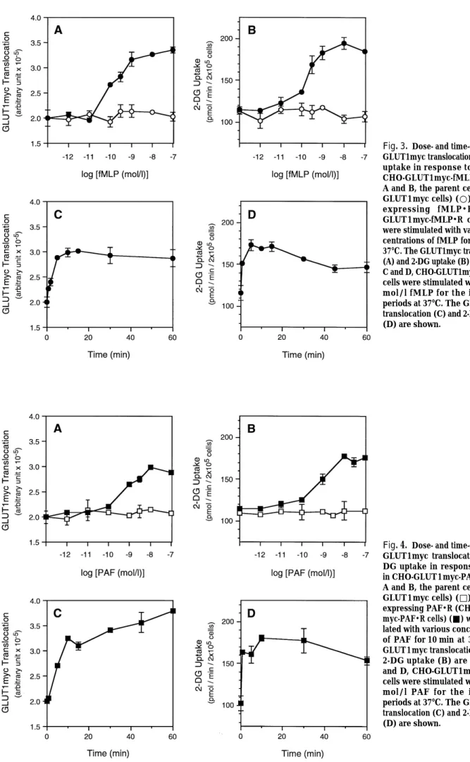
Fig. 3. Dose- and time-dependent GLUT1myc translocation and 2-DG uptake in response to fMLP in CHO-GLUT1myc-fMLP・R cells. A and B, the parent cells (CHO-GLUT1myc cells) (○) and cells expressing fMLP・R (CHO-GLUT1myc-fMLP・R cells) (●) were stimulated with various con-centrations of fMLP for 10 min at 37℃. The GLUT1myc translocation (A) and 2-DG uptake (B) are shown. C and D, CHO-GLUT1myc-fMLP・R cells were stimulated with 1×10⁻⁷ mol/l fMLP for the indicated periods at 37℃. The GLUT1myc translocation (C) and 2-DG uptake (D) are shown.
Fig. 4. Dose- and time-dependent GLUT1myc translocation and 2-DG uptake in response to PAF in CHO-GLUT1myc-PAF・R cells. A and B, the parent cells (CHO-GLUT1myc cells) (□) and cells expressing PAF・R (CHO-GLUT1 myc-PAF・R cells) (■) were stimu-lated with various concentrations of PAF for 10 min at 37℃. The GLUT1myc translocation (A) and 2-DG uptake (B) are shown. C and D, CHO-GLUT1myc-PAF・R cells were stimulated with 1×10⁻⁷ mol/l PAF for the indicated periods at 37℃. The GLUT1myc translocation (C) and 2-DG uptake (D) are shown.
23
The Journal of Medical Investigation Vol. 47 2000 23
due to the translocation of GLUT1 in CHO cells expressing chemoattractant-receptors.
We reported that phosphatidylinositol 3-kinase (PI 3-kinase) is essential for insulin-triggered GLUT4 translocation (27), and that GLUT4 translocation is triggered not only by insulin but also by acti-vations of G-protein coupled receptors (10, 11). To determine the molecular mechanisms of fMLP- or PAF-induced GLUT1 translocation, effects of IAP (Gi inhibitor) and wortmannin (PI 3-kinase inhibitor) on the chemoattractant-stimulated GLUT1 trans-location were examined using CHO cells. The IAP-pretreatment completely inhibited the fMLP-stimulated GLUT1 translocation in CHO-GLUT1myc-fMLP・R cells, but the PAF-stimulated GLUT1 translocation was only slightly inhibited by IAP in CHO-GLUT1myc-PAF・R (Fig. 5). Because the GLUT1 translocation by fMLP is transmitted through Gi coupling to the receptor, and PAF-triggered GLUT1 translocation is mediated through both Gi and Gq coupling to the receptor, IAP is not able to inhibit the PAF-triggered translocation completely (10, 17). Pretreatment of wortmannin at 1×10⁻⁷mol/l completely inhibited
the insulin-stimulated GLUT1 translocation, but only partly inhibited the fMLP-stimulated GLUT1 trans-location (Fig. 5A). The insulin-induced GLUT1 translocation is more sensitive to wortmannin than is the fMLP-induced GLUT1 translocation. The PAF-stimulated GLUT1 translocation was not inhibited by wortmannin (Fig. 5 B). The PAF receptor appar-ently transmitted the signal for GLUT1 translocation via Gq, which is an insulin-independent and PI 3-kinase-independent pathway (11). However, the pathway of fMLP-stimulated GLUT1 translocation via Gi may be partly mediated through PI 3-kinases (see DISCUSSION). Next, we asked whether serine/ threonine protein kinase Akt (also called PKB or RAC-PK) existing downstream of PI 3-kinase would be activated by fMLP- or PAF-treatment. Akt kinase activity is regulated by PI 3-kinase products, phosphatidylinositol 3,4-diphosphate and phos-phatidylinositol 3,4,5-triphosphate, and activation of the kinase coincides with phosphorylations at Thr308 and Ser473 by 3-phosphoinositide (PI 3-kinase products)-dependent protein kinase (PDK) 1/2 (28, 29), detectable from the slower migration, compared to non-activated Akt, on western analysis (30). As shown in Fig.6, fMLP activated Akt kinase in both CHO cells and PMNs, but PAF activated only a very small amount of this kinase. PAF slightly transmitted the signal of Akt phosphorylation via Gi because the PAF receptor is partly coupled to
Gi and may be mainly coupled to Gq. While the phosphorylated Akt in CHO cells continued to be activated for 5~10 min, at maximal levels, Akt in phagocytes showed a maximum of activation at 30~90 sec (Fig. 6). The Akt activation process in phagocytes was more transient than that in CHO cells. In phagocytes, the receptor activated by fMLP or PAF triggers activation or phosphorylation of mitogen-activated protein kinase (MAPK) families which include extracellular signal-regulated kinases (ERKs), p38 MAPK, and c-Jun N-terminal kinases (31, 32). We also found that both fMLP and PAF in CHO cells cause phosphorylation of ERKs, using western blots and phospho-specific antibody against ERK (data not shown). The same protein kinases Fig. 5. Effects of IAP and wortmannin on GLUT1myc trans-location in response to fMLP or PAF in CHO-GLUT1myc cells expressing its receptor. A, the CHO-GLUT1myc-fMLP・R cells were stimulated with 1×10⁻⁷mol/l fMLP, 1×10⁻⁷mol/l insulin, or buffer alone after pretreatment with 100 ng/ml IAP or the indicated concentration of wortmannin for 20 min at 37℃, and GLUT1myc translocation was measured. B, the CHO-GLUT1myc-PAF・R cells were stimulated with 1×10⁻⁷mol/l PAF or buffer alone after pretreatment with 100 ng/ml IAP or the indicated concentration of wortmannin for 20 min at 37℃, and GLUT1myc translocation was measured.
A. Hagi et al. GLUT 1 translocation
24 A. Hagi et al. GLUT 1 translocation
were activated by fMLP- or PAF-stimulation in phagocytes and in CHO cells.
DISCUSSION
As a first approach to elucidating the mechanism(s) of glucose uptake in phagocytes, investigators used the principles of enzyme kinetic analysis and the Michaelis-Menten equation, similar to a previous study of the mechanism by which insulin increases glucose uptake by adipocytes (33). Several groups of researches found that the fMLP-induced increase in glucose uptake by PMNs was associated with a decrease in the Kmfor this process, but the Vmax remained unchanged (21, 22, 34). These results indicated that the enhanced glucose uptake in chemoattractant-stimulated PMNs involves a mech-anism other than an increase in the number of cell surface transporters. They reported that the rapid glucose uptake on fMLP-stimulation in PMNs was consistent with an increased intrinsic activation of glucose transporter molecules, but the possibility that transporter translocation contributes to the stimulated glucose uptake has to be considered. On the other hand, Okuno and Gliemann reported that both an increase in the Vmaxand a decrease in Km are important for chemoattractant-stimulated glucose uptake (13). Malide et al used confocal laser
scanning microscopy to define the subcellular localization of GLUT1 in cultured macrophages. In response to long term treatment with PMA or fMLP, GLUT1 was translocated to the cell surface (26). However, the molecular mechanism related to the acute regulation of glucose uptake in phagocytes is poorly understood. In the present study we used CHO cells stably expressing both GLUT1myc and chemoattractant-receptors to elucidate the molecular mechanisms of acute regulation of glucose uptake in response to chemoattractants, because of technical difficulties in estimating the amount of endogenous GLUT1 on the cell surface of phagocytes. We found that in CHO cells stimulated with fMLP or PAF, glucose uptake is increased through GLUT1 trans-location, and it seemed that the same intracellular events are triggered by fMLP or PAF in phagocytes. fMLP- and PAF-induced GLUT1 translocation are initiated by ligands binding to the receptors. fMLP receptors couple with the heterotrimeric G-protein, Gi. PAF receptors couple with Gi and Gq. Upon acti-vation of Gq by the binding ligands, phospholipase C (PLC)-β is activated and PLC-β activation results in the generation of two secondary messengers, inositol 1,4,5-triphosphate (IP3) and diacylglycerol
(DAG) (1,2). IP3triggers an increase in the cytosolic
Ca2+
concentration ([Ca2+
]i) from intracellular pools, and DAG acts in conjunction with calcium to activate various isoforms of protein kinase C (PKC) (2). The Fig. 6. Time course of Akt kinase activation in the CHO cells and rat PMNs. fMLP・R cells, CHO-GLUT1myc-PAF・R cells, and rat PMNs were stimulated with 1×10⁻⁷mol/l fMLP or 1×10⁻⁷mol/l PAF for the indicated periods at 37℃. Cell lysates (10µg) were separated on 7% SDS-PAGE gels and activation of Akt was detected on western blots using an anti-Akt antibody. Akt-P and Akt indicates phosphorylated and non-phosphorylated Akt, respectively.
25
The Journal of Medical Investigation Vol. 47 2000 25
activated PKC catalyzes various protein phosphorylations that finally result in the activation of various phagocyte functions (2). In PMNs, increased [Ca2+
]iin the presence of fMLP or PAF triggered activation of glycogen phosphorylase a activity (data not shown) (3). But Ca2+
ionophores did not trigger GLUT1 translocation (10), and phorbol 12,13-dibutyrate (PDBu) pretreatment which induced downregu-lation of PKC, had practically no effect on fMLP-and PAF-induced GLUT1 translocation in CHO cells (unpublished data). Therefore, fMLP- and PAF-stimulated GLUT1 translocation seems to be mediated by the Gα subunit (Gi or Gq) through some unknown pathway(s), following G-protein activation but probably is not a secondary phenomenon as the result of phosphatidylinositol 4,5-diphosphate breakdown.
Tan et al reported that a PKC inhibitor, calphostin C, and a tyrosine kinase inhibitor, genistein, inhibited fMLP-stimulated 2-DG uptake in human peripheral blood PMNs (21). In CHO cells expressing the fMLP receptor, calphostin C and genistein at same concentrations also inhibited fMLP-stimulated GLUT1 translocation (unpublished data). Inhibitor studies suggest that the acute activation of glucose transport in response to fMLP involves tyrosine and serine/ threonine kinase.
Recently, several investigators suggested that Akt kinase activation is closely related to GLUT4 translocation (35, 36). We also found that Gi-activation but not Gq-activation induced 3-phosphoinositide accumulation via PI 3-kinase activation (unpublished data). Therefore, the mechanism(s) of Gi-triggered GLUT1 translocation by fMLP may involve Akt activation by PDK 1/2 (28, 29). Several isoforms of PI 3-kinase have been characterized in PMNs, for example, the classical typeα (p85/p 110) and type γ PI 3-kinase (p101/p110), which is activated by βγ subunits of fMLP-stimulated G-protein, but only PI 3-kinase typeα was expressed in CHO cells (37-39). Differences in the expression of the PI 3-kinase subtype and numbers of the receptors may be related to differences in the response time of Akt activation processes between phagocytes and CHO cells. On the other hand, Gq activation did not induce activation of PI 3-kinase, and Gq-triggered GLUT1 translocation by PAF was not related to Akt activation.
The rapid response of glucose uptake to fMLP or PAF in phagocytes is not due to activation of GLUT1 gene expression. Although the possibility that increased transporter affinity for glucose may contribute to the observed increase in glucose
uptake in activated phagocytes cannot be excluded, we propose that phagocytes activated by fMLP or PAF can trigger translocation of GLUT1 from the internal pool to the cell surface.
ACKNOWLEDGMENTS
We thanks Drs. J. Miyazaki, P. Vignais, and T. Shimizu, for providing pCXN2 plasmid, cDNAs encoding fMLP receptor and the PAF receptor. M. Ohara provided comments on the manuscript.
REFERENCES
1. Thelen M, Dewald B, Baggiolini M : Neutrophil signal transduction and activation of the respi-ratory burst. Physiol Rev 73 : 797-821, 1993 2. Rossi F : The O
2--forming NADPH oxidase of the phagocytes : nature, mechanisms of activation and function. Biochim Biophys Acta 853 : 65-89, 1986
3. Borregaard N, Herlin T : Energy metabolism of human neutrophils during phagocytosis. J Clin Invest 70 : 550-557, 1982
4. Weisdorf DJ, Craddock PR, Jacob HS : Granulocytes utilize different energy sources for movement and phagocytosis. Inflammation 6 : 245-256, 1982
5. Pessin JE, Bell GI : Mammalian facilitative glucose transporter family : structure and mo-lecular regulation. Annu Rev Physiol 54 : 911-930, 1992
6. Yang J, Holman GD : Comparison of GLUT4 and GLUT1 subcellular trafficking in basal and insulin-stimulated 3T3-L1 cells. J Biol Chem 268 : 4600-4603, 1993
7. Ishii K, Hayashi H, Todaka M, Kamohara S, Kanai F, Jinnouchi H, Wang L, Ebina Y : Possible domains responsible for intracellular targeting and insulin-dependent translocation of glucose transporter type 4. Biochem J 309 : 813-823, 1995
8. Kanai F, Nishioka Y, Hayashi H, Kamohara S, Todaka M, Ebina Y : Direct demonstration of insulin-induced GLUT4 translocation to the surface of intact cells by insertion of a c-myc epitope into an exofacial GLUT4 domain. J Biol Chem 268 : 14523-14526, 1993
9. Todaka M, Hayashi H, Imanaka T, Mitani Y, Kamohara S, Kishi K, Tamaoka K, Kanai F, A. Hagi et al. GLUT 1 translocation
26 A. Hagi et al. GLUT 1 translocation
Shichiri M, Morii N, Narumiya S, Ebina Y : Roles of insulin, guanosine 5’-[gamma-thio] triphosphate and phorbol 12-myristate 13-acetate in signalling pathways of GLUT4 translocation. Biochem J 315 : 875-882, 1996
10. Kishi K, Hayashi H, Wang L, Kamohara S, Tamaoka K, Shimizu T, Ushikubi F, Narumiya S, Ebina Y : Gq-coupled receptors transmit the signal for GLUT4 translocation via an insulin-independent pathway. J Biol Chem 271 : 26561-26568, 1996
11. Kishi K, Muromoto N, Nakaya Y, Miyata I, Hagi A, Hayashi H, Ebina Y : Bradykinin directly triggers GLUT4 translocation via an insulin-independent pathway. Diabetes 47 : 550-558, 1998
12. Baron EJ, Proctor RA : Elicitation of peritoneal polymorphonuclear neutrophils from mice. J Immunol Methods 49 : 305-313, 1982
13. Fukuzumi M, Shinomiya H, Shimizu Y, Ohishi K, Utsumi S : Endotoxin-induced enhancement of glucose influx into murine peritoneal macro-phages via GLUT 1. Infect Immun 64 : 108-112, 1996
14. Niwa H, Yamamura K, Miyazaki J : Efficient selection for high-expression transfectants with a novel eukaryotic vector. Gene (Amst.) 108 : 193-199, 1990
15. Boulay F, Tardif M, Brouchon L, Vignais P : The human N-formylpeptide receptor. Charac-terization of two cDNA isolates and evidence for a new subfamily of G-protein-coupled receptors. Biochemistry 29 : 11123-11133, 1990
16. Nakamura M, Honda Z, Izumi T, Sakanaka C, Mutoh H, Minami M, Bito H, Seyama Y, Matsumoto T, Noma M, Shimizu T : Molecular cloning and expression of platelet-activating factor receptor from human leukocytes. J Biol Chem 266 : 20400-20405, 1991
17. Okuno Y, Gliemann J : Effect of chemotactic factors on hexose transport in polymorphonuclear leucocytes. Biochim Biophys Acta 941 : 157-164, 1988
18. Ebina Y, Edery M, Ellis L, Standring D, Beaudoin J, Roth RA, Rutter WJ : Expression of a functional human insulin receptor from a cloned cDNA in Chinese hamster ovary cells. Proc Natl Acad Sci USA 82 : 8014-8018, 1985 19. Laemmli UK : Cleavage of structural proteins
during the assembly of the head of bacteriophage T4. Nature 227 : 680-685, 1970
20. Konishi H, Kuroda S, Tanaka M, Matsuzaki
H, Ono Y, Kameyama K, Haga T, Kikkawa U : Molecular cloning and characterization of a new member of the RAC protein kinase family : association of the pleckstrin homology domain of three types of RAC protein kinase with protein kinase C subspecies and beta gamma subunits of G proteins. Biochem Biophys Res Commun 216 : 526-534, 1995
21. Tan AS, Ahmed N, Berridge MV : Acute regu-lation of glucose transport after activation of human peripheral blood neutrophils by phorbol myristate acetate, fMLP, and granulocyte-macrophage colony-stimulating factor. Blood 91 : 649-655, 1998
22. Weisdorf DJ, Craddock PR, Jacob HS : Glycogenolysis versus glucose transport in human granulocytes : differential activation in phagocytosis and chemotaxis. Blood 60 : 888-893, 1982
23. Seow WK, Smith SE, McCormack JG, Thong YH : Uptake of 3
H-deoxyglucose as a microassay of human neutrophil and monocyte activation. J Immunol Methods 98 : 113-118, 1987
24. Walker BA, Seiler AJ, Owens CA, Hagenlocker BE, Ward PA : Absence of FMLP receptors on rat macrophages. J Leukoc Biol 50 : 600-606, 1991
25. Ahmed N, Kansara M, Berridge MV : Acute regulation of glucose transport in a monocyte-macrophage cell line : Glut-3 affinity for glucose is enhanced during the respiratory burst. Biochem J 327 : 369-375, 1997
26. Malide D, Davies-Hill TM, Levien M, Simpson IA : Distinct localization of GLUT-1,-3, and -5 in human monocyte-derived macrophages : effects of cell activation. Am J Physiol 274 : E516-E 526, 1998
27. Kanai F, Ito K, Todaka M, Hayashi H, Kamohara S, Ishii K, Okada T, Hazeki O, Ui M, Ebina Y : Insulin-stimulated GLUT4 trans-location is relevant to the phosphorylation of IRS-1 and the activity of PI3-kinase. Biochem Biophys Res Commun 195 : 762-768, 1993 28. Stephens L, Anderson K, Stokoe D,
Erdjument-Bromage H, Painter GF, Holmes AB, Gaffney P, Reese CB, McCormick F, Tempst P, Coadwell J, Hawkins PT : Protein kinase B kinases that mediate phosphatidylinositol 3,4,5-trisphosphate-dependent activation of protein kinase B. Science 279 : 710-714, 1998 29. Alessi DR, Deak M, Casamayor A, Caudwell
FB, Morrice N, Norman DG, Gaffney P, Reese CB, MacDougall CN, Harbison D, Ashworth
27
The Journal of Medical Investigation Vol. 47 2000 27
A, Bownes M : 3-Phosphoinositide-dependent protein kinase-1 (PDK 1) : structural and func-tional homology with the Drosophila DSTPK 61 kinase. Curr Biol 7 : 776-789, 1997
30. Tilton B, Andjelkovic M, Didichenko SA, Hemmings BA, Thelen M : G-Protein-coupled receptors and Fcgamma-receptors mediate activation of Akt/protein kinase B in human phagocytes. J Biol Chem 272 : 28096-28101, 1997
31. Rane MJ, Carrither SL, Arthur JM, Klein JB, McLeish KR : Formyl peptide receptors are coupled to multiple mitogen-activated protein kinase cascades by distinct signal transduction pathways : role in activation of reduced nicotinamide adenine dinucleotide oxidase. J Immunol 159 : 5070-5078, 1997
32. Thompson HL, Marshall CJ, Saklatvala J : Characterization of two different forms of mitogen-activated protein kinase kinase induced in polymorphonuclear leukocytes following stimu-lation by N-formylmethionyl-leucyl-phenylalanine or granulocyte-macrophage colony-stimulating factor. J Biol Chem 269 : 9486-9492, 1994 33. Olefsky JM : Effects of fasting on insulin binding,
glucose transport, and glucose oxidation in isolated rat adipocytes : relationships between insulin receptors and insulin action. J Clin Invest 58 : 1450-1460, 1976
34. Abughali N, Dubyak G, Tosi MF : Impairment of chemoattractant-stimulated hexose uptake in neonatal neutrophils. Blood 82 : 2182-2187, 1993
35. Calera MR, Martinez C, Liu HZ, ElJack AK, Birnbaum MJ, Pilch PF : Insulin increases the association of Akt-2 with Glut4-containing vesicles. J Biol Chem 273 : 7201-7204, 1998 36. Cong LN, Chen H, Li YH,. Zhou LX, McGribbon
MA, Taylor SI, Quon MJ : Physiological role of Akt in insulin-stimulated translocation of GLUT4 in transfected rat adipose cells. Mol Endocrinol 11 : 1881-1890, 1997
37. Thelen M, Uguccioni M, Bosiger J : PI 3-kinase-dependent and in3-kinase-dependent chemotaxis of human neutrophil leukocytes. Biochem Biophys Res Commun 217 : 1255-1262, 1995
38. Stephens L, Smrcka A, Cooke FT, Jackson TR, Sternweis PC, Hawkins PT : A novel phos-phoinositide 3 kinase activity in myeloid-derived cells is activated by G protein beta gamma subunits. Cell 77 : 83-93, 1994
39. Stephens LR, Eguinoa A, Erdjument-Bromage H, Lui M, Cooke F, Coadwell J, Smrcka AS, Thelen M, Cadwallader K, Tempst P, Hawkins PT : The G beta gamma sensitivity of a PI3K is dependent upon a tightly associated adaptor, p101. Cell 89 : 105-114, 1997
A. Hagi et al. GLUT 1 translocation
28 A. Hagi et al. GLUT 1 translocation