総 説 東女医大誌 90(1): 1-13, 2020.2
炎症性疾患
(1)炎症とは
東京女子医科大学医学部微生物学免疫学教室 カ ト ウ ヒデヒト加藤 秀人
(受理 2020 年 1 月 10 日) Inflammatory Disease (1) What Is Inflammation? Hidehito KatoDepartment of Microbiology and Immunology, Tokyo Women s Medical University, Tokyo, Japan
Stress responses are part of an important system in maintaining the homeostasis of a living organism. After acute inflammation, if tissue repair is incomplete or stress is not completely removed with an excessive and prolonged inflammatory response, damage is accumulated in living organs and chronic inflammation persists. Thus, irre-versible tissue and organ damage occurs, which is accompanied by fibrosis. Further, inflammatory diseases, such as fibrotic diseases, autoimmune diseases, allergies, and arteriosclerosis, account for most causes of deaths in hu-mans; therefore, it is extremely important to understand mechanisms of inflammation and to establish a control method. Inflammation is caused by a complex influence of various molecules and cells that form networks through inflammatory cytokines. Since various factors influence each other, the onset time, degree, and duration of inflammation may differ. This article explains the mechanism of inflammation, presents findings on its regula-tory factors and methods, and enumerates the innate immune system receptors (pattern recognition receptors) and responsible cells (neutrophils, innate lymphoid cells, natural killer T cells) that cause inflammation. This arti-cle thus aims to understand the complex networks involved in inflammation.
Key Words: inflammation, pathogen-associated molecular patterns (PAMPs), damage-associated molecular pat-terns (DAMPs), neutrophils, natural killer T (NKT) cells
はじめに
生体は,微生物感染による生物学的ストレスや,
温度変化や打撃等による物理的ストレス,酸やアル
カリ,毒物等の化学的ストレスのようなストレス侵
襲を常に受けている.このストレス侵襲によって体
に異変が生じる過程を炎症と呼ぶ.炎症部位は発熱,
発赤,腫脹,疼痛を生じ,結果として機能障害が生
じる.このような症状は,体の侵襲を受けた組織が
ストレスに応答した結果生じる.炎症部位では,血
管の拡張や血流の増加,血管からの血液成分の漏出,
白血球の炎症組織への浸潤,局所的に産生された物
質による神経への刺激などが観察され,次第に炎症
:加藤秀人 〒162―8666 東京都新宿区河田町 8―1 東京女子医科大学医学部微生物学免疫学教室 Email: [email protected] doi: 10.24488/jtwmu.90.1_1Copyright Ⓒ 2020 Society of Tokyo Women s Medical University. This is an open access article distributed under the terms of Creative Commons Attribution License (CC BY), which permits unrestricted use, distribution, and reproduction in any medium, provided the original source is properly credited.
症状が進行する.しかし,これらの症状は生体防御
システムにより時間経過とともに収束する.このよ
うな炎症反応の進行過程において,死滅した細胞や
感染した微生物は除去され,創傷を受けた組織は再
生し,失われた組織は線維組織に置き換わり,創傷
は修復されて治癒する.ストレスに応答するシステ
ムは,生体の恒常性を維持するための重要なシステ
ムであるが,急性炎症が生じた後,組織修復が不完
全な場合や,ストレスが完全に除去されず過剰な炎
症反応が長期間持続した場合は,生体組織にダメー
ジが蓄積され,慢性炎症状態が持続し,結果的に線
維化などを伴う不可逆的な組織・臓器障害を生じ
る.線維化疾患,自己免疫疾患,アレルギー,動脈
硬化等の炎症性疾患は,ヒトの死因の大半を占める
ため,炎症のメカニズムの理解,およびその制御法
の確立は,極めて重要である.本稿では,炎症を惹
起する自然免疫系の受容体および担当細胞を列挙
し,炎症のメカニズムを解説するとともにその制御
因子,および制御法における最近の知見を検証する
ことで,炎症への理解を深める.
1.急性炎症のメカニズム(炎症を惹起する因子)
1)自然免疫系受容体
生体には非自己を認識するセンサーが備わってい
る.そのセンサーが感知する刺激を Danger Signal
と呼ぶ.刺激物質は大きく分けて 2 種類あり,細菌
やウイルスの構成成分を pathogen-associated
mo-lecular patterns(PAMPs),ダメージを受けた細胞や
細 胞 外 基 質 か ら 放 出 さ れ る 成 分 を
damage-associated molecular patterns(DAMPs)と 呼 ぶ.
PAMPs と DAMPs は,センサーにより認識され防
御反応(炎症)が始まる(Figure 1)
1).認識するセン
サーを pattern recognition receptors(PRRs)と呼
ぶ.生体はこの PRRs の認識機構により,炎症性サイ
トカインを分泌して感染部位や損傷部位へ免疫担当
細胞を遊走して炎症応答を誘導する.PAMPs の中
でも nod-like receptor(NLR)family や
hematopoie-tic interferon(IFN)-
γ inducible nuclear protein with
the 200-amino-acid repeat(HIN-200)family が,非自
己 を 認 識 し て apoptosis-associated speck-like
pro-tein containing a caspase activation and
recruit-ment domain(ASC)や caspase-1 などと結合すると
inflammasome
と呼ばれる複合体が形成される.in-flammasome では,活性型 caspase-1 が炎症性サイ
トカインを誘導することで炎症が惹起される
2).in-flammasome の形成に基づく炎症応答は,感染防御
に有効であり
3),腸内細菌叢の制御および腸管上皮の
バリアを保護し,腸管における恒常性を維持すると
考えられている.一方,過剰な inflammasome の形成
は慢性炎症を惹起し,動脈硬化,痛風,2 型糖尿病,
アルツハイマー病などの疾患の発症に起因すると考
えられている
4)5).これらの一連の応答を自然免疫応
答と呼び,遊走してきたマクロファージや好中球に
よる病原体の直接的な排除を惹起するのみならず,
後に起きる獲得免疫の確立や障害を受けた組織の治
癒にも必須の反応である
6).
2)好中球
好中球は生体防御において最初に稼働する細胞
で,非常に重要な機能を担っている.白血球中の約
70% を占め,体内に非自己(微生物等)の侵入を感
知すると,血管壁をすり抜け,侵入物の局在する場
所へ移動(遊走)し活性化する.好中球走化因子と
して,細菌由来のペプチド,補体成分(C5a),アラ
キドン酸代謝産物(ロイコトリエン B4),ケモカイン
(NAP-2,MIP-2),サイトカイン[interleukin(IL)-8]
等が知られている.活性化好中球は非自己(細菌)を
貪食し,活性酸素等で感染を防御する.活性化好中
球は,自身のクロマチンを細胞外に放出することで
感染を防御する
7).このクロマチンネットは
neutro-phil extracellular traps(NETs)と呼ばれ,この細胞
死の過程はネクローシスやアポトーシスとはタイプ
が異なる為に,NETosis と呼ばれる.ネット状の
NETs は細菌を捉える.捉えられた細菌は好中球や
マクロファージに貪食されやすくなり,また,NETs
そのものにも殺菌作用がある
8).peptidylarginine
de-iminase(PAD)4 の遺伝子欠損マウスでは NETs
形成が強く抑制されており,そのマウスは易感染性
になることから,PAD4 が NETs 形成に重要であ
る
9).さらに,NETs は血小板をトラップして活性化
し,血栓形成に重要な働きをしていること
10)や癌転
移に重要
11)なこと,慢性かつ難治性の自己免疫疾患
の一つである systemic lupus erythematosus(SLE)
の発症に重要であること
12)が明らかとなり,NETs
が感染防御や血栓・癌転移・自己免疫疾患等に重要
な役割を果たしていることが明らかとなっている.
NETosis は,各種菌体成分[2 本鎖 RNA,フラジェ
リン,リポテイコ酸,lipopolysaccharide(LPS)等]
菌体,生体物質[活性化内皮細胞,カルシウムイオ
ン,フィブリノーゲン,IL-1,IL-8,IFN-α,壊死組織,
血小板活性化因子,血小板,toll-like receptor(TLR),
tumor necrosis factor(TNF)-
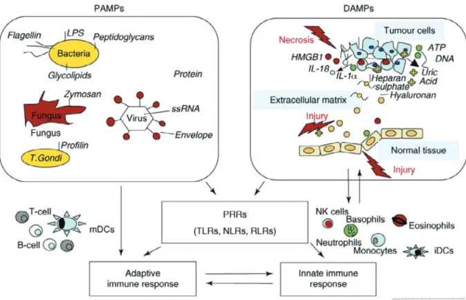
α,myeloperoxidase-Figure 1 Classes of molecules that initiate innate and adaptive immune responses1).
The initiating events in inflammation and the resultant adaptive immune response include both pathogen-associated molecular patterns (PAMPs) and damage-associated molecular patterns (DAMPs). These are protein (e.g. HMGB1) or non protein (e.g. ATP, uric acid) molecules which in healthy conditions are confined within cells but are delivered outside upon cell damage, such as necrosis, and in some instances secreted by cells. The levels of released DAMPs depend on the abundance of intracellular DAMPs and are often higher and enduring in neoplastic tissues than in normal tissues. This leads to persistent inflam-mation. Other sources of DAMPs include degraded extracellular matrix, such as heparan sulphate and hyaluronan. Many of the receptors so far identified for DAMPs and PAMPs are shared, and belong to the family of Pattern Recognition Receptors, PRRs. Therefore, both sterile and non sterile inflammation signals converge on a common pathway. Matz-inger has suggested a unique integrating hypothesis that all molecules signaling danger or damage are DAMPs, with hydrophobic portions of molecules (HYPPOs) derived either from dead and dying host cells or pathogens68) 69).
LPS, lipopolysaccharide; ssRNA, single strand RNA; T. gondii, Trypanasoma gondii (proto-zoan parasite); HMGB1, high mobility group box 1 protein; PRRs, pattern recognition recep-tors; TLRs, toll-like receprecep-tors; NLRs, NOD1-like receprecep-tors; RLRs, RIG-I-like receptors.
anti-neutrophil cytoplasmic antibody(MPO-ANCA)
等],各種ウイルス,化学物質(ピロリン酸カルシウ
ム,カルシウムイオノフォア,活性酸素,PMA,ス
タ チ ン,尿 酸,一 酸 化 窒 素 等)の よ う な 様 々 な
PAMPs や DAMPs により誘導される.したがって,
好中球は,自身の産生する顆粒成分や抗原提示能,
貪食抗原の放出等により,獲得免疫の主役である T
細胞を抑制したり活性化したりして,免疫系を調整
することが可能であり,その証拠も
いつつある
(Figure 2)
13)∼26).
3)Innate lymphoid cell(ILC)
ILC は,皮膚,肺,腸などの哺乳類のバリア表面,
および脂肪や粘膜関連のリンパ組織に存在し,炎症
の開始,調節,解消に重要である.サイトカインお
よび微生物シグナルに迅速に応答し,炎症性および
免疫調節性サイトカインの供給源であり,獲得免疫
の調節因子としても重要な役割を果たしている
27).
表面マーカーの発現パターン,サイトカイン発現パ
ターン,転写因子の発現パターンに基づいて,グルー
プ 1,2,3 の 3 つのサブセットに分類される
28).ILC
Figure 2 Mechanisms involved in T-cell inhibition (left panel) and activation (right panel) by neutrophils13).
Neutrophils can establish T-cell inhibition by (1) degranulation of granular constituents. The serine proteases elastase and cathepsin G inactivate T-cell stimulating cytokines, IL-2 and IL-6, and catalyze shedding of cytokine receptors for IL-2 and IL-6 on T-cells14) 15). (2)
Production of reactive oxygen species (ROS) and release of arginase. Both agents can result in downregulation of TCR ζ on T-cells, thereby arresting the cell in the G0-G1 phase16) ∼ 21).
(3) Expression of PD-L1. Upregulation of this ligand is associated with interferon-dependent PD1-mediated T-cell apoptosis22) 23). T-cell activation by neutrophils is attained by (4)
indi-rect antigen presentation. Dendritic cells take up antigens from apoptotic neutrophils and serve as APC for T-cells24). (5) Direct antigen presentation. Neutrophils possess the capacity
to cross-prime CD8+ T-cells directly in a MHCI-dependent manner25). (6) Release of
microbi-al metabolites (HMB-PP). Neutrophils release bacterimicrobi-al products after ingestion to activate γδ-T-cells26).
の研究は比較的新しく,現在,免疫制御に関する成
果が続々と発表されている
29)∼31).
4)Natural killer T(NKT)細胞
CD1d 拘束性 NKT 細胞は,自然免疫システムと
獲得免疫システムの境界に存在し,NK 細胞と同様
に刺激にすばやく反応し,様々なサイトカインを産
生する
32)33)ことによって,自然免疫と獲得免疫の橋渡
しをしている
34).NKT 細胞は脂質抗原を認識し,T
細胞の T cell receptor(TCR)が major
histocompati-bility complex(MHC)に提示されたペプチドを抗原
として認識するのに対して,NKT 細胞の TCR は
CD1d 分子に提示された糖脂質を認識し認識範囲を
T 細胞と分担している.NKT-TCR により 2 種類に
大別され,Va24Ja18(ヒト)という均一な可変部位
を保持する TCR を発現しているものを I 型,それ以
外を II 型と呼ぶ.大部分の NKT 細胞は I 型であり
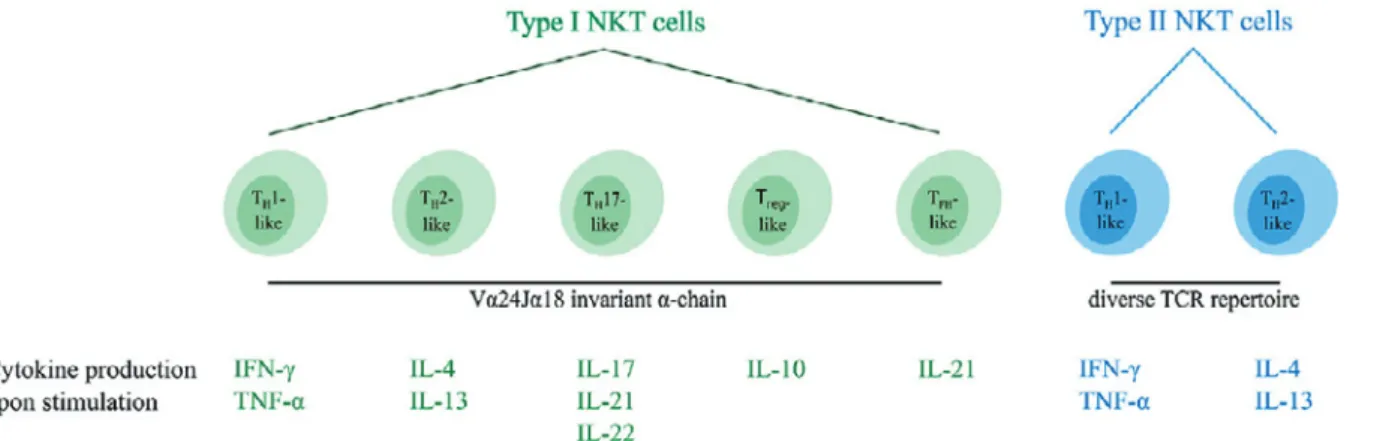
Figure 3 Overview of the different functional human NKT cell subsets41).
CD1d-restricted human NKT cells can be divided into subsets based on their TCR reper-toire and cytokine profile. Type I NKT cells express the invariant Vα24Jα18 TCR α-chain and can be subdivided into five distinct functional subsets (indicated in green). In addi-tion, type II NKT cells express a diverse TCR repertoire and can be subdivided into two functional subsets (indicated in blue). Upon activation, NKT cells secrete a unique pattern of cytokines, indicated for each subtype. Type I and type II NKT cells are able to switch between different functional subsets upon interactions within the TME.
NKT, natural killer T; TCR, T cell receptor; TH, helper T; T reg, regulatory T; TFH, fol-licular helper T; TME, tumor microenvironment.
I 型の TCR には多様性がないため,invariant NKT
(iNKT)細 胞 と 呼 ば れ る.I 型 NKT 細 胞 は
α-galactosyl ceramide(α-GalCer)のほか,微生物の糖
脂質と自己抗原も認識する
35)∼39).α-GalCer はすべて
の I 型 NKT 細胞の強力な活性化因子であり,大量
の IFN-γ を 産 生 さ せ,CD8 T 細 胞 と antigen
pre-senting cell(APC)の両方を活性化する
40).
一方,CD1d 拘束性 II 型 NKT 細胞は,invariant
TCR を発現しない.このサブセットは,I 型 NKT
細胞によって認識されるものとは異なる糖脂質抗原
を認識し,I 型ほど詳細に調べられていない.I 型
NKT 細胞は ILC と同様に,T helper(Th)1 のマス
ター遺伝子である T-bet を発現し,IL-4 と IL-13 を
産 生 す る NKT1,Th2 の マ ス タ ー 遺 伝 子 で あ る
Gata-3 を 発 現 し IFN-γ と IL-4 の 両 方 を 産 生 す る
NKT2,retinoid-related orphan receptor(ROR)γt
を 発 現 し IL-17A や IL-22 を 産 生 す る NKT17 の 3
種類のグループに分類される(Figure 3)
41).さらに,
IL-10 を産生する NKT10(NKT reg)も確認されて
いる
42).NKT 細胞はほとんどの臓器でマイナーな免
疫細胞サブセットを構成するが,炎症性あるいは抗
炎症性サイトカインを分泌することにより,免疫応
答を短時間で炎症または寛容に傾けることができる
ため,免疫調節に大きな影響を及ぼす.
2.炎症を抑制する因子
1)Leukocyte cell-derived chemotaxin-2(LECT 2)
LECT 2 は 1996 年にヒト T 白血病細胞株 SKW3
の培養上清から,ヒト好中球走化性を指標にして山
越らが精製単離した
43)多機能蛋白である.最初にヒ
ト骨髄性白血病細胞株 ML-1 の培養上清からヒト好
中球走化性因子 LECT として精製されたが,この
LECT は IL-8 であることが分かったため,2 番目に
精製された LECT という意味で LECT 2 と命名さ
れた.LECT 2 は主に肝臓で産生されるため,肝機能
と密接に関係していると考えられている
44).多方面
の研究により多機能な蛋白であることが示唆され,
以下に示すように炎症の抑制を中心に多数の研究が
なされている.
(1)Concanavalin A(Con A)誘導性肝炎の抑制
肝臓は自然免疫を担う NK 細胞や NKT 細胞が多
く存在し,免疫応答の場である.Con A 投与により
誘 導 さ れ る 肝 傷 害 は,エ フ ェ ク タ ー 細 胞 と し て
NKT 細胞や好中球が重要な役割を担っていると考
えられている.Saito らは,Con A により誘導される
肝炎が野生型マウスに比べ LECT 2 knock out(KO)
マウスにおいて増悪することを見出した
44).肝臓の
NKT 細胞が LECT 2 KO マウスで増加しており,
LECT 2 が肝臓の NKT 細胞の活性化を抑制するこ
とでCon A誘導性肝炎を制御していると考えられる.
Figure 4 Plasma cytokine/chemokine levels are increased in a Staphyiococcal entero-toxin A (SEA)-dose dependent manner, thereby influencing prognosis46).
(A) B6mice were injected with 0.1 and 1.0 μg SEA/D-GalN, and the plasma levels of the indicated cytokines and a chemokine were followed for 72 h after treatment. Data are pre-sented as the mean ± SD of 3 samples at each time point. (B) B6 mice were injected with 1.0 μg SEA/D-GalN, and blood samples were collected at 12 h thereafter. The mice were then observed, and blood samples were again collected from the mice still alive at 72 h thereaf-ter. The indicated plasma cytokine/chemokine levels were compared among the control, surviving and dead mice. Data are presented as the mean ± SD of the indicated number of samples. *p<0.05, **p<0.01, ***p<0.005.
(2)抗コラーゲン抗体誘導性関節炎の抑制
マ ウ ス に お け る collagen-induced arthritis
(CIA)は,ヒト慢性関節リウマチの実験モデルとし
て幅広く利用されているが,関節炎の発症には長時
間かかり,発症頻度もマウスの系統によって異なる.
一方,抗コラーゲン抗体を使用した抗体誘導性関節
炎モデルは,CIA とは異なりマウスによる系統差が
なく,短時間で関節炎を発症する.Okumura らは,
抗コラーゲン抗体誘導関節炎において LECT 2 KO
マウスが炎症性サイトカインの発現増加を伴い増悪
化することを見出した
45).この関節炎の増悪化は,
LECT 2 遺伝子導入により改善した.
(3)Staphyiococcal enterotoxin A(SEA)誘導性
ショックの抑制
D―ガラクトサミン(D-GalN)は軽微な肝障害を誘
発する.D-GalN と SEA や LPS を同時投与すると
ショックが誘導されることが知られている.
我々は,LECT 2 KO マウスは,正常マウスと比較
し SEA と D-GalN の 同 時 投 与 に よ り 誘 導 さ れ る
ショックの致死率が上昇すること,血中 LECT 2 濃
度と炎症性サイトカイン濃度は逆相関することを見
出した(Figure 4)
46).SEA ショック誘導時に
recom-binant LECT 2 を投与すると,生存率が改善し炎症
性サイトカイン産生が抑制された(Figure 5)
46).
(4)敗血症患者における血漿 LECT 2 濃度の低下
我々は,ヒトの炎症の重症度と血漿 LECT 2 濃度
との関連を統計学的に調べたところ,敗血症患者の
ICU 入室時と退室時では,入室時に低下し,退室時
には上昇することを見出した
47).好中球,幼若白血
球,C-reactive protein(CRP),IL-6 の血漿中濃度は,
入室時は高く,退室時に低下するが,LECT 2 の変動
はこれらの炎症性パラメーターと有意に逆相関し
た.
(5)発癌との関係
LECT 2 は Wnt/β―カテニンシグナル伝達系を抑
制することが知られている
48).Anson M らは,LECT
Figure 5 Administration of leukocyte cell-derived chemotaxin-2 (LECT 2) reduces lethal-ity after treatment with Staphyiococcal enterotoxin A (SEA)/D-GalN in a manner that is associated with theinhibition of cytokines in plasma46).
(A) B6 mice were injected with 5 μg of rLECT2 protein (Treated, n=15) or PBS (Untreated, n=15) as acontrol at 0.5 h and 6 h after treatment with 1.0 μg SEA/D-GalN, and the surviv-al rate was examined 72 h later. (B) Indicated plasmacytokine/chemokine levels in B6 mice compared at 12 h after treatment. Data are presented as the mean ± SD of 3 samples. * p<0.05.
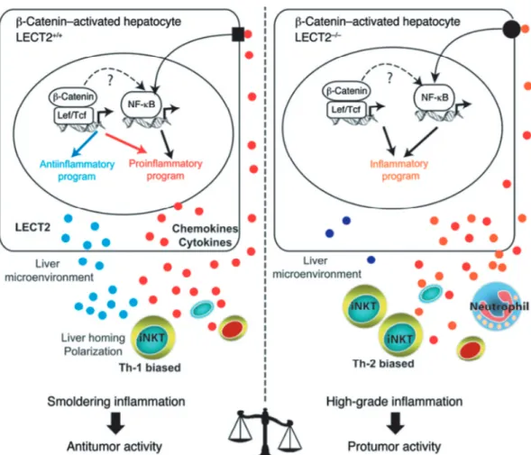
2 KO マウスや NKT KO マウスで悪性度の高い肝
細胞癌が形成をされることを見出し
49),抗炎症性メ
ディエーターにより作り出される微小環境が,腫瘍
の形成に影響を与えるという炎症と肝細胞癌との関
連モデルを提唱した(Figure 6).通常は,LECT 2
の作用による NKT 細胞数の増殖抑制によって Th1
優勢環境に傾き,前癌段階である炎症が抑制されて
いるが,急性炎症等がきっかけとなり LECT 2 が減
少すると,NKT 細胞が増加して Th2 優勢環境に傾
きその結果産生されるサイトカインにより炎症が亢
進し前癌段階が前進する.
(6)肥満との関係
Lan F らは,LECT 2 が,ヒトの肥満とインスリン
抵抗性の両方の重症度と正の相関があることを見出
した
50).肥満者では,過剰に産生された LECT 2 が,
筋肉でインスリン抵抗性を誘導することで糖尿病を
発症しやすくなると考えられる.組換え LECT 2 を
C2C12 筋細胞に添加したところ,Jun NH2 末端キ
ナーゼのリン酸化を介してインスリンシグナル伝達
を障害した.肥満は慢性炎症の結果であるという考
え方が定着しつつあり,炎症を抑えるために LECT
2 が肥満者で過剰発現したと考えられる.過剰発現
した LECT 2 を抑制することで,肥満者が糖尿病を
発症するリスクを軽減できる可能性がある.
2)Alternatively activated(M2)マクロファージ
マウスの脂肪組織には少なくとも 2 種類の性質の
異なる極性を有するマクロファージが存在すること
が明らかになった.非肥満の脂肪組織では,M2 マク
ロファージが抗炎症性サイトカイン IL-10 や nitric
oxide(NO)生合成を抑制するアルギナーゼを産生す
ることによって炎症性変化を抑制する.これに対し
て,肥満に伴い増加する classically activated(M1)マ
クロファージは,細胞死に陥った脂肪細胞を取り囲
む特徴的な組織像(crown-like structure)を呈し,
多くの炎症性サイトカインを分泌して脂肪組織の炎
症性変化を促進する
51).
3 ) Receptor activator of NF-kappa B ligand
(RANKL)
近年,免疫系において重要な役割を果たすサイト
カインや転写因子の幾つかが骨代謝系を制御してい
ることが明らかになり,
「骨免疫学」が創生された
52).
Maruyama らは感染により血中 RANKL 値が速や
か に 低 下 す る こ と や,RANKL KO マ ウ ス は LPS
シ ョ ッ ク の 致 死 率 が 上 昇 す る こ と,マ ウ ス に
Figure 6 Liver inflammation is critical for β-catenin-induced liver tumorigenesis49).
Oncogenic activation of β-catenin in hepatocytes triggers an intrinsic inflammatory pro-gram with both pro- and anti-inflammatory mediators that together construct an inflam-matory microenvironment that controls tumor progression. In Apc-deficient (Apc-/-) hepatocytes, β-catenin signaling is constitutively activated and induced: (a) the expression of a proinflammatory program resulting from both a direct control by the Wnt/β-catenin signaling and an indirect control by NF-κB that is not yet understood and (b) the expres-sion of an anti-inflammatory program including at least the direct leukocyte cell-derived chemotaxin-2 (LECT2) target gene. 2 interconnected factors relay the anti-inflammatory response, the chemokine-like factor LECT2 and the iNKT cells. iNKT cell homeostasis is controlled by LECT2 at the level of liver homing and cytokine polarization. Together, the β-catenin-induced liver microenvironment exhibits a low grade of chronic inflammation that preserves an immune response with antitumor activity. In mice deficient in Apc and LECT2 (Apc-/-LECT2-/-), the lack of LECT2 causes highgrade inflammation in the liver microenvironment, which strongly potentiates the tumoral process and results in lung me-tastases.
RANKL を前投与しておくと LPS ショックによる
マウス致死率が改善されることを発見し,RANKL
は敗血症性ショックのマーカーであると共に予防因
子であることを見出した
53).欠損するとショックの
致死率が上昇し,RANKL 値が炎症反応と逆相関す
る現象は LECT 2 の場合と同様である.
4)合成 CRP
合成 CRP は CRP の活性化部位である 174∼185
までの 12 個のアミノ酸残基を合成したものであり,
本来の CRP と同様の生物学的活性を有するとされ
る.
Inatsu らは合成 CRP を前投与したマウスに LPS
を静脈内投与すると,TNF-α と IL-12 の上昇が抑制
されることを見出した.さらに LPS 投与時のクッ
パー細胞の貧食能が CRP 投与マウスで亢進した
54).
炎症の際に CRP が高値となり炎症の指標になって
いるのは周知の事実であるが,病態生理学的な役割
は現在明らかにされていない.この研究は CRP の意
義を理解するうえで貴重な研究である.
5)Myeloid associated Ig like receptors-I
(MAIR-I)
MAIR-I(CD300a)は肥満細胞や NK 細胞などに発
現する分子量 60 kDa の細胞表面分子である.小田
らは,MAIR-I がアポトーシスによって細胞表面に
生じた phosphatidylserine(PS)を認識する免疫グロ
ブリン様受容体であることを見出した
55).さらに,
MAIR-I KO マウスを作成しその機能を調べたとこ
ろ,MAIR-I KO マ ウ ス は cecal ligation and
punc-ture(CLP)による細菌性敗血症ショックに抵抗性を
示した.ショック抵抗性を示した MAIR-I KO マウ
スの腹腔内では,顆粒球が増加し,細菌数が減少し
ていた.MAIR-I KO マウスの肥満細胞を,LPS およ
び MAIR-I と結合する死細胞と一緒に培養したとこ
ろ,好中球遊走能を持つケモカインの産生が増加し
た.したがって,肥満細胞に存在する MAIR-I は死細
胞の PS と結合し,好中球遊走能を持つケモカイン
の産生を抑制している可能性が示唆された
56).
6)Regulatory dendritic cells(DC regs)
Sato らは未熟樹状細胞に各種の刺激を加えるこ
とにより,regulatory T(T reg)細胞誘導能等の T
細胞制御機能を持つ DC regs を開発した
57).DC regs
の腹腔内投与により,LPS ショックや CLP により
誘導された敗血症炎症マウスの血中サイトカイン濃
度の上昇や致死率が改善された
58).
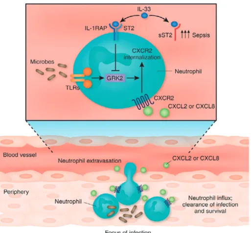
7)IL-33
細菌が生体内に侵入すると,マクロファージ,単
球や血管内皮細胞が好中球を呼び寄せるために(C-X-C motif chemokine)ligand(CXCL)2 を 産 生 す る.
CXCL2 は 好 中 球 に 存 在 す る 受 容 体 で あ る C-X-C
motif chemokine receptor(CXCR)2 に結合して好中
球を遊走する.一方,LPS 等の細菌由来物質は TLR
を 介 し て 好 中 球 の G protein-coupled receptor
kinase(GRK)2 の発現を増強する.GRK2 は CXCR
2 のシグナルを抑制して好中球の遊走を減少させる
ため,感染症を発症しやすくすることが示唆される.
Alves-Filho JC らは,IL-33 が GRK2 の発現を抑制
することを 見 出 し,そ の 結 果 CLP に よ る 敗 血 症
ショックが抑制された
59).通常の敗血症患者では好
中球の遊走能を維持するために IL-33 が膜結合型の
supression of tumorigenicity(ST)2 に 結 合 し て
GRK2 の発現を抑制しているが,致死的な敗血症患
者では IL-33 の受容体である soluble ST2(sST2)が
誘導されて敗血症が増悪することが見出されている
(Figure 7)
60).
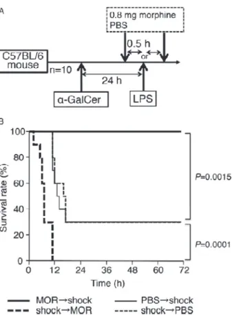
8)Morphine
Systemic
inflammatory
response
syndrome
(SIRS)は,細菌に対する免疫応答によって産生され
るサイトカインにより惹起される.敗血症ショック
時にモルヒネを投与することはショック応答を増悪
するため禁忌とされるが,モルヒネ投与はサイトカ
インの産生を抑制し,ショック応答を抑制するとい
う報告もあり,神経細胞や消化管以外への作用はあ
まり解析されていない.我々はマウスモデルにおい
て,LPS ショック誘導前にモルヒネを投与すると
ショックが抑制され,誘導後に投与すると増悪する
ことを見出した(Figure 8)
61).この結果は,モルヒ
ネが生体内の免疫応答の変化により異なる作用機序
を示すことを示唆している.さらに,マウスの肝臓
浸潤細胞をモルヒネで処理したところ NKT 細胞が
特異的に消滅していた
未発表).この結果を基に,モルヒ
ネ投与時期による相反した症状を説明するため,モ
ルヒネは NKT17 と NKT reg を消滅させるという
仮説を立てた.すなわち,通常のショック応答は,
ショックが誘導(LPS の投与)され,NKT17 が活性
化されると次に好中球が活性化されショックが惹起
される.その際,NKT reg が作用してショックが中
程度は抑制される.ショック誘導前にモルヒネを投
与した場合は,NKT17 が消滅してしまうため好中
球は活性化されず,結果としてショックは誘導され
ない.ショック誘導後にモルヒネを投与した場合は,
好中球は活性化された後であり,さらに,NKT reg
が消滅してしまうために,ショックが増強する.
NKT が好中球を制御するメカニズムはまだ解明さ
れていないが,この仮説を立証することで,好中球
制御の一端を解明したいと考えている.
9)Anaphase-promoting complex(APC)
Xu J らは,細胞外ヒストンを抗ヒストン抗体によ
りブロックしたり, APC で分解したりすることで,
LPS,TNF-α,CLP で誘発したマウス敗血症ショッ
クによる致死率が低下することを見出した
62).
10)ILC3
新生児において,長期的な抗生物質の使用は,好
中球の減少が原因とされる遅発型敗血症のリスクを
上昇させる.妊娠した母マウスに抗生物質を長期投
与すると,母体から新生仔への微生物の移行が減少
する.Deshmukh らは,抗生物質を投与された新生
仔マウスおよび無菌新生仔マウスの血中や骨髄中で
の好中球数や,骨髄中の顆粒球/マクロファージに拘
束された前駆細胞数が減少することを見出した
63).
Figure 7 IL-33 promotes the migration of neutrophils into the focus of infection60).
Detection of microbes by Toll-like receptors (TLRs) expressed by neutrophils increases the expression of G protein-coupled receptor kinase (GRK) 2. GRK2 mediates the internalization of (C-X-C motif chemokine) receptor (CXCR) 2 and desensitizes neutrophils to the action of the chemokines (C-X-C motif chemokine) ligand (CXCL) 2 and CXCL8, produced by mono-cytes/macrophages, epithelial cells and endothelial cells at the focus of infection. The cy-tokine IL-33, expressed by fibroblasts and epithelial, endothelial, mast and innate immune cells in response to infection, signals through a receptor complex composed of soluble su-pression of tumorigenicity (ST) 2 and IL-1 receptor accessory protein (IL-1RAP). IL-33 also binds a soluble isoform of ST2 (sST2) that acts as a decoy receptor. sST2 is upregulated in people with sepsis and functions as a modulator of IL-33. Alves-Filho et al. report that IL-33 lowers the expression of GRK2, overriding the downregulation of CXCR2 and the inhibition of neutrophil chemotaxis induced by TLR agonists59). This signaling cascade enhances
anti-microbial host defenses.
母マウスへの抗生物質投与は,新生仔マウスの腸管
の IL-17 産 生 細 胞 の 数 や,granulocyte
colony-stimulating factor(G-CSF)の産生を低下させた.新
生仔マウスは,顆粒球減少によって大腸菌 K1 株や
肺炎桿菌(klebsiella pneumoniae)による敗血症に対す
る免疫応答が障害され,この疾患に対する感受性が
上昇したが,これらは G-CSF の投与によって部分的
に回復した.抗生物質を投与した新生仔マウスへ正
常な細菌叢を移入すると,腸管で ILC3 による IL-17
産生が誘導され,TLR4 および myeloid
differentia-tion factor 88(MyD88)依存的に血漿中 G-CSF レベ
ルが上昇し,好中球数が増加して,敗血症に対する
IL-17 依存的な抵抗性が回復した.ILC を特異的に除
去すると,IL-17 と G-CSF に依存して起こる顆粒球
増多や敗血症に対する抵抗性が生じなくなった.こ
れらの結果は,新生仔の顆粒球増多や好中球恒常性,
敗血症に対する宿主抵抗性の調節に腸管微生物叢お
よび ILC が重要な役割を担っていることを裏付け
ている.
3.慢性炎症
慢性炎症は,悪性腫瘍,動脈硬化,肥満,アルツ
ハイマー病などの種々の疾患の発症や進展に関与す
ることが分かっている
64)65).中でもヒト の 癌 の 約
20% は,慢性炎症が原因だと考えられる.Pikarsky
Figure 8 Timing of morphine administration affects the survival rate of mice with LPS-mediated lethal endotoxic shock61).
The administration schedule is shown in (A). Mice were sensitized with α-GalCer (2 mg/mouse) 24 h before the LPS (1.5 mg/mouse) challenge. A, Mice were administered 0.8 mg of morphine or PBS at 0.5 h before or after the LPS challenge. B, The survival rate was observed every hour for 72 h. Each group comprised 10 mice. The significance of the difference between the survival rates of each test group and the respective control group was evaluated us-ing the Wilcoxon test.
LPS, indicates lipopolysaccharide; PBS, phosphate-buffered saline.
E らは,nuclear factor-kappa B(NF-κB)が炎症と癌
との関連物質であることを見出した
66).肝炎と肝臓
癌 を 自 然 発 症 す る multidrug resistance gene 2
(Mdr2)-KO マウスを使って実験したところ,症状の
悪化と共に炎症による NF-
κB の活性化が促進され
ることを見出し,NF-κB の発現を抑制すると癌化を
防ぐことを発見した.その結果,NF-κB は炎症関連
癌の促進に不可欠であることが分かった.大島らは,
浸潤性大腸がんを自然発生するモデルマウスにおい
て,浸潤癌組織で炎症反応が強く誘導されているこ
とを見出した
67).癌 抑 制 に 関 連 す る transforming
growth factor-β(TGF-β)受容体を欠損したマウス
に,潰瘍性大腸炎を誘発させると浸潤性大腸癌が発
生することを観察した.その結果,癌の悪性化進展
は特定の遺伝子変異と慢性炎症の相互作用により誘
導されることが明らかとなった.
おわりに
1 と 2 の項に記述したように,急性炎症は免疫学
の進歩により詳細な仕組みが明らかになってきた.
一方,炎症が慢性化するメカニズムについては不明
な点が多い.過剰な inflammasome の形成が慢性炎
症を惹起する一因となっていると考えられている
4)5)が,慢性化を誘導,維持する因子は同定されていな
い.慢性炎症は急性炎症よりも多くの要素が複雑に
作用しているため,特定の因子を同定することは困
難を極めると考えられる.しかしながら,慢性炎症
の起因は,急性炎症であることに間違いはなく,急
性炎症の制御は結果的に慢性炎症の制御につながる
と考えられる.
開示すべき利益相反状態はない. 文 献1)Rubartelli A, Lotze MT : Inside, outside, upside down: damage-associated molecular-pattern mole-cules (DAMPs) and redox. Trends Immunol 28 (10): 429―436, 2007
2)Schroder K, Tschopp J: The inflammasomes. Cell 140: 821―832, 2010
3)Fang R, Tsuchiya K, Kawamura I et al: Critical roles of ASC inflammasomes in caspase-1 activation and host innate resistance to Streptococcus pneu-moniae infection. J Immunol 187: 4890―4899, 2011 4)Vandanmagsar B, Youm YH, Ravussin A et al:
The NLRP 3 inflammasome instigates obesity-induced inflammation and insulin resistance. Nat Med 17: 179―188, 2011
5)Davis BK, Wen H, Ting JP: The inflammasome NLRs in immunity, inflammation, and associated diseases. Annu Rev Immunol 29: 707―735, 2011 6)Takeuchi O, Akira S: Pattern recognition
recep-tors and inflammation. Cell 140: 805―820, 2010 7)Brinkmann V, Reichard U, Goosmann C et al:
Neutrophil extracellular traps kill bacteria. Science 303: 1532―1535, 2004
8)Parker H, Albrett AM, Kettle AJ et al: Myeloper-oxidase associated with neutrophil extracellular traps is active and mediates bacterial killing in the presence of hydrogen peroxide. J Leukoc Biol 91 : 369―376, 2012
9)Li P, Li M, Lindberg MR et al: PAD4 is essential for antibacterial innate immunity mediated by neu-trophil extracellular traps. J Exp Med 207 : 1853 ― 1862, 2010
10)Fuchs TA, Brill A, Duerschmied D et al: Extracel-lular DNA traps promote thrombosis. Proc Natl Acad Sci U S A 107: 15880―15885, 2010
Neutrophil extracellular traps sequester circulating tumor cells and promote metastasis. J Clin Invest 123: 3446―3458, 2013
12)Knight JS, Zhao W, Luo W et al: Peptidylarginine deiminase inhibition is immunomodulatory and vas-culoprotective in murine lupus. J Clin Invest 123 : 2981―2993, 2013
13)Leliefeld PH, Koenderman L, Pillay J: How neu-trophils shape adaptive immune responses. Front Immunol 6: 471 (1-8), 2015
14)Bank U, Reinhold D, Schneemilch C et al: Selec-tive proteolytic cleavage of IL-2 receptor and IL-6 receptor ligand binding chains by neutrophil-derived serine proteases at foci of inflammation. J Interferon Cytokine Res 19: 1277―1287, 1999 15)Bank U, Ansorge S : More than destructive :
neutrophil-derived serine proteases in cytokine bio-activity control. J Leukoc Biol 69: 197―206, 2001 16)Pillay J, Kamp VM, van Hoffen E et al: A subset
of neutrophils in human systemic inflammation in-hibits T-cell responses through Mac-1. J Clin Invest 122: 327―336, 2012
17)Rodriguez PC, Quiceno DG, Ochoa AC: L-arginine availability regulates T-lymphocyte cell-cycle pro-gression. Blood 109: 1568―1573, 2007
18)Makarenkova VP, Bansal V, Matta BM et al: CD 11b+/Gr-1+myeloid suppressor cells cause T cell dysfunction after traumatic stress. J Immunol 176: 2085―2094, 2006
19)Yachimovich-Cohen N, Even-Ram S, Shufaro Y et al: Human embryonic stem cells suppress T cell responses via arginase I-dependent mechanism. J Immunol 184: 1300―1308, 2010
20)Gelderman KA, Hultqvist M, Holmberg J et al: T cell surface redox levels determine T cell reactivity and arthritis susceptibility. Proc Natl Acad Sci U S A 103: 12831―12836, 2006
21)Malmberg KJ, Arulampalam V, Ichihara F et al: Inhibition of activated/memory (CD45RO+) T cells
by oxidative stress associated with block of NF-kappaB activation. J Immunol 167: 2595―2601, 2001 22)de Kleijn S, Langereis JD, Leentjens J et al:
IFN-γ-stimulated neutrophils suppress lymphocyte prolif-eration through expression of PD-L1. PLoS One 8: e 72249, 2013
23)Hotchkiss RS, Monneret G, Payen D : Sepsis-induced immunosuppression: from cellular dysfunc-tions to immunotherapy. Nat Rev Immunol 13: 862― 874, 2013
24)Schuster S, Hurrell B, Tacchini-Cottier F: Cros-stalk between neutrophils and dendritic cells : a context-dependent process. J Leukoc Biol 94: 671― 675, 2013
25)Hufford MM, Richardson G, Zhou H et al: Influenza-infected neutrophils within the infected lungs act as antigen presenting cells for anti-viral CD8+T cells. PLoS One 7: e46581, 2012
26)Davey MS, Lin CY, Roberts GW et al: Human neutrophil clearance of bacterial pathogens trig-gers anti-microbialγδ T cell responses in early in-fection. PLoS Pathog 7: e1002040, 2011
27)Sonnenberg GF, Artis D: Innate lymphoid cells in the initiation, regulation and resolution of inflamma-tion. Nat Med 21 (7): 698―708, 2015
28)Spits H, Artis D, Colonna M et al: Innate lymphoid cells--a proposal for uniform nomenclature. Nat Rev Immunol 13: 145―149, 2013
29)Halim TY, MacLaren A, Romanish MT et al: Retinoic-acid-receptor-related orphan nuclear re-ceptor alpha is required for natural helper cell de-velopment and allergic inflammation. Immunity 37: 463―474, 2012
30)Salimi M, Barlow JL, Saunders SP et al: A role for IL-25 and IL-33-driven type-2 innate lymphoid cells in atopic dermatitis. J Exp Med 210: 2939―2950, 2013
31)Halim TY, Steer CA, Mathä L et al: Group 2 in-nate lymphoid cells are critical for the initiation of adaptive T helper 2 cell-mediated allergic lung in-flammation. Immunity 40: 425―435, 2014
32)Matsuda JL, Naidenko OV, Gapin L et al: Track-ing the response of natural killer T cells to a glycol-ipid antigen using CD1d tetramers. J Exp Med 192 (5): 741―754, 2000
33)Godfrey DI, Kronenberg M: Going both ways: im-muneregulation via CD1d-dependent NKT cells. J Clin Invest 114 (10): 1379―1388, 2004
34)Brennan PJ, Brigl M, Brenner MB : Invariant natural killer T cells: an innate activation scheme linked to diverse effector functions. Nat Rev Immu-nol 13 (2): 101―117, 2013
35)Brutkiewicz RR: CD1d ligands: the good, the bad, and the ugly. J Immunol 177 (2): 769―775, 2006 36)Tupin E, Kinjo Y, Kronenberg M: The unique role
of natural killer T cells in the response to microor-ganisms. Nat Rev Microbiol 5 (6): 405―417, 2007 37)Bendelac A, Savage PB, Teyton L: The biology of
NKT cells. Annu Rev Immunol 25: 297―336, 2007 38)Chang DH, Deng H, Matthews P et al:
Inflammation-associated lysophospholipids as ligands for CD1 d-restricted T cells in human can-cer. Blood 112: 1308―1316, 2008
39)Zhou D, Mattner J, Cantu C 3rd et al: Lysosomal glycosphingolipid recognition by NKT cells. Sci-ence 306: 1786―1789, 2004
40)La Cava A, Van Kaer L, Fu-Dong-Shi : CD 4+ CD25+Tregs and NKT cells: regulators regulating regulators. Trends Immunol 27 (7): 322―327, 2006 41)Gonzalez-Aseguinolaza G, Van Kaer L,
Bergmann CC et al: Natural killer T cell ligand alpha-galactosylceramide enhances protective im-munity induced by malaria vaccines. J Exp Med 195: 617―624, 2002
42)Sag D, Krause P, Hedrick CC et al: IL-10-producing NKT10 cells are a distinct regulatory in-variant NKT cell subset. J Clin Invest 124 (9): 3725― 3740, 2014
43)Yamagoe S, Yamakawa Y, Matsuo Y et al: Purifi-cation and primary amino acid sequence of a novel neutrophil chemotactic factor LECT 2. Immunol Lett 52: 9―13, 1996
in hepatic NKT cells in leukocyte cell-derived che-motaxin 2-deficient mice contributes to severe con-canavalin A-induced hepatitis. J Immunol 173: 579― 585, 2004
45)Okumura A, Saito T, Otani I et al: Suppressive role of leukocyte cell-derived chemotaxin 2 in mouse anti-type II collagen antibody-induced ar-thritis. Arthritis Rheum 58: 413―421, 2008
46)Dang MH, Kato H, Ueshiba H et al: Possible role of LECT2 as an intrinsic regulatory factor in SEA-induced toxicity in d-galactosamine-sensitized mice. Clin Immunol 137 (3): 311―321, 2010
47)Ando K, Kato H, Kotani T et al: Plasma leukocyte cell-derived chemotaxin 2 is associated with the se-verity of systemic inflammation in patients with sepsis. Microbiol Immunol 56: 708―718, 2012 48)Phesse TJ, Parry L, Reed KR et al: Deficiency of
Mbd2 attenuates Wnt signaling. Mol Cell Biol 28 : 6094―6103, 2008
49)Anson M, Crain-Denoyelle AM, Baud V et al: On-cogenic β-catenin triggers an inflammatory re-sponse that determines the aggressiveness of hepa-tocellular carcinoma in mice. J Clin Invest 122 (2): 586―599, 2012
50)Lan F, Misu H, Chikamoto K et al: LECT2 func-tions as a hepatokine that links obesity to skeletal muscle insulin resistance. Diabetes 63 ( 5 ) : 1649 ― 1664, 2014
51)Mantovani A, Sica A, Sozzani S et al: The chemokine system in diverse forms of macrophage activation and polarization. Trends Immunol 25 : 677―686, 2004
52)Takayanagi H, Ogasawara K, Hida S et al: T-cell-mediated regulation of osteoclastogenesis by signal-ling cross-talk between RANKL and IFN-gamma. Nature 408: 600―605, 2000
53)Maruyama K, Takada Y, Ray N et al: Receptor activator of NF-kappa B ligand and osteoprotegerin regulate proinflammatory cytokine production in mice. J Immunol 177: 3799―3805, 2006
54)Inatsu A, Kinoshita M, Nakashima H et al: Novel mechanism of C-reactive protein for enhancing mouse liver innate immunity. Hepatology 49: 2044― 2054, 2009
55)Nakahashi-Oda C, Tahara-Hanaoka S, Honda S et al: Identification of phosphatidylserine as a ligand for the CD300 a immunoreceptor. Biochem Biophys Res Commun 417 (1): 646―650, 2012 56)Nakahashi-Oda C, Tahara-Hanaoka S, Shoji M et
al: Apoptotic cells suppress mast cell inflammatory responses via the CD300a immunoreceptor. J Exp Med 209 (8): 1493―1503, 2012
57)Sato K, Yamashita N, Yamashita N et al: Regula-tory dendritic cells protect mice from murine acute graft-versus-host disease and leukemia relapse. Im-munity 18: 367―379, 2003
58)Fujita S, Seino K, Sato K et al: Regulatory den-dritic cells act as regulators of acute lethal systemic inflammatory response. Blood 107: 3656―3664, 2006 59)Alves-Filho JC, Sônego F, Souto FO et al:
Interleukin-33 attenuates sepsis by enhancing neu-trophil influx to the site of infection. Nat med 16 (6): 708―712, ONLINE METHODS, 2010
60)Roger T, Calandra T : Interleukin-33 safeguards neutrophils in sepsis. Nat Med 16 (6): 638―639, 2010 61)Fukada T, Kato H, Ozaki M et al: Impact of the
timing of morphine administration on lipopolysaccharide-mediated lethal endotoxic shock in mice. Shock 45 (5): 564―569, 2016
62)Xu J, Zhang X, Pelayo R et al: Extracellular his-tones are major mediators of death in sepsis. Nat Med 15: 1318―1321, 2009
63)Deshmukh HS, Liu Y, Menkiti OR et al: The mi-crobiota regulates neutrophil homeostasis and host resistance to Escherichia coli K1 sepsis in neonatal mice. Nat Med 20: 524―530, 2014
64)Tabas I, Glass CK: Anti-inflammatory therapy in chronic disease: challenges and opportunities. Sci-ence 339 (6116): 166―172, 2013
65)Landskron G, De la Fuente M, Thuwajit P et al: Chronic inflammation and cytokines in the tumor microenvironment. J Immunol Res 2014 : 149185, 2014
66)Pikarsky E, Porat RM, Stein I et al: NF-kappa B functions as a tumour promoter in inflammation-associated cancer. Nature 431: 461―466, 2004 67)Oshima H, Nakayama M, Han TS et al:
Suppress-ing TGFβ signaling in regenerating epithelia in an inflammatory microenvironment is sufficient to cause invasive intestinal cancer. Cancer Res 75 (4): 766―776, 2015
68)Matzinger P: The danger model: a renewed sense of self. Science 296 (5566): 301―305, 2002
69)Seong SY, Matzinger P : Hydrophobicity : an an-cient damage-associated molecular pattern that in-itiates innate immune responses. Nat Rev Immunol 4 (6): 469―478, 2004