Title
Two poorly known species of the genus Hyastenus White, 1847
(Decapoda: Brachyura: Epialtidae) from the Ryukyu
Archipelago
Author(s)
Ohtsuchi, Naoya; Takeda, Masatsune; Ashida, Akira
Citation
Fauna Ryukyuana, 58: 1-14
Issue Date
2020-12-04
URL
http://hdl.handle.net/20.500.12000/47426
Fauna Ryukyuana
ISSN 2187-6657 http://w3.u-ryukyu.ac.jp/naruse/lab/Fauna_Ryukyuana.htmlTwo poorly known species of the genus Hyastenus White, 1847 (Decapoda:
Brachyura: Epialtidae) from the Ryukyu Archipelago
Naoya Ohtsuchi
1, Masatsune Takeda
2& Akira Ashida
31International Coastal Research Center, Atmosphere and Ocean Research Institute, The University of Tokyo,
1-19-8 Akahama, Iwate 028-1102, Japan (ohtsuchi@g.ecc.u-tokyo.ac.jp)
2Department of Zoology, National Museum of Nature and Science, 4-1-1, Amakubo, Tsukuba,
Ibaraki 305-0005, Japan (takeda@kahaku.go.jp)
3School of Agricultural Sciences, Nagoya University, 6 Furou-cho, Chikusa-ku, Nagoya,
Aichi 464-8601, Japan (Ashigram9@gmail.com) Abstract. This paper reports the first records of
Hyastenus ambonensis Griffin & Tranter, 1986 from
the Ryukyu Archipelago and H. uncifer Calman, 1909 from Okinawa Island. Hyastenus brockii De Man, 1888 is excluded from the carcinological fauna of Japan, because the only Japanese specimen from Kuro Island, Yaeyama Group, deposited at the Wakayama Prefectural Museumm of Natural History, is found to represent H. ambonensis. Diagnostic characters of closely allied H. ambonensis and H.
baru Windsor & Ahyong, 2013 are reassessed. Introduction
Hyastenus White, 1847 is a large spider crab genus
of the family Epialtidae MacLeay, 1838, including 36 species distributed throughout Indo-West Pacific waters (Griffin & Tranter 1986; Yang & Dai 1994; Ng et al. 2008; Windsor & Ahyong 2013; Lee et al. 2018; Lee & Ng, 2019). In Japan, Hyastenus is represented by 10 species: H. ambonensis Griffin & Tranter, 1896, H. borradailei (Rathbun, 1907), H.
brockii De Man, 1904, H. convexus Miers, 1884, H. cornigerus Sakai, 1938, H. diacanthus (De
Haan, 1837), H. elatus Griffin & Tranter, 1986, H.
elongatus Ortmann, 1893, H. kyusyuensis (Yokoya,
1933), and H. uncifer Calman, 1909 (Sakai 1938, 1976; Griffin & Tranter 1986; Marumura & Kosaka 2003; Komatsu 2011; Takeda et al. 2019).
In Japan, Hyastenus brockii is known only by one male collected from off Ishigaki Island, which is a part of the Nagai Collection deposited at the Wakayama Prefectural Museum of Natural History (WMNH) (Marumura & Kosaka 2003). Marumura & Kosaka (2003) also listed a female specimen from Okinawa Island which was left unidentified (as
Hyastenus sp.). Our reexamination of the Hyastenus
specimens in the Nagai Collection revealed that their “H. brockii” from Ishigaki Island as well as
Hyastenus sp. from Okinawa Island are actually H. ambonensis (type locality: Ambon). Hyastenus ambonensis was recorded from Ogasawara Islands
for the first time from Japan (Komatsu 2011), and the present study represents the second records from the country and the first records from the Ryukyu Archipelago.
Hyastenus uncifer is also poorly known in Japan,
with only two records by Nomura et al. (1996) from Aka Island, Kerama Group and by Marumura & Kosaka (2003) from Kuro Island, Yaeyama Group. Nomura et al. (1996) did not provide any accessible materials (see Discussion), and Marumura & Kosaka (2003) only listed the specimen data in the catalogue with a small photograph (Marumura & Kosaka 2003: pl.7 fig. 38). Recently, we newly obtained a male specimen of H. uncifer from Okinawa Island, which is the first record from the island.
This paper also discusses possible ontogenetic changes on taxonomically important characters based on the specimens in the Nagai Collection and our recent collection.
Materials and methods
Most of the Nagai Collection deposited at WMNH are dried specimens. The specimens examined in this study were rehydrated by using an aqueous solution of ethylene glycol following Thompson et al. (1966), and then, preserved in 70% ethanol to examine some detailed morphologies such as the male first gonopods. The specimens newly collected in this study are deposited in the Ryukyu University Museum, Fujukan (RUMF), University of the Ryukyus. For the comparative purpose with
H. ambonensis, the holotype of H. baru Windsor &
Ahyong, 2013 deposited in the Muséum national d’Histoire naturelle, Paris (MNHN) was examined by photographs taken at our request.
The morphological terms and abbreviations essentially follow Davie et al. (2015): CW = maximum carapace width excluding epibranchial spines; ChH = maximum chela height; ChL = maximum chela length (above lower margin); CL = carapace length including pseudorostral spines; PCL = maximum carapace length excluding pseudorostral spines; PRL = pseudorostral spine length. Other abbreviations used in the text: P2–5 = second to fifth pereiopods (first to fourth ambulatory legs), respectively; G1 = first gonopod.
Recently, Ohtsuchi et al. (2016, 2018) revealed that the post-megalopal growth of two kelp crab species of the genus Pugettia Dana, 1851 can be divided into at least three ontogenetic stages in both sexes, which can be distinguished by the different combination of chelal, pleopodal, and pleonal morphologies (also see Ohtsuchi & Kawamura 2019; Ohtsuchi et al. 2020). Griffin & Tranter (1986) recognized two types of tubular (= functional as spermatic duct) G1 in some Hyastenus. For example, smaller male specimens of H. diacanthus (11.5–32.0 mm, one exception 45 mm) have gradually tapering G1 (Griffin & Tranter 1986: fig. 48c), whereas larger individuals (33.5–70.5 mm) have distally filiform G1 (Griffin & Tranter 1986: fig. 48a, b). We recently found in H. diacanthus that there are medium-sized males with a combination of distally filiform G1 and relatively narrow, enlarged but not inflated chela between smaller males with gradually tapering G1 and proportionally short, simple and slender chela and larger males with distally filiform G1 and relatively wide, enlarged and inflated chelae, (Ohtsuchi et al. in prep.). In this study, the ontogenetic stages defined for the species of Pugettia (Ohtsuchi et al. 2016, 2018) were adjusted for the species of Hyastenus. The ontogenetic stage of each specimen was evaluated in a comprehensive manner by using the following terms:
Full-grown males: male individuals estimated to
be at terminal anecdysis. They are characterized by their enlarged chelipeds, chelae robust, with fingers widely gaping, movable finger bearing only a strong tooth, and fully developed G1.
Adolescent male: male individuals which did
not yet experience the terminal molt. They are characterized by their relatively small chelipeds, chelae slender, with fingers narrowly gaping, movable finger bearing a few teeth including large, rectangular, subproximal tooth, and less developed G1.
Full-grown female: female individuals estimated
to be at terminal anecdysis. They are characterized by having expanded pleon and fully opened (= not slit-like) gonopores.
Each specimen examined in this study belong to one of the stages defined above. Ohtsuchi et al. (2016, 2018) recognized additional 1 and 2 ontogenetic stages for males and females of Pugettia species, respectively, and these can also be applied for
Hyastenus species (Ohtsuchi et al. in prep.). Taxonomy
Superfamily Majoidea Samouelle, 1819 Family Epialtidae MacLeay, 1838 Genus Hyastenus White, 1841
Hyastenus ambonensis Griffin & Tranter, 1986
[Japanese name: Ambon-tsuno-gani] (Figs. 1, 2, 3A–C, 4)
Hyastenus ambonensis Griffin & Tranter, 1986: 125–
127, fig. 39d–f. — Komatsu 2011: 241, figs. 8F–I, 13A.
Hyastenus brocki [sic] Marumura & Kosaka 2003:
33. [not Hyastenus Brockii De Man, 1888]
Hyastenus sp. Marumura & Kosaka 2003: 33
(catalogue).
Material examined. WMNH-Na-Cr 0352, 1 female
(8.7 × 5.6 mm), Hirase, Okinawa, 77–83 m, 10 May 1991, coll. Tsuchida (dried). WMNH-Na-Cr 0348, 1 male (7.2 × 4.6 mm), Ishigaki Island, Okinawa, 100 m, 1990, coll. S. Nagai (rehydrated).
Additional materials. NSMT-Cr S 957, 3 males (4.8 × 3.2–7.1 × 5.4 mm), 1 ovigerous female (6.4 × 4.5 mm), KY-08-15, West of Chichi Island, 83–81 m, 28 Oct. 2008; NSMT-Cr S 958, 1 male (6.7 × 4.8 mm), KY-08-20, East of Chichi Island, 54–52 m, 29 Oct. 2008; NSMT-Cr S 959, 1 ovigerous female (8.0 × 5.8 mm), KY-10-31, West of Chichi Island, 96.8– 96.5 m, 9 Jul. 2010. See Komatsu (2011: 241, table 1) for details.
Comparative material. Hyastenus baru Windsor & Ahyong, 2013. Photographs of holotype: MNHN-IU-2012-994, male (7.8 × 4.7 mm), Banda Sea, Kei Islands, west of Kei-Besar, KARUBAR stn DW22, 5°22′S 133°01′E, 85-124 m, 25 October 1991.
Description of the specimens from the Ryukyu Archipelago. Carapace (Figs. 1A, 2A) pyriform,
length 1.6 as long as width, surface covered with tomentum (mostly abraded in male specimen), regions not clearly defined. Gastric region (Figs. 1A, C, 2A, C) elevated, with three mesogastric
tubercles increasing in size posteriorly (largest one slightly anterior to summit); protogastric region with faint, small tubercles mesially, distinct but low tubercle laterally; urogastric region flat, smooth. Hepatic region (Fig. 1A, 2C) moderately expanded,
laterally with small, pointed tubercle. Cardiac region (Fig. 1A, C) strongly elevated, subacute triangular in lateral view (Fig. 1C), as high as gastric region in males, lower than gastric region in female (Fig. 2C). Branchial region (Fig. 1A, C) not markedly
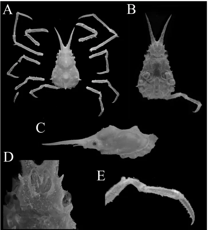
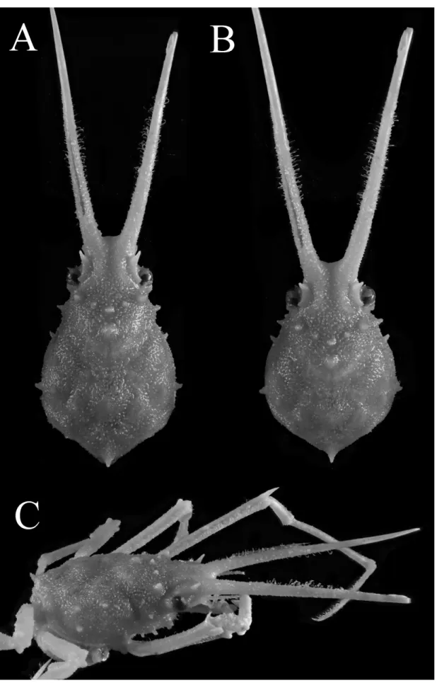
Fig. 1. Hyastenus ambonensis Griffin & Tranter, 1986. Adolescent male (7.2 × 4.6 mm, WMNH-Na-Cr 0348), Ishigaki Island, Ryukyu. A, overall habitus in dorsal view; B, C, cephalothorax in ventral view (B) and left lateral view (C, left P5 not shown); D, anterior part of cephalothorax in ventral view; E, dactylus, propodus, carpus, and disal half of merus of right fifth pereiopod.
図 1. アンボンツノガニ . 最終脱皮前の雄 (7.2 × 4.6 mm, WMNH-Na-Cr 0348), 石垣島 . A, 全体 , 背面観 ; B, C, 頭胸部 , 腹面観 (B) および側面観 (C); D, 頭胸部前方 , 腹面観 ; E, 右第 4 歩脚 , 指節 , 前節 , 腕節 , および長節 外端部 .
expanded, with two small spines on anterolateral subsurface; prebranchial region slightly elevated, dorsolaterally with two, acute tubercles, dorsally with slightly larger protuberance just behind gastric region; epibranchial region with lateral tubercle just dorsal to epibranchial spine; metabranchial region with low protuberance. Intestinal region (Figs. 1A, C, 2A) with median spine slightly curved dorsally, just above posterior carapace margin.
Pseudorostrum (Figs. 1A, B, 2) well divergent anteriorly, distance between tips as broad as (in male), or slightly narrower than (in female) CW; pseudorostral spines 0.6 as long as PCL in male, 0.5 in female. Supraorbital eave (Figs. 1A, 2A)
weakly concave on lateral margin; preorbital angle produced into short spine, acuminate at tip, directed anterolaterally; antorbital angle produced into small, blunt lobe with rounded apex. Eyestalk slender, cornea diameter less than quarter of front width. Postorbital lobe (Fig. 1D) cup-like, dorsal anterior margin with low, rounded, proximal lobe, lateral surface weakly keeled medially, ventral anterior margin almost straight. Epibranchial spine (Figs. 1A, C, 2) short, subacute, slightly curved dorsally.
Subhepatic region (Figs. 1B, 2B) weakly inflated, with two similar-sized blunt tubercles on pterygostomial ridge (anterior slightly larger than posterior). Subprebranchial region (Figs. 1B, 2B)
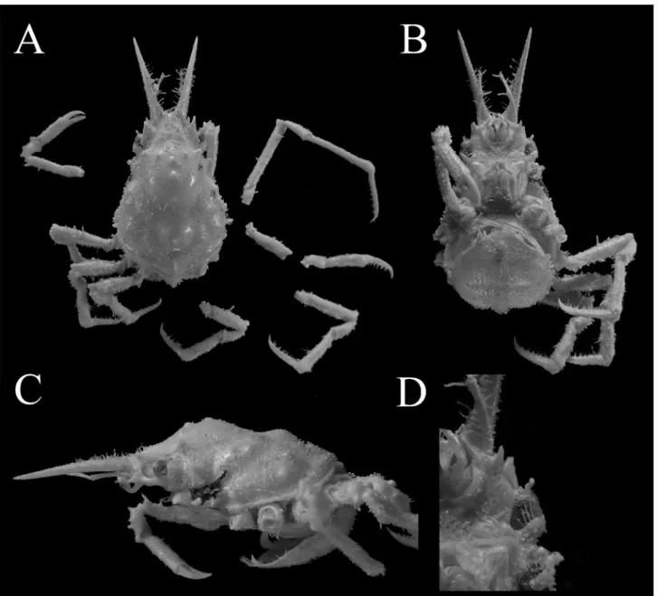
Fig. 2. Hyastenus ambonensis Griffin & Tranter, 1986. Full-grown female (8.7 × 5.6 mm, WMNH-Na-Cr 0352), Okinawa Island, Ryukyu. A, overall habitus in dorsal view; B, C, cephalothorax in ventral view and lateral view; D, orbital region in ventral view.
図 2. アンボンツノガニ . 最終脱皮後の雌 (8.7 × 5.6 mm, WMNH-Na-Cr 0352), 沖縄島 . A, 全体 , 背面観 ; B, C, 頭胸部 , 腹面観 (B) および側面観 (C); D, 眼窩周辺部 , 腹面観 .
with strong spine directed anterolaterally just above cheliped basis-ischium.
Basal antennal article (Fig. 1D) smooth, slender, with ridge along mesial margin; distolateral angle produced into strong spine projecting laterally, slightly incurved apically, only distally visible in dorsal view (Fig. 1A); lateral margin expanded into rounded, triangular lobe (anterior margin as long as posterior margin) in proximal half, faintly bent dorsally, with small tubercle basally.
Third maxilliped (Figs. 1A, 2B) ischium with broad depression medially. Merus anterolateral angle moderately elongated, rounded triangular. Distolateral angle of buccal cavity frame produced anterolaterally into rounded lobe with entire margin.
Cheliped (Figs. 1A, 2) merus dorsally with
three low tubercles (proximal one largest), long, acute spine on distal end; low tubercle in distal half ventrally on outer edge; carpus with small lateral tubercle dorsally at about distal third. Chela (Figs. 1A, 2B) slender 6.0 longer than height in male, 4.9 in female; fingers uniformly dentate, contiguous in distal two-thirds, narrowly gaped in proximal one-third.
Ambulatory legs (Figs. 1A, E, 2) slender, decreasing in length posteriorly, P4 as long as P5. Merus subcylindrical, extensor surface dorsomesially with 4, 2, 2, 1 small spines in proximal half in P2, 3, 4, 5, respectively; distoextensor projection strong, triangular spine in male P2, blunt tubercle in female P2, blunt, subrectangular tubercle in P3–5; upper flexor margin with 0–3 faint tubercles in P2, 2–4 in
Fig. 3. Male first gonopods (left). A–C, Hyastenus ambonensis Griffin & Tranter, 1986. A, B, adolescent male (7.2 × 4.6 mm, WMNH-Na-Cr 0348), Ishigaki Island, Ryukyu (A, abdominal; B, sternal); C, full-grown male (7.1 × 5.4 mm, NSMT-Cr S 957), off Ogasawara Islands (sternal). D, Hyastenus uncifer Calman, 1909, adolescent male (8.5 × 5.5 mm, RUMF-ZC 5892), Okinawa Island (abdominal).
図 3. 第一雄性生殖肢 ( 左 ). A–C, アンボンツノガニ . A, B, 最終脱皮前の雄 (7.2 × 4.6 mm, WMNH-Na-Cr 0348), 石垣島 ; C, 最終脱皮後の雄 (7.1 × 5.4 mm, NSMT-Cr S 957), 小笠原諸島沖 . D, ナガツノガニ . 最終脱皮前の雄 (8.5 × 5.5 mm, RUMF-ZC 5892), 沖縄島 .
P3–5; lower flexor margin with 6 faint tubercles in P2, 0–2 in P3–5. Carpus (Fig. 1E) extensor surface subproximally with low, subacute tubercle, medially with shallow, elongate depression, upper, lower margins each with small tubercle. Dactylus (Fig. 1A, E) weakly clawed, longer than half length of propodus, flexor surface with row of calcareous spines (11 or 12 spines in P2, 7 or 8 spines in P3–5), increasing in size, interval distally.
Pleon (Figs. 1B, 2B) with six pleomeres and telson in both sexes. In adolescent male (Fig. 1B), pleomeres 3–6 functionally fused, medially with weak, discontinuous keel; pleomere 3 with large protuberance on both sides; pleomere 6 dilated laterally; telson elongate, 1.2 as long as width, with rounded apex. In full-grown female (Fig. 2B), pleomeres 3–6 functionally fused, pleomere 3 to telson medially with low, blunt keel; telson broad triangular with rounded apex.
G1 (Fig. 3A, B) shaft broad at base, gradually curved, distal half gradually twisted, tapering toward narrow apex.
Female gonopore as oval aperture, narrowed laterally, opening anteriorly.
Remarks. The specimens of Hyastenus ambonensis at hand include an adolescent male
from Ishigaki Island and a full-grown female from Okinawa Island. The former and the latter were previously listed as “Hyastenus brockii” and
“Hyastenus sp.”, respectively, in the catalogue of the Nagai Collection (Marumura & Kosaka 2003). However, Nagai’s specimens differ from H. brockii in many morphological characters including the G1 structure (cf. Griffin & Tranter 1986: 134, figs. 38c, d, h, 42c, d). For example, 1) H. brockii is a large-eyed species, in which the cornea diameter is almost as long as one-third of the front width, but in our specimens, the cornea width is one quarter or less; 2) the relative length of pseudorostral spine (PRL/PCL) of H. brockii varies 0.7–1.0 (cf. Griffin & Tranter 1986: 134), but it is 0.5–0.6 in our specimens; 3) the antorbital angle is not markedly produced in
H. brockii as in our specimens; 4) the epibranchial
spine is shorter and more robust in H. brockii than in our specimens; 5) the intestinal region is armed with a minute tubercle rather than a spine in H.
brockii, but in our specimens there is a strong spine
slightly curved dorsally; 6) the lateral lobe of basal antennal article is more rounded and less produced in H. brockii than in our specimens; and 7) the two pterygostomial tubercles are different in size, and the anterior one is larger than the posterior one in H.
brockii, but they are similar in size in our specimens.
The morphological characters of our specimens generally agree well with the original description of
Hyastenus ambonensis based on full-grown females
(Griffin & Tranter 1986). Komatsu (2011) examined some male specimens from the Ogasawara Islands
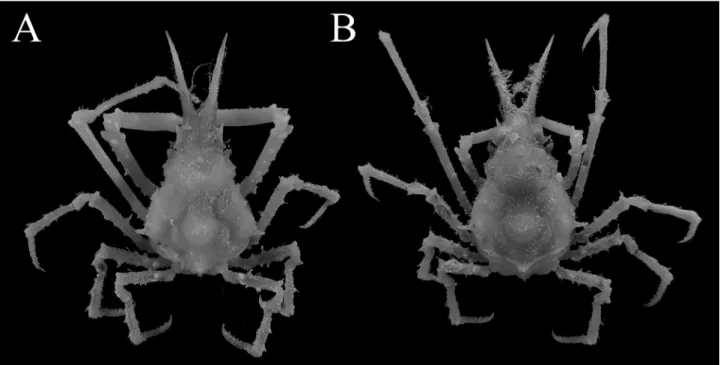
Fig. 4. Hyastenus ambonensis Griffin & Tranter, 1986. Overall habitus in dorsal view, A, full-grown male (7.1 × 5.4 mm, NSMT-Cr S 957); B, full-grown female (8.0 × 5.8 mm, NSMT-Cr S 959), off Ogasawara Islands.
図 4. アンボンツノガニ . 全体 , 背面観 . 最終脱皮後の成熟雄 (7.1 × 5.4 mm, NSMT-Cr S 957); B, 最終脱皮後の 雌 (8.0 × 5.8 mm, NSMT-Cr S 959), 小笠原諸島沖 .
and illustrated the G1 morphologies. It should be remarked that distal part of G1 in our male specimen is only narrowed toward the apex and not broadly dilated as figured by Komatsu (2011) (Fig. 3A (7.2 × 4.6 mm) versus Komatsu 2011: fig. 8F–H (7.1 × 5.4 mm)). However, direct comparison between our and the Ogasawara specimens confirmed that the G1 as well as chelipeds of our specimen are not fully developed, but essentially similar in general structure to those of the Ogasawara specimens (Figs. 1A, 3B vs. 3C, Komatsu 2011, fig. 13A). These differences are probably due to ontogenetic differences between examined materials. Although their sizes are close to each other, the Ogasawara specimens are probably in terminal anecdysis, whereas our material has not experienced the terminal molt. Indeed, the drastic change of G1 structure by a single molting was observed in H. diacanthus in the captive condition (Ohtsuchi & Takeda, unpublished). Griffin & Tranter (1986) also indicated that the G1 structure differs between larger and smaller specimens in some
Hyastenus species, such as H. aries (Latreille, 1825), H. diacanthus, and H. elatus, and in H. diacanthus.
Hyastenus ambonensis is relatively more similar
to H. baru Windsor & Ahyong, 2013 (type locality: Banda Sea, Indonesia) rather than to H. brockii.
Hyastenus baru was described based on the male
holotype (MNHN-IU-2012-994, 11.5 mm CL, 7.8 mm PCL, 4.7 mm CW), which can be considered as an adolescent male. On the other hand, the paratype
H. ambonensis examined by Windsor & Ahyong
(2013) was a full-grown female (AM P34609, 20.0 mm CL, 14.6 mm PCL, 9.3 mm CW). Our examination of male H. ambonensis shows that the following two diagnostic characters raised by Windsor & Ahyong (2013) need to be amended. Windsor & Ahyong (2013) observed that the female
H. ambonensis has the cardiac region being lower
than gastric region (Fig. 2C), but the cardiac region of male is strongly elevated and obviously higher than gastric region, which closely resembles H. baru in lateral view (Fig. 1C; Windsor & Ahyong 2013: fig. 1B). The distoextensor projection of P2 merus of female H. ambonensis is a blunt projection (Fig. 2A; Windsor & Ahyong 2013: 721), but those of males are strong spine in both species (Figs. 1A, 5A; Windsor & Ahyong 2013: figs. 1A, 2F).
Comparison with the photographs of the adolescent male holotype of H. baru and our adolescent male of H. ambonensis shows some additional distinguishing characters between the two species: the front is more elongated in H. baru than
in H. ambonensis (Figs. 5C vs. 1D); the distolateral spine of the basal antennal article is more elongated and fully visible in the dorsal view in H. baru, but it is less elongated and only distally visible in dorsal view in H. ambonensis (Figs. 5A, C vs. 1A, D); the anterior margin of basal antennal article is as long as the posterior margin in H. ambonensis, whereas it is longer than the posterior margin in H. baru (Fig. 1D vs. 5C). In addition, the G1 is more dilated and twisted in the distal half in H. baru than in H.
ambonensis (Windsor & Ahyong 2013: fig. 2K, L vs.
Fig. 3C; Komatsu 2011: fig. 8F–H).
Distribution. Ambon, Indonesia (type locality),
and the Ogasawara Islands, and Ishigaki and Okinawa Islands, Ryukyu, Japan, at depth of 52–90 m (Griffin & Tranter 1986; Komatsu 2001; this study). This is the second record of this species from Japan.
Hyastenus uncifer Calman, 1909
[Japanese name: Naga-tsuno-gani] (Figs. 3D, 6–8)
Hyastenus uncifer Calman, 1909: 712, pl. 72 figs.
8, 9. — Griffin &Tranter 1986: 156–157, figs. 38e, f, i, 39c, 42e–g. — Nomura et al. 1996: 15 (list), table 1. — Marumura & Kosaka 2003: 33 (catalogue), pl. 7 fig. 38. — Poupin et al. 2018: 18–19, fig. 8C.
Material examined. WMNH-Na-Cr 0347, 1 male
(8.8 × 5.7 mm, voucher specimen designated for new standard Japanese name), 1 female (12.5 × 7.9 mm), east of Kuro Island, Okinawa, 10 m, 1977, coll. S. Nagai (rehydrated). RUMF-ZC-5892, 1 male (8.5 × 5.5 mm), on mooring rope, Motobu Fishery Port, Okinawa Island, Okinawa, 5 Nov. 2018, coll. A. Ashida.
Description of the specimens from the Ryukyu Archipelago. Carapace (Figs. 6A, 7)
pyriform, length 1.5–1.6 as long as width, regions not clearly defined. Gastric region (Figs. 6A, 7) elevated, as high as cardiac region (Fig. 7C), with broad, Y-shaped line of six subacute tubercles, two mesogastric (posterior one larger than anterior), two lined obliquely anteriorly on either side of midline (lateral one larger than sublateral); anterior slope with long, laterally concaved rows of hooked setae on either side of midline. Hepatic region (Figs. 6A, 7) weakly expanded, with transverse rows of hooked setae between anterior hepatic spines. Cardiac region (Figs. 6A, 7) weakly elevated (Fig. 7C). Branchial
Fig. 5 Hyastenus baru Windsor & Ahyong, 2013. Holotype, male (7.8 × 4.7 mm, MNHN-IU-2012-994). A, overall dorsal habitus in dorsal view; B, anterior part of cephalothorax in ventral view. Photographs courtesy of Laure Corbari and Paula Martin-Lefèvre (MNHN).
図 5. Hyastenus baru Windsor & Ahyong, 2013. ホロタイプ , 雄 (7.8 × 4.7 mm, MNHN-IU-2012-994), A, 全体 , 背面 観 ; B, 頭胸部の前方部の腹面観 . 写真は Laure Corbari, Paula Martin-Lefèvre 両氏 ( フランス国立自然史博物館 ) のご厚意による .
region (Figs. 6A, 7C) not markedly expanded, with three, subacute tubercles (excluding epibranchial spine) increasing in size posteriorly, two on mesobranchial region, one on epibranchial region (above epibranchial spine); weakly curved rows of hooked setae on anterolateral surface. Intestinal region (Fig. 7C) with acute, dorsally curved spine, just above posterior carapace margin.
Pseudorostrum (Figs. 6, 7) widely divergent anteriorly, distance between tips as broad as CW; pseudorostral spine 1.3, 1.1 as long as PCL in adolescent males, full-grown female, respectively. Supraorbital eave (Fig. 6A) slightly upturned laterally, distinctly concave on lateral margin, demarcated from frontal region by shallow but distinct groove; preorbital spine acuminate, directed anterolaterally; antorbital lobe acute, triangular. Postorbital lobe (Figs. 6A, 7C) cup-like, projecting laterally, dorso-anterior, ventro-anterior margins entire. Hepatic spine (Figs. 6, 7) strong, projected laterally, acute, posteriorly with minute, subacute tubercle subdorsally. Epibranchial spine (Figs. 6A, 7A, C) short, subacute, tip slightly curved anteriorly
(Fig. 7C).
Subhepatic region (Figs. 6B, 7C) weakly inflated, with two incurved spines on pleural suture (anterior one much larger than posterior one), another large, triangular, incurved spine on anterolateral subsurface of carapace (above cheliped basis-ischium).
Basal antennal article (Fig. 6B) slender, anterior margin oblique, distolateral angle produced into sharp spine, directed anterolaterally, slightly incurved; lateral margin sinuous, proximal half extended into low, broad, rounded lobe. Antenna slender, 0.5 as long as pseudorostral spine; penultimate article 0.6 as long as ultimate article.
Third maxilliped (Fig. 6B) ischium with deep, longitudinal groove medially. Merus anterolateral angle elongated, rounded triangular. Distolateral angle of buccal cavity frame produced anterolaterally, apically with two or three small, sharp spines (absent in female specimen probably due to abrasion) (Fig. 6B).
Cheliped (Fig. 6A) merus subcylindrical, extensor surface with long, acute spine on distal end; carpus moderately inflated, inner surface scattered
Fig. 6. Hyastenus uncifer Calman, 1909. Adolescent male (8.8 × 5.7 mm, WMNH-Na-Cr 0347), Kuro Island, Okinawa. A, overall dorsal habitus; B, cephalothorax in ventral view.
図6 ナガツノガニ. 最終脱皮前の雄 (8.8 × 5.7 mm, WMNH-Na-Cr 0347), 黒島, 沖縄. A, 全体, 背面観; B, 頭胸部, 腹面観 .
Fig. 7 Hyastenus uncifer Calman, 1906, carapace. Full-grown female (12.5 × 7.9 mm, WMNH-Na-Cr 0347), Kuro Island. A, horizontal dorsal view; B, dorsal view with anterior side being tilted up about 30°; legs not shown. C, right diagonally lateral view.
図 7. ナガツノガニ , 甲 . 雌 (12.5 × 7.9 mm, WMNH-Na-Cr 0347), 黒島 . 甲 , 水平に撮影 (A), および前方を背側 に 30 度傾けて撮影した背面観 (B); やや上方から撮影した右側面観 (C).
with microscopic spines, outer surface unarmed, with obliquely lined three spines increasing in size distally from (proximalmost rudimentary). Chela (Fig. 6A) quite slender, 7.6–9.1 as long as height in adolescent males, 7.9 as long as height in full-grown female.
Ambulatory legs (Figs. 6A, 7C) slender, decreasing in length posteriorly, P4 as long as P5; Merus subcylindrical, longer than PCL in P2, extensor surface with strong spine subproximally, triangular, upper distal lobe, longest, acuminate, curved anteriorly in P2, triangular lobe in P3, subrectangular in P4, 5 (Fig. 6A, 7C). Carpus (Figs. 6A, 8A) subproximally with small, rounded tubercle, medially depressed on extensor surface. Dactylus (Figs. 6A, 8A) weakly clawed, longer than half length of propodus, flexor surface with row of 13–15 long, sharp teeth in P2, 6–8 in P3–5, those teeth curving toward propodi.
Pleon (Fig. 6B) with six pleomeres and telson in both sexes. In adolescent males, pleomeres 3–6 functionally fused, medially with weak, discontinuous keel; pleomere 3 with large protuberance on both sides; pleomere 6 dilated laterally; telson elongate, 1.5 as long as width, with rounded apex. In full-grown female, pleomeres 3–6
functionally fused, pleomeres 3 to telson medially with low, blunt keel; telson broad triangular with rounded apex.
G1 (Fig. 3D) shaft broad at base, weakly curved, twisted >90° in distal half, distal one-fifth dilated, with broad, subrectangular lobe on pleonal surface; aperture almost lateral.
Female gonopore as oval aperture, opening anteriorly.
Coloration in life. Carapace generally light
brown; pseudorostral spines olive proximally, white distally, with broad olive band subdistally, medially with long, dark brown, broken line, which is continued to sub-lateral dark brown blotch of supraorbital eave. Basal antennal article generally white, with medial dark brown mottling. Epistome dark brown. Cheliped generally white, with olive irregular mottling dorsally. Ambulatory legs generally white, with olive irregular mottling in extensor surface. Pleon generally light brown, telson medially with large, white spot. See Fig. 8A and also Poupin et al. (2018: fig. 8C).
Remarks. Hyastenus uncifer was originally
described based on one male and one female from Christmas Island in the Indian Ocean (Calman 1909). Griffin & Tranter (1986) compared their three
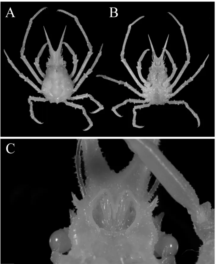
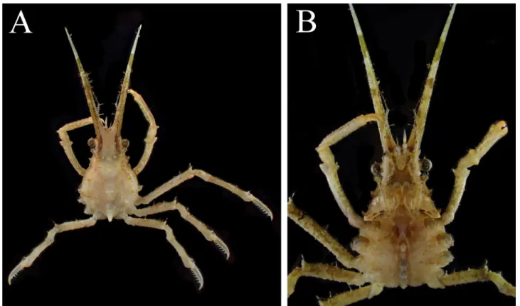
Fig. 8. Hyastenus uncifer Calman, 1909, coloration in life. adolescent male (8.5 × 5.5 mm, RUMF-ZC 5892), Okinawa Island, Okinawa. A, B, dorsal habitus; C, cephalothorax in ventral view.
図 8. ナガツノガニ , 生時の体色 . 最終脱皮前の雄 (8.5 × 5.5 mm, RUMF-ZC 5892), 沖縄島 . A, 全体 , 背面観 ; B, 頭胸部 , 腹面観 .
male and two female specimens from South Timor and Banda with one female syntype.
Rathbun (1911) showed a full-grown male with regenerating left cheliped as ‘type’ of “Halimus
uncifer” in her plate, but the locality of her only
examined material is Diego Garcia, Indian Ocean (Rathbun 1911: 252, pl. 20 fig.7). Based on her photograph, the pseudorostral spines of the ‘type’ male looks much longer than PCL, whereas those of syntype males are “equal to, or a very little shorter than the carapace” (Calman 1909: 712). Consequently, Rathbun’s (1911) ‘type’ seems to be different specimens from the syntypes.
According to Griffin & Tranter (1986), Rathbun’s (1911) specimen differs only in G1 morphology from their specimens and was rather similar to those recorded by Guinot (1962: fig. 16a, b) from Aldabra as “Hyastenus uncifer”. As Griffin & Tranter (1986) suggested, it is possible there is another species closely resembles Hy. uncifer in the western Indian Ocean. Recently Poupin et al. (2018) also recorded many specimens of Hy. uncifer from the Mayotte Island, western Indian Ocean.
The specimens at hand essentially agree with the original description, but the cardiac region of the carapace is weakly elevated in our specimens, whereas it appears to be provided with longitudinally aligned two median protuberances in the drawing of the female syntype of Calman (1909: pl. 72 fig. 8). Griffin & Tranter (1986: 157) reexamined the female syntype and found that the cardiac region was indeed only weakly elevated; Calman’s drawing (1909: pl. 72 fig. 8) seems to be inaccurate at least on this point.
Our specimens also agree well with many sporadic notes by Griffin & Tranter (1986: 13, 134, 157) in many morphological characters, including the G1 structure, but the carapace appears to be slightly different from their drawing. The carapace drawn in Griffin & Tranter (1986: fig. 38i, based on non-type male) looks proportionally shorter than those of our specimens, and the hepatic spines and subacute tubercles on anterolateral subsurface on both sides are noticeably curved anteriorly. However, these differences are probably due to different setting angle of the specimen during observation, because our specimens showed close resemblance with the specimen figured by Griffin & Tranter (1986: fig. 38i) only when their anterior sides were tilted slightly upward (Fig. 7B).
In Japanese water, the first record of H. uncifer was provided in the list of Nomura et al. (1996),
which recorded one individual from 0–10 m depth off Aka Port, Aka Island in the Kerama Group. However, this preliminary report did not include any other information, such as sex, size, and deposition of their specimen. Nomura et al. (1996) noted that “A part of the specimens examined will be deposited in the Natural History Museum and Institute, Chiba [CBM]” but the first author could not locate their specimens in the CBM despite kind support from Dr. T. Komai. The materials examined in the present study are, at present, the only available specimens collected from Japanese waters.
Distribution. Indo-West Pacific: from
Mozambique, Mayotte (Poupin et al. 2018) to Indonesia, Sumatra, Christmas Island (Calman 1909), Banda, South Timor (Griffin & Tranter 1986), and the Ryukyus, southwest Japan (Marumura & Kosaka 2003; this study). Our specimens greatly extend the distribution of the species northward.
Habitat. Sand, coral, at depth of 0–30 m (Griffin
& Tranter 1986; Poupin et al. 2018). One male from Okinawa Island was obtained from mooring rope that was encrusted with some coral species. According to Poupin et al. (2018), the range of habitat depth is “subtidal to 300 m”, but, based on their own examined material, the deepest depth recorded of H.
uncifer was 30 m.
Acknowledgements
We deeply thank Laure Corbari and Paula Martin-Lefèvre (MNHN) for supporting the examination of type specimens. We would like to express our gratitude to Yusuke Yamana and Akira Kosaka (WMNH) for supporting our research of the Nagai Collection, and to Tomoyuki Komai (CBM) for searching for some specimens from the Kerama Group. Special thanks go to Peter K. L. Ng (Lee Kong Chian Natural History Museum, National University of Singapore) for giving some advice during the progress of this study. We also thank Tohru Naruse (University of the Ryukyus) for depositing a specimen in the RUMF, and two anonymous reviewers for their careful reading and many constructive suggestions.
References
Balss, H., 1938. Die Dekapoda Brachyura von Dr. Sixten Bocks Pazifik-Expedition 1917–1918. Göteborges Kungliga Vetenskaps- och Vitterhets-Samhälles Handlingar, Ser. B 5(7): 1–85.
Buitendjiik, A.M., 1939. Biological results of the Snellius Expedition. V. The Dromiacea, Oxystomata, and Oxyrhyncha of the Snellius Expedition. Temminckia, 4: 223–276, pls. 7–11. Calman, R., 1909. On Decapod crustacea from
Christmas Island, collected by Dr. C.W. Andrews, F.R.S., F.Z.S. Proceedings of Zoological Society of London, 1909: 703–713, pl. 72.
Davie, P.F., D. Guinot & P.K.L. Ng, 2015. Chapter 71-2. Anatomy and functional morphology of Brachyura. In: Castro, P., Davie, P.J.F., Guinot, D., Schram, F.R. & von Vaupel Klein, J.C. (eds.), Treatise on Zoology - Anatomy, Taxonomy, Biology. The Crustacea, Volume 9 Part C (2 vols.). Pp. 11–163, Brill, Leiden.
Griffin, D.J.G. & H.A. Tranter, 1986. The Decapoda Brachyura of the Siboga Expedition. Part 8. Majidae. Siboga-Expeditie, Monographie, 39(c4): 1–335.
Guinot, D., 1962. Sur une collection de Crustacés decapodes brachyoures des iles Maldives et de Mer Rouge (Expedition Xarifa 1957–1958). Kieler Meeresforschungen, 18: 231–244, pls. 1–5.
Komatsu, H., 2011. Crabs dredged off the Ogasawara Islands (Crustacea, Decapoda, Brachyura). Memoirs of the National Museum of Nature and Science, Tokyo, 47: 219–277.
Lee, B.Y. & P.K.L. Ng, 2019. On the identity of Hyastenus inermis (Rathbun, 1911), and description of a new species from Sulawesi, Indonesia (Crustacea: Decapoda: Majoidea: Epialtidae). Raffles Bulletin of Zoology, 67: 490– 497.
Lee, B.Y., S.T. Ahyong, M.E.Y. Low & P.K.L. Ng, 2018. Hyastenus verreauxii A. Milne Edwards, 1872, a synonym of Hyastenus elatus Griffin & Tranter, 1986: lectotype designation and reversal of precedence (Crustacea: Brachyura: Majoidea: Epialtidae). Zootaxa, 4497: 145–150.
Man, J.G., De, 1887. Bericht über die von Herrn Dr. J. Brock im indischen Archipel gesammelten Decapoden und Stomatopoden. Archiv für Naturgeschichte, 1: 215–600, pls. 7–22a.
Marumura, M. & A. Kosaka, 2003. Catalogue of brachyuran and anomuran crabs collection donated by the late Mr. Seiji Nagai to the Wakayama Prefectural Museum of Natural History. Wakayama Prefectural Museum of Natural History, Kainan, 74 pp. [in Japanese] Ng, P.K.L., D. Guinot & J.P.F. Davie, 2008. Systema
Brachyurorum: Part I. An annotated checklist
of extant brachyuran crabs of the world. Ruffles Bulletin of Zoology, Supplement, 17: 1–286. Nomura, K., S. Nagai, A. Asakura & T. Komai,
1996. A preliminary list of shallow water decapod Crustacea in the Kerama Group, the Ryukyu Archipelago. Bulletin of Biogeographic Society of Japan, 51: 7–21.
Ohtsuchi, N. & T., Kawamura, 2019. Redescriptions of Pugettia quadridens (De Haan, 1837) and P.
intermedia Sakai, 1938 (Crustacea: Brachyura:
Epialtidae) with description of a new species. Zootaxa, 4672: 1–68.
Ohtsuchi, N., T. Kawamura, J. Hayakawa H. Kurogi & Y. Watanabe, 2016. Growth patterns and population dynamics of the kelp crab Pugettia
vulgaris (Decapoda, Brachyura, Epialtidae) on
the coast of Sagami Bay, Japan. Crustaceana, 89: 645–667.
Ohtsuchi, N., T. Kawamura, J. Hayakawa, H. Kurogi & Y. Watanabe, 2018. Ontogenetic habitat shift in
Pugettia quadridens on the coast of Sagami Bay,
Japan. Fisheries Science, 84: 211–225.
Ohtsuchi, N., H. Komatsu & X. Li, 2020. A new kelp crab species of the genus Pugettia (Crustacea: Decapoda: Brachyura: Epialtidae) from Shandong Peninsula, Northeast China. Species Diversity, 25: 237–250.
Poupin, J., R. Cleva, J.-M. Bouchard, V. Dinhut & J. Dumas, 2018. The Crabs from Mayotte Island (Crustacea, Decapoda, Brachyura). Atoll Research Bulletin, 617: i–vi, 1–109.
Rathbun, M.J., 1911. The Percy Sladen Trust Expedition to the Indian Ocean in 1905, under the leadership of Mr. J. Stanley Gardiner. Volume III. No. XI. Marine Brachyura. The Transactions of the Linnean Society of London, series 2, Zoology 14: 191–261, pls. 15–20.
Sakai, T., 1938. Studies on the Crabs of Japan. III. Brachygnatha, Oxyrhyncha. Yokendo, Tokyo, pp. 193–364, pls. 20–41.
Sakai, T., 1976. Crabs of Japan and the Adjacent Seas. Kodansha, Tokyo, xxix + 773 pp., 3 maps (English volume); 461 pp. (Japanese volume); 16 pp., 251 pls. (plates).
Takeda, M, H. Komatsu, N. Shikatani, T. Maenosono & T. Naruse, 2019. Annotated list of subtidal crabs in the Shikatani Collection made at Nakagusuku Bay, Okinawa Island, the Ryukyu Islands, Japan. Fauna Ryukyuana, 50: 1–69, pls. 1–20. [in Japanese, with English abstract and figure caption]
1966. A method for restoring dried crustacean specimens to taxonomically usable condition. Crustaceana, 10: 109.
Windsor, A. & S.T. Ahyong, 2013. Hyastenus baru, a new species of spider crab from Indonesia (Brachyura: Majoidea: Epialtidae) with a key to the species of Hyastenus. Crustaceana, 86: 718– 727.
Yang, S.L. & A.Y. Dai, 1994. Further notes on the crabs (Brachyura, Crustacea) from Nansha Islands and its adjacent waters. In: The Multidisciplinary Oceanographic Expedition Team of Academia Sinica to Nansha Islands (eds.), Marine Fauna and Flora and Biogeography of the Nansha Islands and Neighbouring Waters, 1. Pp. 125–148, Ocean Press, Beijing, China.
琉球列島から記録されたツノガニ属 ( 十脚目 : 短尾下目 : モガニ科 ) の 2 稀種 大土直哉1・ 武田正倫2・ 芦田晃3 1〒 028-0035 岩手県上閉伊郡大槌町 1-19-8 東京大学大気海洋研究所国際沿岸海洋研究セ ンター 2〒 305-0005 茨城県つくば市天久保 4-1-1 国立科学博物館動物研究部 3〒 464-8601 名古屋市千種区不老町 6 名古 屋大学農学部 要約 . モガニ科 Epialtidae MacLeay, 1838 ツノガ ニ属 Hyastenus White, 1847 のアンボンツノガニ
Hyastenus ambonensis Griffin & Tranter, 1986 の琉
球列島初記録とナガツノガニ H. uncifer Calman, 1909 の沖縄島初記録を報じる . ブロッキツノガ ニ H. brockii (De Man, 1888) は , 和歌山県立自然 博物館所蔵「永井誠二カニ類標本コレクション」 に含まれる日本産唯一の標本が H. ambonensis の誤同定と判明したため , 日本のカニ類相か ら 除 外 さ れ た . ア ン ボ ン ツ ノ ガ ニ と H. baru Windsor & Ahyong, 2013 の判別形質について再 検討した .
投稿日 : 2020 年 2 月 3 日 受理日 : 2020 年 11 月 3 日 発行日 : 2020 年 12 月 4 日