CLINICAL OUTCOMES IN LUNG CANCER PATIENTS FROM 2009 TO 2013 IN AKITA RED CROSS HOSPITAL
Ryo Morita1), Hirokazu Kurokawa1), Mariko Asano2), Yuji Okuda2), Hidesato Odaka1), Hideki Kawai3), Kazuki Miura4) and Hiroshi Ito2)
(received 1 May 2014, accepted 11 August 2014)
1)Department of Respiratory Medicine, Akita Red Cross Hospital, Japan.
222-1, Nawashirosawa, Saruta, Kamikitate, Akita, 010-1406, Japan
2)Department of Cardiovascular and Respiratory Medicine, Akita University Graduate School of Medicine, Akita, Japan.
Hondo 1-1-1, Akita, 010-8543, Japan.
3)Department of Thoracic Surgery Akita Red Cross Hospital, Japan.
222-1, Nawashirosawa, Saruta, Kamikitate, Akita, 010-1406, Japan
4)Department of Respiratory Medicine, Sotoasahikawa Hospital, Japan.
142, Migota, Sotoasahikawa, Akita, 010-0802, Japan
Abstract
Background: The prevalence of deaths due to malignant tumors in Akita Prefecture was the highest in all of Japan from 1998 to 2013. In 2012, lung cancer caused the greatest number of cancer-related deaths in Akita Prefecture.
Purpose: To investigate the clinical outcomes of patients with lung cancer treated at Akita Red Cross Hospital.
Methods: Patients diagnosed with lung cancer for the first time and treated at Akita Red Cross Hospital from January 2009 to December 2013 were enrolled, and their clinicopathological factors were assessed.
Results and Conclusions: In total, 493 patients were enrolled (328 men, 165 women ; median age=70 years). There were 318 patients with adenocarcinomas, 104 with squamous cell carcino- mas, 12 with adenosquamous cell carcinomas, seven with large-cell carcinomas, and 52 with small-cell carcinomas. Of the total patients, 237 underwent surgical treatment, and their 5-year survival rate was 66.5%. The remaining 256 patients received non-surgical treatment, and their median overall survival was 14 months. The reasoning behind the chemotherapy treatment strategy decisions were largely unclear. These findings highlight the need to develop a clear treatment strategy with regard to relevant objective factors.
Key words : Lung Cancer, Clinical Outcome, Chemotherapy
Address for correspondence : Ryo Morita, MD
Department of Respiratory Medicine, Akita Red Cross Hospital, Japan.
222-1, Nawashirosawa, Saruta, Kamikitate, Akita, 010- 1406, Japan
TEL : 81-18-829-5000 FAX : 81-18-829-5255
E-mail : [email protected]
Introduction
Recently, malignant tumors have become the most common cause of death in Japan1). The situation is simi- lar in Akita Prefecture, where death due to malignant tu- mors has increased every year and peaked from 1998 to 2013. Until 2011, gastric cancer was the most common cause of deaths in Akita Prefecture ; in 2012, however, lung cancer caused the greatest number of deaths2). In-
Clinical outcomes for lung cancer patients (46)
deed, despite surgical resection and the development of new chemotherapy regimens, many lung cancer patients suffer relapses and die3).
In addition, recently patients with lung cancer have been treated differentially according to their histological subtype and/or mutation status. This change has been associated with the development of new drugs for lung cancer, particularly those targeting molecules4). Medical guidelines for lung cancer in Japan now recommend that physicians treat based on the histological subtype and mutation status of epidermal growth factor receptor (EGFR) and translocations involving genes encoding the echinoderm microtubule-associated protein-like 4 and anaplastic lymphoma receptor tyrosine kinase (EML4- ALK)5). Moreover, targeted therapies for mutations of other driver genes, including ROS1, RET, and HER2, are in development6).
The recommended regimens vary according to age and the Eastern Cooperative Oncology Group performance status (ECOG PS). The treatments for lung cancer will further diversify in the future. In this study, we investi- gated the clinical outcomes in lung cancer patients at Akita Red Cross Hospital from 2009 to 2013 in an effort to improve the treatment of lung cancer.
Materials and Methods
Patients diagnosed with lung cancer for the first time and treated at Akita Red Cross Hospital from January 2009 to December 2013 were enrolled in this study. Be- fore treatment, we determined the age, gender, treat- ment received, side of the primary lesion, histological type, driver mutation, and clinical stage of all pa- tients. After the patients underwent surgery, we deter- mined the pathological stage and measured overall sur- vival. Overall survival was also measured in patients who received non-surgical treatments.
We categorized the lung cancer patients who received chemotherapy and/or radiotherapy by histological type. We determined the regimens and measured over- all survival.
All statistical analyses were performed using the JMP software (ver. 8.0 for Windows). Categorical data were compared using Fisher’s exact probability test. Contin-
uous variable data were compared using t-tests between each factor. Correlations of treatment and histological type with overall survival were determined using the Ka- plan-Meier method and the log-rank test. Hazard ratios were determined using the Cox proportional hazards model. P values <0.05 were considered to indicate sta- tistical significance.
Results
In total, 493 patients (328 men, 165 women) with lung cancer were enrolled in the study. Patient characteris- tics are listed in Table 1. The patients had a median age of 70 (range, 31-95) years. In total, 227 patients under- went surgery and 266 patients received non-surgical treatments ; 265 patients had a primary lesion in the right lung and 228 in the left lung.
Regarding cancer type, 318 patients had adenocarcino- mas, 104 had squamous cell carcinomas, 12 had adeno- squamous cell carcinomas, 7 had large-cell carcinomas, and 52 had small-cell carcinomas. Regarding driver mu- tations, 98 patients had EGFR mutations and 6 had EML4-ALK fusion genes.
According to clinical stage, 132 patients were stage IA, 48 were stage IB, 27 were stage IIA, 13 were stage IIB, 43 were stage IIIA, 31 were stage IIIB, and 199 were stage IV. These classifications were made according to the sixth and seventh editions of the General Rules for Clinical and Pathological Records of Lung Cancer.
We measured overall survival according to clinical stage (Fig. 1). Stages IA and IB were not defined. The 5-year survival rate was 81% in patients with stage IA and 56% in patients with stage IB cancer (stage IIA, 38 months ; stage IIB, 32 months ; stage IIIA, 31 months ; stage IIIB, 11 months ; and stage IV, 7 months).
We categorized the lung cancer patients into two groups according to treatment. Patient characteristics are listed in Table 2. We assessed the age, gender, side of the primary lesion, histological type, driver mutation, and clinical or pathological stage in the patients. No sig- nificant difference was seen between the surgical and non-surgical treatment groups in terms of age, gender, side of the primary lesion, or driver mutation.
In the surgical treatment group, there were 139 men
and 98 women, with a median age of 69 (range, 37-87) years. In the non-surgical treatment group, there were 189 men and 67 women, with a median age of 72 (range, 31-95) years.
In the surgical treatment group, 131 patients had a pri- mary lesion in the right lung and 106 in the left lung. In the non-surgical treatment group, 134 patients had a pri- mary lesion in the right lung and 122 in the left lung.
In the surgical treatment group, 174 patients had ade- nocarcinomas, 45 had squamous cell carcinomas, 12 had adenosquamous cell carcinomas, and 6 had large-cell car- cinomas. None of the patients in the surgical treatment group had a small-cell carcinoma. In the non-surgical Table 1. Baseline characteristics of lung cancer patients in our hospital
All patients (n=493)
No. Patients (%)
Age, median (range) 70 (31-95)
Gender
male 328 (66.5)
female 165 (33.5)
Treatment
surgery 227 (46.0)
non-surgery 266 (54.0)
Side of primary lesion
Right lung 265 (53.8)
Left lung 228 (46.2)
Histologic Type
Adeno carcinoma 318 (64.3)
Squamous cell 104 (21.3)
Adenosquamous 12 (2.4)
Large cell 7 (1.4)
Small cell 52 (10.6)
Driver Mutation
EGFR 110 (22.3)
EML4-ALK 6 (1.2) Crinical / Pathologic Stage
IA 132 (26.8)
IB 48 (9.7)
IIA 27 (5.5)
IIB 13 (2.6)
IIIA 43 (8.7)
IIIB 31 (6.3)
IV 199 (40.4)
Fig.1
Overall survival (month)
Probability (%)
IA
IB IIA
IIB IIIA
IIIB IV
Fig. 1 Kaplan-Meier estimates of survival by clinical stage of lung cancer. The 5-year survival rate was 81% in patients with stage IA and 56% in patients with stage IB cancer (stage IIA, 38 months ; stage IIB, 32 months ; stage IIIA, 31 months ; stage IIIB, 11 months ; and stage IV, 7 months).
Clinical outcomes for lung cancer patients
treatment group, 144 patients had adenocarcinomas, 59 had squamous cell carcinomas, 1 had a large-cell carcino- ma, and 52 had small-cell carcinomas. None of the pa- tients in the non-surgical treatment group had an adeno- squamous cell carcinoma. Thus, patients with adenosquamous cell carcinoma were diagnosed only by surgery.
In the surgical and non-surgical treatment groups, 54 and 44 patients had EGFR mutations, respectively, and 3 from each group had EML4-ALK fusion genes.
We measured pathological stage after surgery in the surgical treatment group : 129 patients were stage IA, 47 were stage IB, 23 were stage IIA, 12 were stage IIB, and 26 were stage IIIA. We also measured the clinical stage
prior to non-surgical treatment : 3 patients were stage IA, 1 was stage IB, 4 were stage IIA, 1 was stage IIB, 17 were stage IIIA, and 31 were stage IV.
We also measured overall survival according to treat- ment group (Fig. 2). The patients who underwent sur- gery showed significantly longer overall survival than those who received non-surgical treatments (p<0.001, HR=0.084, 95% CI=0.059-0.119). In the surgical treat- ment group, the 5-year survival rate was 66.5%, but the median survival time had not yet been reached. In the non-surgical treatment group, the median survival time was 14 months.
We also categorized patients by histological type and assessed which anticancer drugs each patient had been (48)
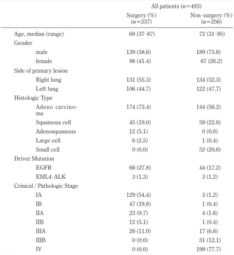
Table 2. Correlation between clinicopathological features and treatment for lung cancer All patients (n=493) Surgery (%)
(n=237) Non-surgery (%)
(n=256)
Age, median (range) 69 (37-87) 72 (31-95)
Gender
male 139 (58.6) 189 (73.8)
female 98 (41.4) 67 (26.2)
Side of primary lesion
Right lung 131 (55.3) 134 (52.3)
Left lung 106 (44.7) 122 (47.7)
Histologic Type Adeno carcino-
ma 174 (73.4) 144 (56.2)
Squamous cell 45 (19.0) 59 (22.8)
Adenosquamous 12 (5.1) 0 (0.0)
Large cell 6 (2.5) 1 (0.4)
Small cell 0 (0.0) 52 (20.6)
Driver Mutation
EGFR 66 (27.8) 44 (17.2)
EML4-ALK 3 (1.3) 3 (1.2) Crinical / Pathologic Stage
IA 129 (54.4) 3 (1.2)
IB 47 (19.8) 1 (0.4)
IIA 23 (9.7) 4 (1.6)
IIB 12 (5.1) 1 (0.4)
IIIA 26 (11.0) 17 (6.6)
IIIB 0 (0.0) 31 (12.1) IV 0 (0.0) 199 (77.7)
administered for first-line chemotherapy listed in Table 3. Among patients with adenocarcinomas, 3 were treat- ed with carboplatin, paclitaxel, and bevacizumab ; 13 with cisplatin and pemetrexed ; 15 with carboplatin and pemetrexed ; 1 with carboplatin and paclitaxel ; 10 with cisplatin and docetaxel ; and 5 with gemcitabine. Gefi- tinib was used in 41 patients with adenocarcinomas who had EGFR mutations, and crizotinib was used in 3 pa- tients with EML4-ALK fusion genes. All patients with driver mutations were treated with targeted therapy as
first-line chemotherapy. The overall survival of the pa- tients who received gefitinib was 19 months. Six pa- tients were treated with radiotherapy alone, and 37 pa- tients chose best supportive care. In patients with squamous cell carcinomas, 18 were treated with carbopla- tin and paclitaxel, 4 with cisplatin and docetaxel, 2 with carboplatin and gemcitabine, 12 with radiotherapy alone, and 21 with best supportive care. In patients with small-cell carcinomas, 12 were treated with cisplatin and etoposide, 11 with carboplatin and etoposide, 8 with cis- platin and irinotecan, 9 with carboplatin and irinotecan, 3 with radiotherapy alone, and 21 with best supportive care.
Discussion
Recently, the number of lung cancer patients has in- creased, and lung cancer has become the most common cause of death in Japan2). This increase has led to im- provements in diagnostic methods and chemotherapeutic regimens for lung cancer.
A previous study reported that 437 patients were diag- nosed with lung cancer in Akita Prefecture in 2002. Of
Probability (%)
Overall survival (month)
Fig. 2 Kaplan-Meier estimates of survival by treat- ment. The median duration of survival for the group non-surgery was 14 months. Corresponding to a haz- ard ratio for death of 0.084 (p<0.0001).
Table 3. Correlation between non-surgical treatment and histlogical type Adenocarcinoma n=144 (%) Small Cell Carcinoma n=52 (%)
CBDCA + PAC + BEV 3 (2.1) CDDP + VP-16 12 (23.1)
CDDP + PEM 13 (9.0) CBDCA + VP-16 11 (21.2)
CBDCA + PEM 15 (10.5) CDDP + CPT-11 8 (15.4)
CBDCA + PAC 1 (0.7) CBDCA + CPT-11 9 (17.3)
CDDP + DTX 10 (7.0) CPT-11 2 (3.8)
CBDCA + GEM 5 (3.5) Radiotherapy 3 (5.7)
Gefitinib 41 (28.5) BSC 7 (13.5)
Crizotinib 3 (2.1)
Others 9 (6.3) Squamous Cell Carcinoma n=59 (%)
Radiotherapy 6 (4.2) CBDCA + PAC 18 (30.5)
BSC 37 (26.1) CDDP + DTX 4 (6.8)
CBDCA + GEM 2 (3.4)
Large Cell Carcinoma n=1 (%) Others 2 (3.4)
CDDP + DTX 1 (100) Radiotherapy 12 (13.6)
BSC 21 (42.3)
CDDP Cisplatin, CBDCA Carboplatin, VP-16 Etoposide, CPT-11 Irinotecan
PAC Paclitaxel, DTX Docetaxel, PEM Pemetrexed, BEV Bevacizumab, GEM Gemcitabine BSC Best Supportive Care
Clinical outcomes for lung cancer patients
these patients, 35% received surgical treatment and 65%
received chemotherapy and/or radiotherapy7). In our study, 46% of the patients received surgical treat- ment. Thus, the proportion of patients who underwent surgical resection was increased compared with the pre- vious report. In 2002, according to the same report, 42% of patients with lung cancer were classified as hav- ing stage I, II, or IIIA disease7).
The number of early-stage patients was increased compared with the 2002 report. One factor contributing to this is improvements in CT technology, which today provides highly detailed, abnormal shadows, allowing for early detection. When we observe abnormal shadows, particularly ground-glass opacities (GGOs), we follow up with reference to the guidelines for the management of pulmonary nodules every few months8). Those guide- lines state that any pulmonary nodules greater than 5 mm detected on the first CT scans should be classified as solid, part-solid, or pure ground-glass nodules (GGNs). It is recommended that a definite diagnosis be made when a solid nodule measures more than 10 mm in maximal diameter on a CT scan. When the maximal di- ameter of a solid nodule on CT is in the >5- but <10- mm range and the patient is a smoker, follow-up CT ex- aminations are performed after 3, 6, 12, 18, and 24 months. If the patient is a non-smoker, follow-up CT examinations are performed at 4, 12, and 24 months8). The reason for the different follow-up examination inter- vals between smokers and non-smokers is that the lung cancer doubling time is shorter in smokers9). Part-solid nodules have a high probability of becoming malignant tumors10). However, inflammatory lesions also look like part-solid nodules ; therefore, CT is performed 3 months later to exclude inflammatory lesions. If a pure GGN is 15 mm or larger in maximal diameter on CT scans, then a definitive diagnosis should be made. If a pure GGN is less than 15 mm in maximal diameter, follow-up CT is performed at 3, 12, and 24 months8).
The Japanese Lung Cancer Society recommends surgi- cal treatment for patients in clinical stages I, II, and/or IIIA5). The 5-year survival rate for patients with lung cancer undergoing surgical treatment was 66.5% in this study. In general, in lung cancer cases undergoing sur- gery in Japan, the percentages of female patients, adeno-
carcinomas, small tumors, and aged patients have in- creased, whereas the rate of surgery-related deaths has decreased. With such changes, the 5-year survival rate for all lung cancer patients who underwent surgery was 69.6% in 200411). Moreover, the 5-year survival rates were : stage IA, 82.0% ; stage IB, 66.1% ; stage IIA, 54.5% ; and stage IIB, 46.1%11). In our study, the 5-year survival rates were : stage IA, 81.2% ; stage IB, 56.6% ; stage IIA, 35.4% ; and stage IIB, 20.8%. The reason for the slightly lower (~3%) 5-year survival rate in this study may be the high number of elderly sub- jects. The Japanese lung cancer registry study con- tained 11,663 patients, whose mean age was 66.7 years11). In our study, the median age of the 237 patients who underwent surgical treatment was 68.5 years. A previous study suggested that the 5-year survival rate for older patients was worse than that for younger pa- tients12). However, it was also reported that the 5-year survival rate for elderly patients has improved in gener- al13). It seems likely that the number of elderly lung cancer patients will continue to increase in the fu- ture. We must adapt our surgical approach for elderly people and provide careful follow-up treatment.
From 2009 to 2013, 256 patients received non-surgical therapy ; of these, 65 received palliative therapy only and 191 received chemotherapy and/or radiotherapy. It is recommended that a treatment strategy be selected with regard to the ECOG-PS, patient age and histologic type of disease5). Among patients with a PS of 0 or 1, the treatment strategy was different for those patients 75 years old or older. Patients with a PS of 2 often received chemotherapy with a single agent. Patients with a PS of 3 or 4 received best supportive care. However, the ap- plied treatment strategy was different for patients with a driver mutation. Patients with an adenocarcinoma and an EGFR mutation who were classified as ECOG-PS 3 or 4 received targeted molecular therapy14). However, careful follow up is necessary to prevent the onset of side effects for cases with a poor PS15,16).
All patients diagnosed at our hospital were examined for genetic mutations. For all lung cancer patients with EGFR mutations, we used EGFR tyrosine kinase inhibi- tors (EGFR-TKIs) as first-line chemotherapy in our hos- pital from 2009 to 2013. PFS can be extended by the (50)
use of EGFR-TKIs during primary therapy17). However, in deciding when to use EGFR-TKIs for patient treat- ment, there is some discussion about whether combina- tion therapy with chemotherapy and EGFR-TKIs or monotherapy with an EGFR-TKI is more appropri- ate. In the NEJ005 clinical trial, the North East Japan Study Group examined whether concurrent or sequential therapy with chemotherapy and an EGFR-TKI was more effective. Presently, combination therapy with chemo- therapy and an EGFR-TKI and monotherapy with an EGFR-TKI are being examined in the NEJ009 clinical trial.
In recent years, tumor angiogenesis has come to be viewed as important in lung cancer proliferation and me- tastasis18). Overall survival is extended when an angio- genesis inhibitor is combined with conventional chemo- therapy19). The most common chemotherapeutic regimens for adenocarcinomas include pemetrexed. Re- cently, maintenance therapy using pemetrexed has been recommended. Paz-Arez et al.20) showed that overall survival was extended in patients receiving maintenance therapy. However, no clear policy has yet been made in our hospital as to whether maintenance therapy should be provided or whether angiogenesis inhibitors should be used.
It is recommended to select a treatment strategy with regard to the patient’s ECOG-PS, age and smoking histo- ry5). Because we referred to past hospitalization medi- cal records in this study, we did not examine the ECOG- PS or smoking history of the patients, which is a limitation of our study. It is necessary for us to reach a clear policy on drug regimens for lung cancer patients with reference to objective factors, including the ECOG- PS, age, histological type of disease, mutations and smok- ing history.
Conclusions
In conclusion, 493 patients (328 men and 165 women, median age=70 years) with lung cancer were enrolled in this study from 2009 to 2013. Those patients who un- derwent surgical treatment showed significantly longer overall survival than those who received non-surgical treatment. Indeed, their 5-year survival rate was
66.5%. In total, 255 patients received non-surgical treatment. However, the reasoning behind the chemo- therapy treatment strategy decisions was unclear. We must develop a clear treatment strategy with regard to various objective factors, including the ECOG-PS and age of the patient.
References
1) Health, Labour and Welfare Statistics Association, Japan. Journal of health and welfare statistics 2013.
2) Annual Health Statistics of Akita 2013.
3) Gridelli, C., Rossi, A. and Maione, P. (2003) Treat- ment of non small cell lung cancer state of the art and development of new biologic agent. Oncogene, 22, 6629-6638.
4) Yatabe, Y. (2013) Recent changes in the therapeutic strategy for NSCLC in association with new anti- cancer agents. Rinsho Byori, 61, 328-333.
5) The Japan Lung Cancer Society, Medical guidelines for lung cancer 2012.
6) Mitsudomi, T. (2013) Driver gene mutation and tar- get therapy of lung cancer. Jpn. J. Cancer Che- mother., 40, 285-290.
7) Sasaki, M., Miura, M., Watarai, J., et al. (2006) A statistical analysis of lung cancer in Akita Prefecture in 2002. Akita Med. J., 56, 73-81.
8) The Japanese Society of CT Screening. (2013) Guidelines for the management of pulmonary nodules detected by low-dose CT lung cancer screening version 3.
9) Hasegawa, M., Sone, S., Takashima, S., Li, F., Yang, Z.G., Maruyama, Y. and Watanabe, T. (2000) Growth rate of small lung cancers detected on mass CT screening. Brit. J. Radiol., 73, 1252-1259.
10) Li, F., Sone, S., Abe, H., Macmahon, H. and Doi, K.
(2004) Malignant versus benign nodules at CT screening for lung cancer : comparison of thin-sec- tion CT findings. Radiology, 233, 793-798.
11) Sawabata, N., Miyaoka, E., Asamura, H., et al. (2011) Japanese lung cancer registry study of 11,663 surgi- cal cases in 2004. J. Thorac. Oncol., 6, 1229-1235.
12) Asamura, H., Goya, T., Koshiishi, Y., et al. (2008) Japanese Joint Committee of lung cancer registry. A Japanese lung cancer registry study : prognosis of 13,010 resected lung cancers. J. Thorac. Oncol., 3,
Clinical outcomes for lung cancer patients
46-52.
13) Koike, T., Yamato, Y., Asamura, H., et al. (2009) Improvements in surgical results for lung cancer from 1989 to 1999 in Japan. J. Thorac. Oncol., 4, 1364-1369.
14) Inoue, A., Kobayashi, K., Usui, K., et al. (2009) First-line gefitinib for patients with advance non- small-cell lung cancer harboring epidermal growth factor receptor mutations without indication chemo- therapy. J. Clin. Oncol., 271, 394-400.
15) Ando, M., Okamoto, I., Yamamoto, N., et al. (2006) Predictive factors for interstitial lung disease, antitu- mor response, and survival in non-small-cell lung cancer patients treated with gefitinib. J. Clin. Ocol., 24, 2549-2556.
16) Kudoh, S., Kato, H., Nishiwaki, Y., et al. (2008) Interstitial lung disease in Japanese patients with lung cancer : a cohort and nested case-control study.
Am. J. Respir. Crit. Care Med., 177, 1348-1357.
17) Maemondo, M., Inoue, A., Kobayashi, K., et al. (2010) Gefitinib or chemotherapy for non-small cell lung cancer with mutated EGFR. N. Engl. J. Med., 362, 2380-2388.
18) Sandler, A (2007) Bevacizumab in non-small cell lung cancer. Clin. Cancer Res., 13, 4613-4616.
19) Sandler, A., Gray, R., Perry, MC., Brahmer, J., Schil- ler, JH., Dowlati, A., Lilenbaum, R. and Jounson, DH. Paclitaxel-carboplatin alone or with bevaci- zumab for non-small-cell lung cancer. N. Engl. J.
Med., 355, 2542-2550.
20) Paz-Arez, L., de Marinis, F., Dediu M., et al. (2012) Maintenance therapy with pemetrexed plus best supportive care versus placebo plus best supportive care after induction therapy with pemetrexed plus cisplatin for advanced non-squamous non-small-cell lung cancer (PARAMOUNT) : a double-blind, phase 3, randomised controlled trial. Lancet. Oncol., 13, 247-255.
(52)