Association of clinical and radiographic signs of femoroacetabular impingement in the general population
Ryota Yamauchi1, MD, Ryo Inoue1, MD, PhD, Daisuke Chiba1,2, MD, PhD, Yuji Yamamoto1, MD, PhD, Yoshifumi Harada1, MD, PhD, Ippei Takahashi2, MD, PhD, Shigeyuki Nakaji2, MD, PhD, Yasuyuki Ishibashi1, MD, PhD,
1Department of Orthopaedic Surgery, Hirosaki University Graduate School of Medicine
2Department of Social Medicine, Hirosaki University Graduate School of Medicine
Each author certifies that he or she has no commercial associations that might pose a conflict of interest in connection with the submitted article.
This study was approved by the ethics committee of the the Hirosaki University School of Medicine, and all participants gave their written informed consent to participate.
Ryota Yamauchi, MD
Department of Orthopaedic Surgery, Hirosaki University Graduate School of Medicine,
5 Zaifu-cho, Hirosaki, 036-8562 Aomori, Japan
Phone: +81-172-39-5083 Fax: +81-172-36-3826
E-mail: [email protected]
ABSTRACT
Background
The relationships between the clinical and radiographic signs of femoroacetabular impingement (FAI) are unknown. The purpose of this study was to assess the relationship between hip pain, a positive anterior impingement sign (AIS), and radiographic signs of FAI in a general Japanese population.
Methods
A total of 616 individuals participated in this study. Hip pain was assessed using the Japanese Orthopaedic Association Hip-Disease Evaluation Questionnaire (JHEQ) pain category score and the AIS was used as a provocation test. Participants were divided into a positive AIS group (at least one positive hip) and a negative AIS group. Radiographs were assessed for the cross-over sign (COS), ischial spine sign (ISS), posterior wall sign (PWS), and pistol grip deformity (PGD). Then, the relationships between the clinical and radiographic signs of FAI were evaluated.
Results
JHEQ pain scores did not differ between men and women. Seven men (3.4%) and 29 women (7.1%) had a least one hip with a positive AIS. The mean JHEQ pain scores were 22.9 ± 7.2 for the positive and 27.3 ± 2.2 for the negative AIS group (P < 0.01). The prevalences of COS, ISS, PWS, and PGD were 8.9%, 17.2%, 21.8%, and 1.9%, respectively. There were no significant associations between degree of hip
pain, AIS, and each radiographic finding.
Conclusions
Radiographic signs of FAI were not associated with the degree of hip pain or a positive AIS, which suggests that radiographic findings may not be important in the clinical diagnosis of FAI.
Introduction
Femoroacetabular impingement (FAI) was described by Ganz et al. in 2003 [1] and is considered a risk factor for hip osteoarthritis [2]. FAI is strongly suspected in patients with groin discomfort or pain and limited hip motion, particularly during internal rotation and flexion [1]. Generally, other causes of hip pain such as hip dysplasia, rheumatoid arthritis, avascular necrosis of the femoral head, and fractures around the hip joint can be excluded. However, diagnosis of FAI can be difficult because its symptoms are nonspecific and the clinical signs of FAI can resemble those of other hip disorders [3]. The acetabular labral tears that result from FAI are caused by abnormal contact between the proximal femur and acetabular rim. Two types of impingement have been described: anterior acetabular overcoverage (pincer type) and deformity of the femoral head-neck junction (cam type). Many patients suffer from a combination of both forms of impingement (mixed type), and there are few patients who have purely one type of impingement [4].
Several provocation tests for FAI, including the anterior impingement sign (AIS), Faber test, resisted straight leg raise test, and posteroinferior impingement sign, have been described [1, 3]. However, these tests alone are not specific for FAI and radiographic examination is needed to make a definitive diagnosis. Radiographic signs of pincer type impingement include the crossover sign (COS) and ischial spine sign (ISS) when focal anterior acetabular overcoverage is present, while the posterior wall
sign (PWS) indicates focal posterior acetabular overcoverage [5-7]. However, these radiographic signs can be affected by pelvic tilt and rotation, and Siebenrock et al.
reported a high false-positive rate associated with their assessment [5]. Cam type impingement is characterized by pistol grip deformity (PGD), which Gosvig reported may be underestimated based on anteroposterior radiographs of the hip [7].
A diagnosis of FAI requires comprehensive assessment of both clinical examination and radiographic findings. However, few studies have examined the relationships between the AIS provocation test the various radiographic findings associated with FAI in a general population. The purpose of this study was to investigate the correlation between degree of hip pain and positive AIS, and the associations of these clinical signs with radiographic findings in a general population of Japanese people.
Methods Participants
A total of 1167 volunteers participated in the Health Promotion Project in 2014. This annual project has been ongoing since 2005 in a rural region, Japan. It is a community-based program aimed at improving average life expectancy through the regular performance of general health checkups for the population. All participants provided written informed consent, and the study was conducted with the approval of
the ethics committee [8, 9].
All participants filled out questionnaires and underwent physical examinations and radiographic studies of the pelvis. Exclusion criteria for this study were radiographic evidence of excessive pelvic rotation as assessed by asymmetry of the obturator foramina, history of hip joint surgery, hip osteoarthritis (Kellgren-Lawrence ≧ grade 2), and unevaluable radiographs [10]. A total of 616 participants (207 males and 409 females) with a mean (± SD : standard deviation) age of 53.3 ± 14.8 years (range: 19–87 years) were included in this study.
Hip Symptoms
All participants answered the questions in the pain category of the Japanese Orthopaedic Association Hip-Disease Evaluation Questionnaire (JHEQ) [11]. JHEQ is a self-administered questionnaire that is useful for evaluating quality of life in patients with hip joint disease. The pain category consists of a visual analog scale (VAS) and six questions (Table 1). The length of the VAS is divided into five segments and is worth 0–4 points as follows: Up to 20 mm = 4 points, Between 21 mm and 40 mm= 3 points, Between 41 mm and 60 mm = 2 points, Between 61 mm and 80 mm = 1 point, More than 80 mm = 0 points. Each of the six questions has five response categories: “strongly agree”, “agree”, “uncertain”, “disagree”, and “strongly disagree”. Each response was worth 0–4 points, in increasing order starting from
‘‘strongly agree.’’ The total points (0–28 points) were considered indicative of the
degree of hip pain, and higher scores indicated less pain. In this study, we used this JHEQ pain category scores as the evaluation of their degree of hip pain. The previous study indicated that the JHEQ pain subscale correlated with the pain of the Japanese Orthopaedic Association (JOA) hip scores, the short-form 36 health survey (SF-36) bodily pain subscale and the Oxford hip score (OHS) pain subscale [12].
Physical Examination
The AIS was assessed in all patients and was considered positive if groin pain could be reliably reproduced. The participants were divided into two groups based on their AIS results: a positive AIS group (positive AIS in at least one hip joint) and a negative AIS group (negative AIS in both hip joints). Hip range of motion (ROM) (flexion, abduction, internal rotation, and external rotation) was measured with the participants in a supine position. All examinations were performed once by two orthopaedic surgeons (RY, YY).
Radiographic Assessment
Anterior-posterior radiographs of the pelvis were assessed for the COS, ISS, PWS, PGD, center-edge angle (CEA) and acetabular edge angle (AEA). The COS was considered present when the anterior rim line was lateral to the posterior rim in the cranial part of the acetabulum and crossed the rim to become medial in the distal part of the acetabulum (Figure.1-A) [6]. The ISS was defined as when the ischial spine was the most evident prominence along the ilioischial line (Figure.1-B) [7]. The PWS was
defined as when the posterior wall of the acetabulum was lateral to the center of the femoral head (Figure.1-C) [6]. The PGD was defined as a prominent lateral offset of the femoral head-neck junction (Figure.1-D) [2]. The CEA and AEA were measured in all patients. All radiological assessments were performed by a single observer (RY).
The radiographs were reviewed again 6 months after the first assessments by the same observer.
Statistical analysis
Statistical analyses were performed using SPSS ver. 12.0J (SPSS Inc., Chicago, IL, USA). The ages, JHEQ pain category scores, hip ROMs, CEA and AEA of participants in the positive and negative AIS groups were compared using Mann-Whitney U tests. Radiographic findings (COS, ISS, PWS, and PGD) were compared between the positive and negative AIS groups using Chi-square tests.
Multiple regression analysis was performed with the JHEQ pain category score as the independent variable, and age, sex, hip ROM, AIS results, and radiographic findings (COS, ISS, PWS, PDG, CEA and AEA results) as dependent variables. In addition, logistic regression analysis was performed with the presence of AIS as independent variable, and age, sex, and radiographic findings as dependent variables. In all analyses, P-values < 0.05 were considered significant.
Results
Hip Symptoms
The mean total JHEQ pain category scores did not differ between the sexes (27.2 ± 2.9 and 27.0 ± 3.0 for men and women, respectively; p = 0.089; Table 2). The mean JHEQ pain category scores of the positive and negative AIS groups were 22.9 ± 7.2 and 27.3 ± 2.2, respectively (P < 0.01; Table 3).
Physical Examination
There were 7 men (3.4%) and 29 women (7.1%) with at least one AIS positive hip and no difference between the sexes (p = 0.064; Table 2). All examinations of Hip ROM did not significantly differ between the positive and negative AIS groups (Table 3).
Radiographic Assessment
Radiographic findings shows Table 2. The mean CEA for women was significantly smaller than that for men (p < 0.01). Of the 36 participants in the positive AIS group, a few had radiographic findings typical of pincer type FAI; however, none had a positive COS, one participant (2.9%) had a positive ISS, three participants (8.3%) had a positive PWS, and 1 participant (2.9%) had a positive PGD (Table 3).
The relationship between symptoms and radiographic findings
Multiple regression analysis revealed that the JHEQ pain category score was were significantly negatively associated with the mean age (P = 0.008) and having a positive AIS (P < 0.001). There were no significant associations with sex, hip
ROM, or radiographic signs (Table 4).
The relationship between AIS and radiographic findings
Logistic regression analysis was performed to estimate the association of the presence of AIS with radiographic findings. The results indicated that a positive AIS was not associated with radiographic findings of FAI (Table 5).
Discussion
Our results showed no association of radiographic signs of FAI with degree of hip pain and AIS. This suggests that radiographic signs may not be important for the clinical diagnosis of FAI. As far as we know, this is the first study to investigate the association of radiographic signs of FAI with hip pain and provocation test results in a general population.
In the general population represented by our study participants, 3.4% of men and 7.1% of women had a positive AIS. This sign has been reported to be highly specific and to be the test best able to diagnose acetabular labral tears [13]. However, few studies have reported the proportion of the general population with a positive AIS.
Lene et al. did report that the estimated prevalence of anterior impingement in 19 year olds was 7.3% in men and 4.8% in women [14]. In our study, more women than men had a positive AIS; this may be because the pincer type of FAI occurs more commonly in middle-aged women [15].
In our radiographic assessment of FAI signs, 55 hips (8.9%) showed a COS, 106 hips (17.2%) showed an ISS, 134 hips (21.8%) had a PWS, and 12 hips (1.9%) had a PGD. Fujii et al. reported that seven (18%) of the 96 hips in a hip dysplasia group showed the COS [16], and Ezoe et al. found the COS in seven (6%) and a positive PWS in five (5%) of 112 hips in a normal hip group and a positive COS in 13 (18%) and a positive PWS 13 (18%) of the 74 hips in a hip dysplasia group [17]. The positive rate for radiographic signs of FAI is greatly variable among studies because of the different participant populations in each study. However, there have been few reports of the prevalence rate of radiographic findings suggesting pincer-type FAI in the general population.
This study showed no association between radiographic signs of FAI and physical examination findings consistent with FAI, such as a positive AIS and reduced hip ROM. Ranawat et al. reported a poor correlation between pain and radiographic signs of FAI, and patients with greater coxa profunda had less pain [18]. Lene et al.
also reported that radiographic findings of pincer type FAI were not associated with a positive impingement test in young adults [14]. Our results were similar; in our study, almost no patients with a positive AIS had a concurrent COS, PWS, ISS, or any radiographic signs of pincer-type FAI. Clohisy et al. reported reduced internal rotation and hip flexion in symptomatic FAI patients [19]; however, we did not find a significant difference in hip ROM between the positive and negative AIS groups. A
reason for this may be that, in our study population, even when the AIS was positive the FAI was not severe enough to reduce hip ROM.
It is said that the AIS is also described to have labral disorder with hip dysplasia [20]. In this study, the difference was not statistically significant between the positive and negative AIS groups in the CEA and the AEA which was used as the parameter of hip dysplasia. In addition, the JHEQ pain category was also not associated with the CEA and the AEA. However, the number of patients in dysplastic hips was small in this study, and further research will be necessary about the relationship between the AIS and hip dysplasia in a general Japanese population.
This study had several limitations. First, all of the participants in this study were Japanese, and the study was performed in a limited region that may not be representative of Japan as a whole. Second, although JHEQ pain category scores were used to evaluate the degree of hip pain in this study, we do not verify the reliability and validity of JHEQ pain category scores. Third, radiographic findings indicative of cam type FAI, such as PGD, were not directly evaluated since lateral radiographs were not taken. Gosvig et al. suggested that examination of AP radiographs alone results in low rates of detection of PGD [21]. Since this study was based on data obtained during routine health checkups of the general population, performing lateral radiographs of hip was difficult. However, as described earlier, the exclusion criteria for the radiographs used in this study were strict. Fourth, the participants were not assessed
by CT scan or MRI and it is not known whether FAI was actually present. Finally, there was the possibility of a false positive or a false negative AIS. Despite these limitations, the results of this general population-based study clearly showed a lack of association between the radiographic signs of FAI and the degree of hip pain or the AIS. However, future detailed studies utilizing additional imaging techniques and provocation tests are needed to definitively prove this negative association.
In conclusion, radiographic signs of FAI, including COS, ISS, PWS, and PGD, were not associated with hip pain assessed by the JHEQ score or with the results of the AIS provocation test. This suggests that radiographic findings may not be important in the clinical diagnosis of FAI, and may result in its overdiagnosis. When FAI is suspected in physical examinations, particularly with a positive AIS, we propose that MRI and CT should be performed for diagnosis of FAI even if radiographic signs of FAI are negative.
References
1.Ganz R, Parvizi J, Beck M, Leunig M, Nötzli H, Siebenrock KA. Femoroacetabular impingement: a cause for osteoarthritis of the hip. Clin Orthop Relat Res. 2003 Dec;(417):112-20.
2.Gosvig KK, Jacobsen S, Sonne-Holm S, Palm H, Troelsen A. Prevalence of malformations of the hip joint and their relationship to sex, groin pain, and risk of osteoarthritis: a population-based survey. J Bone Joint Surg Am. 2010 May;92(5):1162-9.
3.Martin RL, Enseki KR, Draovitch P, Trapuzzano T, Philippon MJ. Acetabular labral tears of the hip: examination and diagnostic challenges. J Orthop Sports Phys Ther.
2006 Jul;36(7):503-15.
4.Beck M, Kalhor M, Leunig M, Ganz R. Hip morphology influences the pattern of damage to the acetabular cartilage: femoroacetabular impingement as a cause of early osteoarthritis of the hip. J Bone Joint Surg Br. 2005 Jul;87(7):1012-8.
5.Siebenrock KA, Kalbermatten DF, Ganz R. Effect of pelvic tilt on acetabular retroversion: a study of pelves from cadavers. Clin Orthop Relat Res. 2003 Feb;(407):241-8.
6.Tannast M, Siebenrock KA, Anderson SE.: Femoroacetabular Impingement:
Radiographic Diagnosis—What the Radiologist Should Know. AJR Am J Roentgenol.
2007 Jun;188(6):1540-52.
7.Kalberer F, Sierra RJ, Madan SS, Ganz R, Leunig M. Ischial spine projection into the pelvis: a new sign for acetabular retroversion. Clin Orthop Relat Res. 2008 Mar;466(3):677-83.
8.Inoue R, Ishibashi Y, Tsuda E, Yamamoto Y, Matsuzaka M, Takahashi I, Danjo K, Umeda T, Nakaji S, Toh S. Knee osteoarthritis, knee joint pain and aging in relation to increasing serum hyaluronan level in the Japanese population. Osteoarthr Cartilage.
2011 Jan;19(1):51-7.
9.Sasaki E, Tsuda E, Yamamoto Y, Iwasaki K, Inoue R, Takahashi I, Sawada K, Fujita H, Umeda T, Nakaji S, Ishibashi Y. Serum hyaluronan levels increase with the total number of osteoarthritic joints and are strongly associated with the presence of knee and finger osteoarthritis. Int Orthop. 2013 May;37(5):925-30.
10.Kellgren JH, Lawrence JS. Radiological assessment of osteo-arthrosis. Ann Rheum Dis. 1957 Dec;16(4):494-502.
11.Matsumoto T, Kaneuji A, Hiejima Y, Sugiyama H, Akiyama H, Atsumi T, Ishii M, Izumi K, Ichiseki T, Ito H, Okawa T, Ohzono K, Otsuka H, Kishida S, Kobayashi S, Sawaguchi T, Sugano N, Nakajima I, Nakamura S, Hasegawa Y, Fukuda K, Fujii G, Mawatari T, Mori S, Yasunaga Y, Yamaguchi M. Japanese Orthopaedic Association Hip Disease Evaluation Questionnaire (JHEQ): a patient-based evaluation tool for hip-joint disease. The Subcommittee on Hip Disease Evaluation of the Clinical Outcome Committee of the Japanese Orthopaedic Association. J Orthop Sci. 2012 Jan;17(1):25-38.
12.Seki T, Hasegawa Y, Ikeuchi K, Ishiguro N, Hiejima Y. Reliability and validity of the Japanese Orthopaedic Association hip disease evaluation questionnaire (JHEQ) for patients with hip disease. J Orthop Sci. 2013 Sep;18(5):782-7.
13.Troelsen A, Mechlenburg I, Gelineck J, Bolvig L, Jacobsen S, Søballe K. What is the role of clinical tests and ultrasound in acetabular labral tear diagnostics? Acta Orthop.
2009 Jun;80(3):314-8.
14.Lene B, Laborie LB, Lehmann TG, Engesæter IØ, Engesæter LB, Rosendahl K. Is a positive femoroacetabular impingement test a common finding in healthy young adults? Clin Orthop Relat Res. 2013 Jul;471(7):2267-77.
15.Ganz R, Leunig M, Leunig-Ganz K, Harris WH. The etiology of osteoarthritis of the hip: an integrated mechanical concept. Clin Orthop Relat Res. 2008 Feb;466(2):264-72.
16.Fujii M, Nakashima Y, Yamamoto T, Mawatari T, Motomura G, Matsushita A, Matsuda S, Jingushi S, Iwamoto Y. Acetabular retroversion in developmental dysplasia of the hip. J Bone Joint Surg Am. 2010 Apr;92(4):895-903.
17.Ezoe M, Naito M, Inoue T. The prevalence of acetabular retroversion among various disorders of the hip. J Bone Joint Surg Am. 2006 Feb;88(2):372-9
18.Ranawat AS, Schulz B, Baumbach SF, Meftah M, Ganz R, Leunig M. Radiographic predictors of hip pain in femoroacetabular impingement. HSS J. 2011 Jul;7(2):115-9.
19.Clohisy JC, Knaus ER, Hunt DM, Lesher JM, Harris-Hayes M, Prather H. Clinical presentation of patients with symptomatic anterior hip impingement. Clin Orthop Relat Res. 2009 Mar;467(3):638-44.
20. Leunig M, Podeszwa D, Beck M, Werlen S, Ganz R. Magnetic resonance arthrography of labral disorders in hips with dysplasia and impingement. Clin Orthop Relat Res. 2004 Jan;(418):74-80.
21.Gosvig KK, Jacobsen S, Palm H, Sonne-Holm S, Magnusson E. A new radiological index for assessing asphericity of the femoral head in cam impingement. J Bone Joint Surg Br. 2007 Oct;89(10):1309-16.
Figure captions
Figure 1A–D
(A) The cross over sign (COS) was defined as the anterior wall (AW) line being more lateral than the posterior wall (PW) line in the superior part of the acetabulum and crossing medially.
(B) The ischial spine sign (ISS) was defined as an evident prominence of the ischial spine from the ilioischial line.
(C) The posterior wall sign (PWS) was defined as the posterior wall (PW) line running laterally to the center of the femoral head.
(D) The pistol grip deformity (PGD) was defined as a prominent lateral offset of the femoral head-neck junction (arrow).
Table 1. The pain category of the Japanese Orthopaedic Association Hip Disease Evaluation Questionnaire (JHEQ)
Strongly Agree Agree Uncertain Disagree Strongly Disagree
1. Even when I am at rest, my hip is painful.
□ □ □ □ □
2. My hip is painful when I sit in a chair.
□ □ □ □ □
3. I feel pain in my hip when I start to move.
□ □ □ □ □
4. I cannot move my hip joint freely because of the pain.
□ □ □ □ □
5. The pain in my hip joint prevents me from moving with strength.
□ □ □ □ □
6. Because of hip joint pain, I occasionally cannot sleep well.
□ □ □ □ □
Table 2. Age, degree of hip pain, AIS, and radiographic findings in male and female participants.
Age and Hip symptoms Men (n = 207) Women (n = 409)
Age (years) 52.3 ± 15 53.8 ± 15
JHEQ pain score (points) 27.2 ± 2.9 27.0 ± 3.0
Positive AIS (n) 7 (3.4%) 29 (7.1%)
Radiographic Findings
COS (n) 27 (13.0%) 28 (6.8%)
ISS (n) 45 (21.7%) 61 (14.9%)
PWS (n) 34 (16.4%) 100 (24.4%)
PGD (n) CEA (deg) AEA (deg)
10 (4.8%) 30.6 ± 5.9 4.9 ± 5.2
2 (0.5%) 29.6 ± 6.3
4.1 ± 5.4
JHEQ: the Japanese Orthopaedic Association Hip Disease Evaluation Questionnaire.
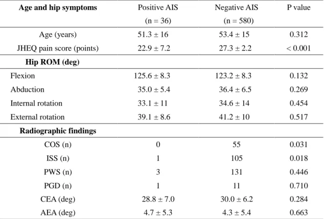
Table 3. Comparisons between the positive and negative AIS groups
Age and hip symptoms Positive AIS (n = 36)
Negative AIS (n = 580)
P value
Age (years) 51.3 ± 16 53.4 ± 15 0.312
JHEQ pain score (points) 22.9 ± 7.2 27.3 ± 2.2 < 0.001 Hip ROM (deg)
Flexion 125.6 ± 8.3 123.2 ± 8.3 0.132
Abduction 35.0 ± 5.4 36.4 ± 6.5 0.269
Internal rotation 33.1 ± 11 34.6 ± 14 0.454
External rotation 39.1 ± 8.6 41.2 ± 10 0.517
Radiographic findings
COS (n) 0 55 0.031
ISS (n) 1 105 0.018
PWS (n) 3 131 0.446
PGD (n) CEA (deg) AEA (deg)
1 28.8 ± 7.0
4.7 ± 5.3
11 30.0 ± 6.2
4.3 ± 5.4
0.710 0.284 0.663
Table 4. Relationship of the JHEQ pain category score with hip ROM, AIS, and radiographic findings
Standard partial
regression coefficient (β) P value 95% confidence interval (CI)
Age −0.112 0.008 −0.039 – −0.006
Sex 0.008 0.833 −0.424 – 0.526
Hip ROM
Flexion −0.063 0.142 −0.052 – 0.007
Abduction −0.042 0.373 −0.061 – 0.023
Internal rotation 0.058 0.241 −0.008 – 0.033
External rotation 0.064 0.126 −0.005 – 0.042
Positive rate of AIS −0.351 < 0.001 −5.336 – −3.457 Radiographic findings
COS 0.044 0.300 −0.403 – 1.306
ISS −0.035 0.413 −0.922 – 0.379
PWS 0.055 0.180 −0.182 – 0.970
PGD −0.019 0.626 −1.997 – 1.203
CEA 0.020 0.639 −0.030 – 0.049
AEA 0.037 0.333 −0.021 – 0.061
Table 5. Relationship of the presence of AIS with radiographic findings
Odds ratio (OR) P value 95% confidence interval (CI)
Age 2.132 0.092 0.884 – 5.140
Sex 0.988 0.330 0.963 – 1.013
Radiographic findings
COS 0.000 0.997 0.000
ISS 0.179 0.094 0.024 – 1.343
PWS 0.642 0.366 0.245 – 1.679
PGD 2.153 0.489 0.245 – 18.946
CEA 0.991 0.777 0.930 – 1.056
AEA 1.008 0.810 0.945 – 1.075