Introduction
Alzheimerʼs disease (AD) is the most common form of dementia in the elderly and it is characterized by progressive impairment in multiple cognitive domains of sufficient severity to interfere with individualsʼ daily living activities. Four articles in the Journal of Alzheimerʼs & Dementia in 2011 1 - 4) described new criteria for AD dementia and mild cognitive
impairment (MCI) due to the AD pathophysiological process (MCI due to AD) as well as their underlying rationale. These new criteria emphasize that the AD pathophysiological process starts years and perhaps decades prior to clinical symptoms, and that biomarkers can be used to detect amyloid beta (Aβ) deposition and the effects of neurodegeneration in the brain. These biomarkers include in vivo findings of brain imaging with positron emission tomography (PET) and
MS#AMN 07268
Reappraisal of the Cerebral Blood Flow Measured Using 123I-I-iodoamphetamine Single-photon Emission Computed Tomography in Normal Subjects and Patients with Alzheimer’s Disease and Dementia with Lewy Bodies
Makiko Seto 1, Naomi FukuShima 1, Takayuki YuaSa 1, Yoko Nakao 1, Katsuhiro ichiNoSe 1, Itsuro tomita 1, Hideyo Satoh 1, Akira Satoh 1, Makoto ochi 2, Mitsuhiro tSujihata 1
1 Section of Neurology, Nagasaki Kita Hospital, 800 Motomuragou, Togitsuchou, Nishisonogigun, Nagasaki 851-2103, Japan
2 Section of Radiology, Nagasaki Kita Hospital, 800 Motomuragou, Togitsuchou, Nishisonogigun, Nagasaki 851-2103, Japan
Purpose: Our aim was to re-evaluate the usefulness of perfusion single-photon emission computed tomography (SPECT) for the diagnosis of AD and DLB.
Methods: A total of 1,232 patients, including 180 cognitively normal subjects (NSs), 148 patients with amnestic mild cognitive impairment (aMCI), 802 patients with AD and 102 patients with dementia with Lewy bodies (DLB) were enrolled. A voxel- based stereotactic extraction estimation analysis was used to assess hypoperfusion areas using the cut-off value of 20% for the “extent %” and >2 hypoperfused lesions.
Results: The top 10 areas with hypoperfusion in AD were the angular gyrus, supramarginal gyrus, precuneus, superior occipital gyrus, inferior parietal lobule, middle temporal gyrus, superior parietal lobule, inferior temporal gyrus, superior temporal gyrus. and posterior cingulate, while DLB had 6 areas in common with AD. The respective sensitivity and specificity for differentiating AD from NSs were 84.2% and 78.9% for 50-59 years old, and 91.8% and 70.0% for 60-69 years old, but those values decreased from 70 years old. The respective sensitivity and specificity in DLB patients were 100% and 75.6%
for 50-69 years old, 84.6% and 70.1% for 70-79 years old and 81.4% and 77.1% for 80-89 years old. In aMCI patients, the 1-year change in the MMSE scores was -2.38±2.02 (n=16) in patients with abnormal SPECT images and -0.67±1.81 (n=18) in those with normal SPECT images (P<0.02).
Conclusions: Our study confirms that perfusion SPECT is still a valid alternative for the diagnosis of AD and DLB when fluorodeoxyglucose-PET is unavailable.
ACTA MEDICA NAGASAKIENSIA 64: 91−100, 2021 Key words: IMP-SPECT, Alzheimerʼs disease, Dementia with Lewy bodies, amnestic mild cognitive impairment,
preferred hypoperfusion areas, 18F-FDG PET
Address correspondence: Mitsuhiro Tsujihata, MD.
Nagasaki Kita Hospital, 800 Motomuragou, Togitsuchou, Nishisonogigun, Nagasaki 851-2103, Japan Tel: +81-95-886-8700, Fax: +81-95-886-8701, E-mail: [email protected]
Received October 7, 2020; Accepted November 10, 2020
magnetic resonance imaging (MRI), imaging of amyloid plaques and biochemical assays of Aβ1-42, Aβ1-40, total tau and phosphorylated tau (p-tau) in the cerebrospinal fluid (CSF).
However, these markers are not available at general hospitals.
In the present study, we investigated the effect of aging on the cerebral blood flow (CBF) measured using N-isopropyl- p-[(123)I]iodoamphetamine (123I-IMP) single-photon emission computed tomography (SPECT) in cognitively normal subjects (NSs) and patients with AD or DLB and determined the preferred areas of hypoperfusion in these diseases. Finally, we discussed the utility of SPECT for the diagnosis of AD, aMCI and DLB.
Methods and Materials
Methods
The SPECT device used in this study was an Infinia Hawkeye 4, General Electronic Company, U.S.A, which had a rotating two-headed gamma camera with a fan beam collimator (64x64). The regional cerebral blood flow (rCBF) changes were acquired for 20 minutes, starting 30 minutes after the administration of 111 MBq of 123I-IMP.
The easy Z-score Imaging System (eZIS) 5), and Voxel- based Stereotactic Extraction Estimation (vbSEE) 6) were used in this study for the quantitative assessment of brain SPECT images. We used the parameter “extent %” with the vbSEE software program, the extent of an abnormal region in each segment (proportion of the coordinates with a Z-value that exceeds the threshold value, among all coordinates within a segment).
We examined 2 indicators of decreased levels (20% and 30% decrease) of the “extent %” in the level 3 of vbSEE software program to decide which “extent %” is appropriate for assessing hypoperfusion for a comparison between NSs and diseased patients.
Materials
We established a “forgetfulness outpatient clinic” in our hospital on May 1, 2000. Thereafter, the hospital was reconstructed, and the medical care was restarted on October 1, 2007, at which point the SPECT device was changed to an Infinia Hawkeye 4, General Electronic Company, USA. For this reason, we recruited subjects based on their medical records starting from October 1, 2007, until December 31, 2017. The number of patients who underwent the mini-mental examination test (MMSE) during this period was 8,566, among whom 4,850
were new patients. We conducted SPECT examination in 2,412 of the new patients.
A total of 1,232 patients were ultimately enrolled in this study, including 180 NSs, 148 patients with aMCI, 802 patients with AD, and 102 patients with DLB.
NSs and AD patients were divided into 4 groups according to age (50-59, 60-69, 70-79 and 80-89 years old), while aMCI and DLB patients were divided into 3 groups according to age (50-69, 70-79, and 80-89 years old) because of small number of patients who were 50-59 years old.
The clinical diagnosis of probable AD was made according to the National Institute of Neurological and Communicative Disorders and Stroke - Alzheimerʼs Disease and Related Disorders Association (NINCDS-ADRDA) criteria 7). aMCI was diagnosed according to the criteria of Petersen et al. 8). Probable DLB was diagnosed according to the criteria of McKeith et al.9, 10). The following subjects were excluded from the study:
patients with AD who had MMSE scores of <14/30 and were associated with widespread leucoaraiosis, cerebral infarction, cerebral hemorrhaging and subarachnoid hemorrhaging; patients with non-amnestic MCI; patients with DLB who had MMSE scores of <14/30 and were associated with cerebral vascular diseases; patients with neurodegenerative diseases, such as multiple system atrophy, progressive supranuclear palsy and corticobasal syndrome; and patients with normal pressure hydrocephalus, severe diabetes mellitus and heart diseases.
Statistical analyses
We used the IBM-SPSS v.24, (U.S.A) and StatMate v.5.
(Atms, Tokyo, Japan) software programs for the data analyses.
Descriptive data are reported as mean and Standard Deviation (SD) or as number and percentage. Differences between each disease were assessed with a one way ANOVA, followed by the Tukeyʼs test for pairwise comparison when ANOVA showed a significant difference.
To estimate the predicative ability, the sensitivity and specificity of the different items were calculated.
Results
Subjects enrolled in this study
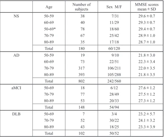
The demographic data of NSs patients with AD, aMCI and DLB are listed in Table 1. As shown in these tables, most patients in our “forgetfulness outpatient clinic” were over 70 years old.
Determination of the prefered areas of hypoperfusion in AD patients
We determined the preferred areas of hypoperfusion by analyzing the “extent %” in AD patients 50-69 years old, and selected the top 10 areas expressing the average “extent %”
value (Table 2) presented in descending order: the angular gyrus, supramarginal gyrus, precuneus, superior occipital gyrus, inferior parietal lobule, middle temporal gyrus, superior parietal lobule, inferior temporal gyrus, superior temporal gyrus and posterior cingulate.
Determination of the cut-off value in NSs and the sensitivity, specificity and efficacy in AD patients
We determined the preferred areas of hypoperfusion in NSs corresponding to the preferred areas obtained in AD patients according to the ages in order to determine whether an
“extent %” of 20% or 30% or 1 or >2 lesions of hypoperfusion was most suitable as the cut-off value (Table 3, 4). The number of AD patients showing hypoperfusion and the sensitivity, specificity and efficacy at each cut-off value compared with the age- and area-mactched NSs were shown in Table 3. The sensitivity was lower while the specificity was higher with an “extent %” of 30% than with 20% and >2 areas of hypoperfusion than with 1 for differenciating AD from NSs (Table 3). We therefore ultimately adopted an
“extent %” of 20% and 2> hypoperfusion areas as the cut-off value in this quantitative analysis of SPECT images, as the respective sensitivity and specificity for differenciating AD patients from NSs were 84.2% and 78.9% for 50-59 years old, 91.8% and 70.0% for 60-69 years old, 76.0% and 75.6%
for 70-79 years old and 53.2% and 71.4% for 80-89 years old.
These results indicate that the sensitivity decreases with aging.
The frequency of hypoperfusion areas in NSs using these cut-off values was 21.1% for 50-59 years old, 32.5% for 60-69 Table 1. Demographic data of enrolled subjects
Age Number of
subjects Sex M/F MMSE scores
mean±SD
NS 50-59 38 7/31 29.6±0.7
60-69 40 11/29 29.3±0.7
50-69* 78 18/60 29.4±0.7
70-79 67 25/42 28.9±1.0
80-89 35 17/18 28.7±1.0
Total 180 60/120
AD 50-59 19 9/10 21.8±3.0
60-69 73 22/51 22.3±3.4
70-79 317 106/211 22.0±3.5
80-89 393 105/288 21.8±3.5
Total 802 242/560
aMCI 50-69 18 6/12 27.6±1.2
70-79 77 28/49 27.5±1.2
80-89 53 20/33 27.3±1.2
Total 148 54/94
DLB 50-69 7 3/4 23.2±5.7
70-79 52 30/22 24.1±3.2
80-89 43 18/25 23.3±3.9
Total 102 50/52
* For comparison with aMCI and DLB patients
Table 2. Frequent areas of hypoperfusion in AD patients (age 50-69)
Brain areas Average Extent %
Angular Gyrus 48.41
Supramarginal Gyrus 38.57
Precuneus 30.60
Superior Occipital Gyrus 29.21
Inferior Parietal Lobule 27.50
Middle Temporal Gyrus 26.95
Superior Parietal Lobule 24.88
Inferior Temporal Gyrus 21.94
Superior Temporal Gyrus 19.97
Posterior Cingulate 19.43
years old, 31.3% for 70-79 years old and 28.6% for 80-89 years old, showing no significant difference among age groups.
Determination of the prefered areas of hypoperfusion in DLB patients
We determined the prefered areas of hypoperfusion by analyzing the “extent %” in 102 patients with probable DLB and selected the 10 main areas expressing the average “extent %”
value (Table 5), presented in descending order: the angular gyrus, superior occipital gyrus, inferior occipital gyrus, middle occipital gyrus, supramarginal gyrus, cuneus, middle temporal gyrus, inferior temporal gyrus, precuneus, and lingual gyrus.
The respective sensitivity and specificity in DLB patients compared with age- and hypoperfusion area-matched NSs were 100% and 75.6% for 50-69 years old, 84.6% and 70.1% for 70-79 years old and 81.4% and 77.1% for 80-89 years old (Table 6). The cut-off value obtained in the AD patients was used to calculate the sensitivity and specificity in DLB patients.
In contrast to our findings in AD patients, the sensitivity in DLB patients did not decrease with age.
The sensitivity and specificity in aMCI patients
The respective sensitivity and specificity in aMCI patients compared with age- and hypoperfusion area-matched NSs were 88.9% and 75.6% for 50-69 years old, 57.1% and 68.7% for 70-79 years old and 30.2% and 71.4% for 80-89 years old (Table 6). The sensitivity in aMCI patients was Table 3. Number of cases showing hypoperfusion areas in AD patients & sensitivity, specificity & efficaxy in each cut-off valuen compared with age- and area-mactched normal subjects
Ages No of
cases Cut-off value
20% No of
cases Sensitivity Specificity Cut-off value
30% No of
cases Sensitivity Specificity
50-59 19 over 1area 19 100.0% 65.8% over 1area 18 94.7% 81.6%
over 2area 16 84.2% 78.9% over 2area 15 78.9% 94.6%
60-69 73 over 1area 70 95.9% 50.0% over 1area 65 89.0% 70.0%
over 2area 67 91.8% 70.0% over 2area 62 84.9% 87.5%
70-79 317 over 1area 271 85.5% 59.0% over 1area 234 73.1% 61.2%
over 2area 241 76.0% 75.6% over 2area 198 62.5% 85.1%
80-89 393 over 1area 266 67.7% 60.0% over 1area 208 52.9% 68.6%
over 2area 209 53.2% 71.4% over 2area 139 35.4% 85.7%
Table 4. Number of cases showing hypoperfusion areas in normal subjects age- and areas-matched to AD
Ages No of
subjects Cut-off
20% No of
subjects Cut-off
30% No of
subjects
50-59 38 over 1area 13 over 1area 7
over 2area 8 over 2area 2
60-69 40 over 1area 19 over 1area 12
over 2area 13 over 2area 5
50-69 78 over 1area 32 over 1area 18
over 2area 21 over 2area 6
70-79 67 over 1area 32 over 1area 26
over 2area 21 over 2area 10
80-89 35 over 1area 14 over 1area 11
over 2area 10 over 2area 5
Table 5. Prefered areas of hypoperfusion in DLB patients
Brain areas Average extent %
Angular Gyrus 40.44
Superior Occipital Gyrus 35.75
Inferior Occipital Gyrus 27.43
Middle Occipital Gyrus 27.26
Supramarginal Gyrus 24.26
Cuneus 23.98
Middle Temporal Gyrus 23.87
Inferior Temporal Gyrus 22.06
Precuneus 20.34
Lingual Gyrus 20.09
considerably lower than in AD patients > 70 years old.
The comparison of the preferred areas of hypoperfusion in AD and DLB patients
The top 10 areas of hypoperfusion in AD and DLB were shown based on the degree of hypoperfusion (Figure 1).
Although there was a difference in the degree of hypoperfusion, 6 of the top 10 areas with hypoperfusion showed a common
decrease in AD and DLB patients. Hypoperfusion in the superior and inferior parietal lobules, precuneus and posterior cingulate suggests a greater likelihood of AD, while that in the middle and inferior occipital gyruses, cuneus, and lingual gyrus suggests a greater likelihood of DLB.
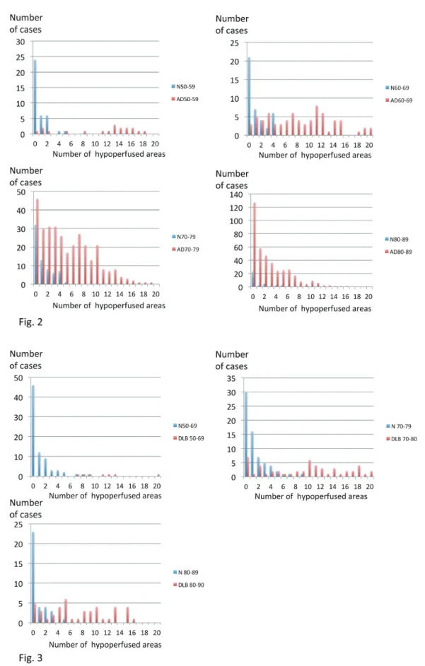
We counted the total number of hypoperfusion areas (maximum number is 20) observed in the top 10 areas of hypoperfusion with an “extent %” over 20%, regardless of the “extent %” in AD and DLB patients (Fig. 2 and 3).
Table 6. Sensitivity and specificity in aMCI, AD, and DLB patients compared with age- and hypoperfusion area-matched normal subjects
Disease Age No of cases sensitivity specificity
MCI 50-69 18 88.9% 75.6%
70-79 77 57.1% 68.7%
80-89 53 30.2% 71.4%
AD 50-59 19 84.2% 78.9%
60-69 73 91.8% 70.0%
70-79 317 76.0% 75.6%
80-89 393 53.2% 71.4%
DLB 50-69 7 100% 75.6%
70-79 52 84.6% 70.1%
80-89 43 81.4% 77.1%
Area of hypoperfusion Diseases AD DLB
Scale showing hypoperfusion High frequency
Low frequency
Superior Temporal Gyrus
Middle Temporal Gyrus
Inferior Temporal Gyrus
Angular Gyrus
Supramarginal Gyrus
Inferior Parietal Lobule
Superior Parietal Lobule
Precuneus
Posterior Cingulate
Superior Occipital Gyrus
Inferior Occipital Gyrus
Middle Occipital Gyrus
Cuneus
Lingual Gyrus
!
Figure 1. The top 10 areas of hypoperfusion in AD patients and DLB patients. Six of the top 10 hypoperfusion areas showed a common decrease in AD and DLB patients. Hypoperfusion in the superior and inferior parietal lobules, precuneus, and posterior cingulate suggests a greater likelihood of AD, while that in the middle and inferior occipital gyruses, cuneus and lingual gyrus suggests a greater likelihood of DLB.
Figure 2. and 3. The distribution of the total number of hypoperfusion areas in AD (Fig.2) and DLB (Fig.3). AD showed a decrease in the high frequency total number of hypoperfusion in older groups, While no such findings were observed in DLB.
N:Normal subjects, AD: Alzheimerʼs disease, DLB: dementia with Lewy bodies
The distribution of these number in AD patients showed a decrease of high frequency number of hypoperfusion in older age groups, while such findings were not observed in DLB patients.
Effects of hypoperfusion of SPECT images on progression of aMCI and AD
In aMCI patients, while the number of cases was small, the 1-year change in the MMSE was -2.38±2.02 (n=16) in
patients with abnormal SPECT images, and -0.67±1.81 (n=18) in those with normal SPECT images (P<0.02, Mann- Whitney test). Eight of 16 patients with abnormal SPECT images eventually developed AD, while 2 of 18 with normal SPECT images eventually developed AD. Although only a small number of cases could be followed, the MMSE scores decreased over time in all AD patients regardless of the normality of their SPECT findings, but the progression in those with normal findings was one year slower than in those with abnormal findings (Fig. 4).
$
Figure 4. Effects of hypoperfusion of SPECT images on progression of AD. The MMSE scores decreased with the passage of time in both AD patients with abnormal SPECT findings as well as in those with normal SPECT findings, although the progression in the latter was about one year slower than that in the former.
SP: SPECT
Discussion
Recently, there have been developments in brain imaging techniques for detecting the core histological features of Alzheimer disease, j.e. amyloid plaques and neurofibrillary tangles. However, the utility of these techniques is still being investigated.
Functional imaging methods, such as 18F-fluorodeoxyglucose [18F-FDG]- PET and perfusion (hexamethylpropyleneamine oxime [HMPAO]) SPECT are advocated as useful for clarifying the diagnosis of dementia when doubt remains. Although specific imaging ligands for amyloid are becoming available, the mainstay of functional brain imaging for the differential diagnosis of dementia will remain 18F-FDG-PET and blood flow SPECT even in general hospitals.
Aging effects of hypoperfuion in NSs
When analyzing the rCBF in patients with neurodegenerative diseases, we need to pay attention to age-related changes in the rCBF. There have been many reports describing the age- related changes in the rCBF in normal subjects, although the main sites of a reduced rCBF have differed among studies.
Indeed, several reports described the anterior cingulate as the main site within which the rCBF is reduced with normal aging11-15), while others failed to demonstrate any marked reduction in the rCBF in this area16, 17). Mozley et.al reported that the reduction in the rCBF was negligible in subjects >40 years of age18). These discrepancies may be attributable to differences in the criteria used to select subjects and the diversity of the image analysis methods.
In the present study, we first determined the preferred
areas of hypoperfusion in AD and DLB patients. Although the degree of hypoperfusion differed, 6 of the 10 main areas of hypoperfusion showed a common decrease in AD and DLB patients. Hypoperfusion in the superior and inferior parietal lobules, precuneus and posterior cingulate suggests a greater likelihood of AD, while that in the middle and inferior occipital gyruses, cuneus and lingual gyrus suggests a greater likelihood of DLB.
Based on these results, the frequency of hypoperfusion in NSs corresponding to the preferred areas of hypoperfuion for AD and DLB was investigated. Our findings indicated no marked difference in the degree of hypoperfusion with age in NSs.
Sensitivity and specificity of SPECT imaging for diagnosing dementia
Studies of the accuracy of SPECT for diagnosing AD reported sensitivities of 65%-85% and specificities of 72%- 87%19). Studies for 18F-FDG PET generally reported higher accuracy, with sensitivities of 75%-99% and specificities of 71%-93% for AD20). OʼBrien et al.21) reported that when performing a visual rating, the key imaging features were a reduced uptake in the precuneus and lateral parietal lobes in both AD and DLB, the relative preservation of the posterior cingulate in DLB, and a more extensive occipital loss in DLB, with a reduced uptake in the temporal and frontal lobe being more likely in AD than DLB. The consensus diagnosis with 18F-FDG PET was superior to that with SPECT for both dementia vs. no-dementia and AD vs. DLB comparisons.
The sensitivity and specificity for dementia/no-dementia was 85% and 90%, respectively, for 18F-FDG PET and 71% and 70%, respectively, for SPECT. The authors recommended that 18F-FDG PET should be performed instead of perfusion SPECT for the differential diagnosis of degenerative dementia if functional imaging was indicated. There have been no reports on the age-related incidence of hypoperfusion on SPECT images in AD patients.
In the present study, the respective sensitivity and specificity for differenciating AD from NSs were 84.2% and 78.9% for 50-59 years old, 91.8% and 70.0% for 60-69 years old, 76.0% and 75.6% for 70-79 years old and 53.2% and 71.4%
for 80-89 years old. These results indicate that sensitivity decreases with aging. The results in aMCI were the same as those in AD; however, in DLB patients, no marked decreese in the sensitivity with aging was observed. While this degree of diagnostic accuracy might still be clinically helpful when comparing dementia and no-dementia patients, the accuracy for differentiating AD from DLB was disappointingly poor13).
Our finding that 6 of the 10 main areas of hypoperfusion showed a common decrease in AD and DLB patients indicates that clear discrimination between AD and DLB is difficult.
To this end, cardiac MIBG scintigraphy is much more useful than SPECT22).
The present study showed that for differentiating AD patients from NSs, SPECT findings were very useful, particularly, among patients <69 years old. Why the sensitivity in aged AD patients, particularly those over 80 years old, was decreased is not clear. The mechanism involving a decreese of rCBF in aged person described in the previous reports is not considered to be aging effects.
Argyrophilic grain disease (AGD) is said to be the second- most common neurodegenerative disease that causes dementia in the elderly after AD23-25). Neuropathologic changes due to AGD may be found in isolation or in association with other neurodegenerative disorders, such as AD, Pickʼs disease, progressive supranuclear palsy (PSP), corticobasal degeneration (CBD), Parkinsonʼs disease, TDP-43 proteinopathies, DLB and multiple system atrophy. Braak et al. reported that patients 81-85 years old had the highest frequency of AGD (40/125 cases), whereas only 5 patients <60 years old were found to have argyrophilic grains on a post-mortem examination24). To date, no study has been able to demonstrate a distinct clinical syndrome associated with AGD. The diagnosis is exclusively based on postmortem findings and no reports on SPECT findings have been published.
Senile dementia of the neurofibrillary tangle (SD-NFT) type is a subset of dementia that primarily occurs in the oldest old patients26, 27). Although SD-NFT is not considered very rare in senile dementia patients, most SD-NFT cases are probably clinically misdiagnosed as AD in the absence of a post-mortem pathological investigation.
These two diseases are clinically impossible to definitely diagnose. We were therefore unable to exclude the possibility that these diseases were present in the aged group of AD patients in our study, which might have reduced the sensitivity in that population.
Prefered areas of hypoperfusion among disease
The pattern of SPECT hypoperfusion and PET hypometabolism usually seen AD patients involves the anterior medial temporal lobes, the posterior cingulate and posterior temporoparietal cortex28-30), Some neuroimaging features of DLB have been proposed to enhance the diagnostic accuracy. Indeed, occipital hypometabolism on 18F-FDG-PET images is a useful biomarker for differentiating DLB from AD31, 32).
In the present study, we determined the top 10 preferred
References
1) Jack CR Jr, Albert M, Knopman D, et al. Introduction to the recommendations from the National Institute on Aging-Alzheimerʼs Association workgroups on diagnostic guidelines for Alzheimerʼs disease. Alzheimers Dement 7:257-62, 2011.
2) Sperling RA, Aisen PS, Beckett LA, et al. Toward defining the preclinical stages of Alzheimerʼs disease: recommendations from the National Institute on Aging-Alzheimerʼs Association workgroups on diagnostic guidelines for Alzheimerʼs disease. Alzheimers Dement 7:280-92, 2011.
3) Albert M, DeKosky ST, Dickson D, et al. The diagnosis of mild cognitive impairment due to Alzheimerʼs disease: report of the National Institute on Aging and the Alzheimerʼs Association workgroup. Alzheimers Dement 7:270-9, 2011.
4) McKhann GM, Knopman DS, Chertkow H, et al. The diagnosis of dementia due to Alzheimerʼs disease: recommendations from the National Institute on Aging and the Alzheimerʼs Association workgroup. Alzheimers Dement 7:263-9, 2011.
5) Matsuda H. “Role of neuroimaging in Alzheimerʼs disease, with emphasis on brain perfusion SPECT,” J Nucl Med 48:1289-1300, 2007.
6) Mizumura S, Kumita S, Cho K, et al. “Development of quantitative analysis method for stereotactic brain image: assessment of reduced accumulation in extent and severity using anatomical segmentation”, Ann Nucl Med 17:289-295, 2003.
7) McKhann G, Drachman D, Folstein M, Katzman R, Price D, Stadlan EM. Clinical diagnosis of Alzheimerʼs disease: report of the NINCDS- ADRDA Work Group under the auspices of Department of Health and Human Services Task Force on Alzheimerʼs Disease. Neurology 34:939- 44, 1984.
8) Petersen RC, Smith GE, Waring SC, Ivnik RJ, Tangalos EG, Kokman E.
Mild Cognitive Impairment; Clinical characterizeation and outcome.
Arch Neurol 56 303-308, 1999.
9) McKeith IG, Dickson DW, Lowe J, et al. Diagnosis and management of dementia with Lewy bodies: third report of the DLB Consortium.
Neurology 65:1863-72, 2005.
10) McKeith IG, Boeve BF, Dickson DW, et al. Diagnosis and management of dementia with Lewy bodies: Fouth concesnsus report of the DLB Consortium. Neurology 89:88-100, 2017.
11) Takahashi K, Yamaguchi S, Kobayashi S, Yamamoto Y. Effects of Aging on Regional Cerebral Blood Flow Assessed By Using Technetium Tc
areas of hypoperfusion using the vbSEE software program.
Despite a marked difference in the hypoperfusion degree, 6 of the top 10 hypoperfusion areas showed a common decrease in AD and DLB patients. Hypoperfusion in the superior and inferior parietal lobules, precuneus and posterior cingulate suggests a greater likelihood of AD, while that in the middle and inferior occipital gyruses, cuneus and lingual gyrus suggests a greater likelihood of DLB.
The pathological hallmarks of DLB are the presence of Lewy bodies and Lewy neurites, but many cases also display an AD pathology postmortem33, 34). However, the importance of concurrent AD pathology in DLB, at what point during the disease it occurs and its relationships to the clinical presentations are unclear35). Considering the previous reports that AD pathology occurs in many cases of DLB, it seems natural for the areas of hypoperfusion to overlap between AD and DLB cases. The cingulate island sign (CIS) on FDG- PET and brain perfusion SPECT was recently proposed as a neuroimaging feature of DLB. This term references the sparing of the posterior cingulate cortex relative to the precuneus plus cuneus and this sign is extremely specific for an accurate diagnosis of DLB36, 37). The CIS is influenced by concomitant AD-type neurofibrillary tangle (NFT) pathology and gradually disappears as DLB progresses38). In the present study, the decreased perfusion in the posterior cingulate in DLB was ranked 16th indicating that posterior cingulate hypoperfusion is not an important finding in the diagnosis of DLB.
Prediction of conversion from aMCI to AD
The concept of MCI is now widely accepted and continues to receive substantial attention in the literature, as it represents a possible treatment target for AD. Its predictive power is reflected in annual conversion rates of up to 15% from MCI to AD, compared to conversion rates of 1%-2% from normal aging to AD39, 8). SPECT has been reported to be valuable for predicting the progression from MCI to AD-dementia40-43). However, differences in the methods of analyzing SPECT images among groups have hindered the comparison of our results with those of previous reports. Our study clearly showed that 50% of aMCI patients with abnormal SPECT findings showed rapid regression of the cognitive function compared to those with normal findings. Identifyiung such at-risk patients at an early time point would enable early intervention and a timely start of treatment, possibly preventing disease progression altogether.
Conclusion
A quantitative SPECT image analysis showed sensitivity and specificity equivalent to those of FDG-PET in AD patients 50-69 years old, suggesting its utility for making an AD diagnosis. A quantitative SPECT image analysis was also shown to be a useful auxiliary diagnostic technique for diagnosing DLB.
Six of the top 10 preferred areas of hypoperfusion were shared between AD and DLB. Hypoperfusion in the superior and inferior parietal lobules, precuneus and posterior cingulate suggests a greater likelihood of AD, while that in the middle and inferior occipital gyruses, cuneus and lingual gyrus suggests a greater likelihood of DLB. Although FDG-PET performs better than perfusion SPECT, our study confirms the utility of perfusion SPECT as a valid alternative when FDG-PET is unavailable.
27) Jellinger KA, Attems J. Neurofibrillary tangle-predominant dementia:
compareson with classical Alzheimer disease. Acta Neuropathol 113:107-117, 2007.
28) Kogure D, Matsuda H, Ohnishi T, Kumihiro T, Uno M, Takasaki M.
Longitudinal evaluation of early Alzheimer disease using brain perfusion SPECT. J. Nucl Med 41, 1155-1162, 2000.
29) Bradley KM, O'Sullivan VT, Soper ND, et al. Cerebral perfusion SPET correlated with Braak pathological stage in Alzheimer's disease. Brain 125, 1772-1781, 2002.
30) Femminella GD, Thayanandan T, Calsolaro V, et al. Imaging and Molecular Mechanisms of Alzheimer's Disease: A Review. Int J Mol Sci online 9:3702, 2018.
31) Ishii K, Imamura T, Sasaki M, et al. Regional cerebral glucose metabolism in dementia with Lewy bodies and Alzheimerʼs disease. Neurology 51:125-30, 1998.
32) Minoshima S, Foster NL, Sima AA, et al. Alzheimerʼs disease versus dementia with Lewy bodies: cerebral metabolic distinction with autopsy confirmation. Ann Neurol 50:358-65, 2001.
33) Nedelska Z, Ferman TJ, Boeve BF, et al. Pattern of brain atrophy rates in autopsy-confirmed dementia with Lewy bodies. Neurobiol Aging 36:452-461, 2015.
34) Ballard C, Ziabreva I, Perry R, et al. Differences in neuropathologic characteristics across the Lewy body dementia spectrum. Neurology 67:1931-1934, 2006.
35) Donaghy P, Thomas AJ, OʼBrien JT. Amyloid PET imaging in Lewy body disorders. Am J Geriatr Psychiatry 23:23-37, 2015.
36) Lim SM, Katsifis A, Villemagne VL, et al. The 18F-FDG PET cingulate island sign and comparison to 123I-beta-CIT SPECT for diagnosis of dementia with Lewy bodies. J Nucl Med 50:1638-45, 2009.
37) Imabayashi E, Yokoyama K, Tsukamoto T, et al. The cingulate island sign within early Alzheimerʼs disease-specific hypoperfusion volumes of interest is useful for differentiating Alzheimerʼs disease from dementia with Lewy bodies. EJNMMI Res 6:67, 2016.
38) Iizuka T, Iizuka R, Kameyama M. Cingulate island sign temporally changes in dementia with Lewy bodies. Sci Rep 7:14745, 2017. Online.
39) DeCarli C. Mild cognitive impairment: prevalence, prognosis, aetiology, and treatment. Lancet Neurol 2:15-21, 2003.
40) Borroni B, Perani D, Broli M, et al. Pre-Clinical diagnosis of Alzheimer disease combining platelet amyloid precursor protein ratio and rCBF spect analysis. J Neurology 252:1359-1362, 2005.
41) Hirao K, Ohnishi T, Hirata Y, et al. The prediction of rapid conversion to Alzheimerʼs disease in mild cognitive impairment using regional cerebral blood flow SPECT. NeuroImage 28: 1014-1021, 2005.
42) Park KW, Yoon HJ, Kang DY, Kim BC, Kim S, Kim JW. Regional cerebral blood flow differences in patients with mild cognitive impairment between those who did and did not develop Alzheimerʼs disease. Psychiatry Res 203:201-206, 2012.
43) Sanchez-Catasus CA, Stormezand GN, van Laar PJ, De Deyn PP, Sanchez MA, Dierckx RA. FDG-PET for Prediction of AD Dementia in Mild Cognitive Impairment. A Review of the State of the Art with Particular Emphasis on the Comparison with Other Neuroimaging Modalities (MRI and Perfusion SPECT). Curr Alzheimer Res 14:127- 142, 2017.
99mHexamethylpropyleneamine Oxime Single-Photon Emission Tomography with 3D Stereotactic Surface Projection Analysis. AJNR 26:2005-2009, 2005.
12) Nakano S, Asada T, Matsuda H, Uno M, Takasaki M. Effects of healthy aging on the regional cerebral blood flow measurements using Tc-ECD SPECT assessed with statistical parametric mapping. Nippon Ronen Igakkai Zasshi 37:49-55, 2000.
13) Van Laere K, Versijpt J, Audenaert K, et al. Tc-ECD brain perfusion SPET: variability, asymmetry and effects of age and gender in healthy adults. Eur J Nucl Med 28:873-887, 2001.
14) Tanaka F, Vines D, Tsuchida T, Freedman M, Ichise M. Normal patterns on Tc-ECD brain SPECT scans in adults. J Nucl Med 41:1456-1464, 2000.
15) Martin AJ, Friston KJ, Colebatch JG, Frackowiak RSJ. Decreases in regional cerebral blood flow with normal aging. J Cereb Blood Flow Metab 11:684-689, 1991.
16) Pagani M, Salmaso D, Jonsson C, et al. Regional cerebral blood flow as assessed by principal component analysis and Tc-HMPAO SPET in healthy subjects at rest: normal distribution and effect of age and gender.
Eur J Nucl Med 29:67-75, 2002.
17) Krausz Y, Bonne O, Gorfine M, Karger H, Lerer B, Chisin R. Age- related changes in brain perfusion of normal subjects detected by Tc- HMPAO SPECT. Neuroradiology 40:428-434, 1998.
18) Mozley PD, Sadek AM, Alavi A, et al. Effects of aging on the cerebral distribution of technetium-99m hexamethylpropylene amine oxime in healthy humans. Eur J Nucl Med 24:754-761, 1997.
19) Dougall NJ, Bruggink S, Ebmeier K. Systematic review of the diagnostic accuracy of 99mTc-HMPAO-SPECT in dementia. Am J Geriatr Psychiatry 12:554-570, 2004.
20) Silverman DH, Small GW, Chang CY, et al. Positron emission tomography in evaluation of dementia: regional brain metabolism and long-term outcome. JAMA 286:2120-2127, 2001.
21) OʼBrien JT, Firbank MJ, Davison C, et al. 18F-FDG PET and Perfusion SPECT in the Diagnosis of Alzheimer and Lewy Body Dementias. J Nucl Med 55;12 1959-1965, 2014.
22) Seto M, Nakata R, Yuasa T, et al. Diagnostic value of 123-I-MIBG cardiac scintigraphy for the prediction of conversion from idiopathic REM sleep behavior disorder to dementia with Lewy bodies, and the differentiatial diagnosis of neurodegenerative diseases. Alzheimers Dis Dement 1:47-55, 2017. [ISSN2578-6490]
23) Sumit D, Abdullah I. Review paper. Argyrophilic grain disease: a clini- copathological review of an overlooked tauopathy. Folia Neuropathol 56:277-283, 2018.
24) Braak H, Braak E. Argyrophilic grain disease: frequency of occurrence in different age categories and neuropathological diagnostic criteria. J Neural Transm 105:801-819, 1998.
25) Saito Y, Nakahara K, Yamanouchi H, Murayama S. Severe involvement of ambient gyrus in dementia with grains. J Neuropathol Exp Neurol 61;9:789-796, 2002.
26) Yamada M, Itoh Y, Otomo E, Suematsu N, Matsushita M. Dementia of the Alzheimer type and related dementias in the aged: DAT subgroups and senile dementia of the neurofibrillary tangle type. Neuropathol 16:89-98, 1996.