Acta Med. Nagasaki 44 : 61-65
Decreasing the Pressure Gradient of the Left Ventricular Outflow Tract by Single-lead VDD Pacing in a Patient with Hypertrophic Obstructive Cardiomyopathy
Satoshi IKEDA 1), Takayuki YOSHINAGA 1), Kikuko OBASE 1), Kouichirou SONODA 1), Kousuke SHIOGUCHI 1), Eisuke KAWAHARA 1), Norihiko SENJYU 1), Kyouji NISHIJIMA 1), Yoshiyuki MIYAHARA 1), Tsuneo FUJIWARA 2), Hironobu KOGA 2), Shigeru KOHNO 1)
1) Second Department of Internal Medicine, Nagasaki University School of Medicine 2) Miyazaki Hospital
A 59-year-old woman with hypertrophic cardiomyopathy
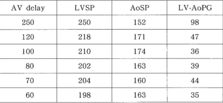
of 8 years duration, who had been taking β‑blocker, was
admitted to our hospital for exertional dyspnea and previ- ous syncope. Cardiac catheterization showed a prominent left-ventricular outflow tract (LVOT) pressure gradient, and hypertrophic obstructive cardiomyopathy (HOCM) was diagnosed. To reduce LVOT obstruction, we implanted a single-lead VDD-mode pacemaker. Cardiac catheterization after the implantation revealed a remarkable decrease in the LVOT pressure gradient with short atrioventricular delay, 80 msec, and her symptoms disappeared. A single- lead VDD pacemaker is also a useful treatment for an HOCM patient due to the relative ease with which it can be implanted.
valve replacement'-'), or atrioventricular sequential pacing10"5. But surgery frequently fails to reduce LVOT obstruction and leads to a 2-11 % possibility of early mortality". Dual-chamber pacing has been shown to induce the reduction of the LVOT pressure gradient and to lead to improvement in symptoms of hypertrophic obstructive cardiomyopathy (HOCM).
Recently single-lead VDD pacing",") has been used widely to patients with advanced atrioventricular block. We report a decrease in the LVOT pressure gradient by single-lead VDD pacing in an HOCM pa- tient.
Case Report
Key words: single-lead VDD pacing, hypertrophic obstructive cardiomyopathy (HOCM), left-ventricular outflow
tract obstruction
Introduction
The degree of obstruction of the left ventricular out- flow tract (LVOT) is an important determining factor in the clinical course of patients with hypertrophic
cardiomyopathy(HCM). To relieve the LVOT obstruc- tion, the initial therapeutic approach is pharmacothe- rapy such as 1 -blocker and verapamil1-4'. However, many patients develop a resistance to such therapies.
The next approach is either surgery involving left ventricular septal myectomy or mitral
Address Correspondence: Satoshi Ikeda M.D.
Second Department of Internal Medicine, Nagasaki University School of Medicine, Sakamoto 1-7-1, Nagasaki 852-8501, Japan TEL: +81-95-849-7280 FAX: +81-95-849-7280
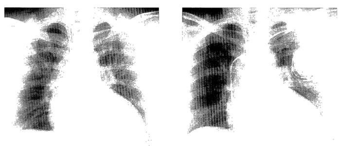
On 25 December 1998, a 59-year-old woman was ad- mitted to our hospital for further examinations due to exertional dyspnea, chest pain and a history of syn- cope. She had been diagnosed as having HCM in an- other hospital about 8 years before and had received medicinal treatment, (9 -blocker, and antiplatelet ther- apy. On admission, electrocardiography (ECG) showed T inversion in leads I, II, aVL, aVF, and V4-V6; ST ele- vation in leads V 1 and V2; and high voltage (S V 1 + RV5 = 8.59 mV). Cardiomegaly (cardiothoracic ratio:
63.7 %) was observed by thoracic radiography (Fig. 1,
left). Concentric left-ventricular hypertrophy with 17-
to 19-mm wall thickness and LVOT narrowing were
shown by echocardiography (Fig. 2). Hematology and
biochemistry results showed iron deficiency anemia
and high LDH (679 IU/1). We performed cardiac
catheterization on the day of admission. Pressure
study revealed a left ventricule (LV) -aorta (Ao) pres-
sure gradient of 112 mmHg, and left ventriculography
of right-anterior, oblique view showed a spade-like
shape, which was compatible with HCM, during sys-
tolic phase. Coronary angiography showed no signifi-
cant stenosis.