Acta Med. Nagasaki 39: 119-124
CORONARY ARTERY MORPHPLOGY AND REACTIVITY TO ACUTE HYPOXIA IN CHRONIC PULMONARY DISEASE
Kazuo MATSUNAGA, Yoshihisa MIYAHARA, Takashi HARADA, Toshihiko YAMASA and Toshiyuki IMAMURA
The Second Department of Internal Medicine, Nagasaki University School of Medcine, Japan.
In patients with chronic pulmonary disase (CPD), myocar- dial infarction is rare. To elucidate why this is so, we investigated the morphological changes and the reactivity of the coronary artery to acute hypoxia in patients with CPD.
Sixty patients with CPD and 28 normal subjects were studied. Measurements of pulmonary homodynamics and coronary angiography were undertaken before and after inhalation of 13%O2 for 15 minutes. The size of the coronary arteries was measured using a densitometric method, and a coronary narrowing score was calculated according to the WHO criteria.
The size of the left anterior descending artery of patients with low %VC and hypoxia was larger than that of the normal subjects. In patients with CPD, the coronary narrow- ing score was low and the atherosclerotic change was mini- mal. The reactivity of the coronary arteries to acute hypoxia was reduced in patients with CPD when compared with normal subjects.
KEY WORDS : Cor pulmonale, myocardial infarction, Atherosclerosis, Hypoxic challenge.
INTRODUCTION
Myocardial infarction in patients with chronic pulmo- nary disease (CPD) has been a relatively infrequent clini- cal finding in our experience.
Recently, the increased availability of cardiac catheteri- zation has stimulated studies on the hemodynamics in cor pulmonale. Little is known, however, about the morphol- ogy of the coronary artery in cor pulmonale.
In this study, the diameters of the left and right coro- nary arteries, coronary sclerosis, and the responses of the coronary artery diameters to hypoxic loading in patients with chronic lung diseases were compared with those in a control group to clarify the morphological characteristics of the coronary arteries of patients with chronic pulmo- nary diseases.
SUBJECTS AND METHODS
Sixty patients who underwent coronary angiography for various reasons between 1988 and 1992 were regarded as the CPD group, and 28 patients who complained of chest pain but showed no organic changes by coronary angio- graphy served as controls. The patients with CPD con- sisted of 9 with chronic bronchitis, 9 with bronchiectasis, 19 with chronic pulmonary emphysema, 4 with diffuse panbronchiolitis, and 19 with pulmonary fibrosis. Inform- ed consent was obtained from all subjects. All of these patients underwent cardiac catheterization in a chronic stable condition.
Blood gas analysis and measurement of the cardiac output were performed simultaneously by the Fick method or the thermodilution method. The results of standard pulmonary function tests obtained at about the same time were also evaluated. The right and left coronary artery diameters were measured in both groups by densitometry using a CATHEX (CCIP-310) system for measurement of the coronary artery stenosis rate and the cardiac function, and their changes before and after hypoxic loading (inha- lation of 13 % Oxygen for 10 minutes) were compared (Fig. 1).
Coronary stenosis was scored for each artery as 5 for 100 % obstruction, 4 for 75 % stenosis, 3 for 50 % stenosis, 2 for 25 % stenosis and 1 for mild stenosis, according to the WHO criteria, and the sum of the maximum scores in the three coronary arteries was used as a stenosis index.
RESULTS
1. Pre-challenge coronary angiography
(1) Standard pulmonary function, hemodynamics and blood gas concentrations were compared between 60 patients and 28 normal controls. Significant differences were observed in PaO2, %VC, FEV1.0 %, mean pulmonary artery pressure, and cardiac output between the two groups.
Fig. 2 : Comparison of the coronary artery diameter between the CPD group and the control group
Control = Normal subjects, CPD = Chronic pulmonary disease, NS = Not Significant.
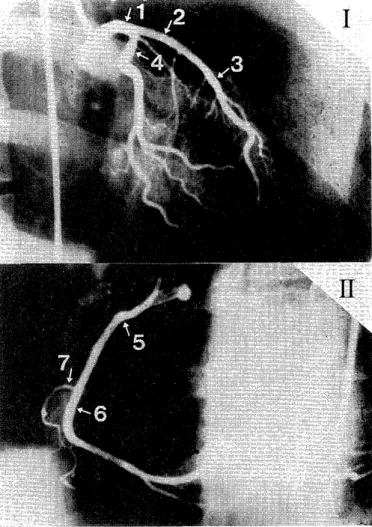
Fig. 1 : The sites of measurement in coronary angiograms.
The sites of measurement of the various coronary arteries are shown for the left coronary artery in the right anterior oblique (RAO) (I ), and for the right coronary artery in the left anterior oblique (LAO) (1 1).
(1) LMT : Left main trunk
(2) p-LAD : proximal left anterior descending coronary artery
(3) d-LAD : distal left anterior descending coronary artery (4) Lcx : left circumflux coronary artery
(5) p-RCA : proxismal right coronary artery (6) d-RCA : distal right coronary artery (7) RV-branch : right ventricular branch
Fig. 3: The ratio of the diameter of the proximal rifht coronary artery (p-RCA) and the diameter of the distal right coronry artery (d-RCA) to the diameter of the main trunk of the left coronary artery.
Table 1 : Characteristics of the subjects
Coronary artery diameters were measured in the same subjects. Comparison of the coronary diameters at various sites reveales no significant difference between the two groups (Fig. 2).
Next, the ratios of the diameter of the proximal right coronary artery (p-RCA) and the diameter of the distal right coronary artery (d-RCA) to the diameter of the main trunk of the left coronary artery were examined, but no significant difference was observed between the two groups (Fig. 3).
Data Control CPD Patients
No. of patients 28 60
Age (yrs) 57±13 59±10
Path (Torr) 93± 7.5 83±15.6 *
%VC (%) 100±16.6 78±24.0 *
FEV,.o % (%) 80± 7.7 65±21.5 *
PAm (mmHg) 12± 3.3 17±10.3 *
CO (L/min) 6.6± 2.3 5.1± 1.3 *
PAm : mean pulmonary pressure Co : cardiac output
* Values are mean ± SD. Significance taken as p < 0.05.
* Significantly different from control values.
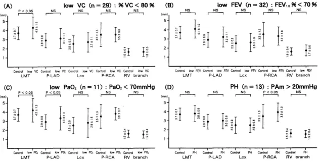
Fig. 4 : Impairment of the ventilatory and blood oxygen concentration, pulmonaary artery pressure, and coronary artery diameter.
(A) Comparison of restrictive ventilatory disease patients (low VC) and normal subjects (Contrl n = 28).
(B) Comparison of obstructive pulmonary disease patients (low FEV,.0) and Control.
(C) Comparison of CPD patients showing hypoxia (low Pa02) and Control.
(D) Comparison of pulmonary hypertension patients (PH) and Control.
Table 2 : Hemodynamics before and after hypoxic challenge
Data Chronic b pulmonary desease (n = 21) Control (n = 10)
efore hypoxia before hypoxia
Pa02 (torr) 88±15.6 47±9.8 96±8.6 48±5.6
Sa02 ( %) 96±1.8 82±8.4 97±0.6 84±4.7
SV02 (Torr) 74±7.1 63±10.1 77±7.7 62±6.3
PAm (mmHg) 13±4.2 19±8.0 12±4.5 16±3.7
TPR (unit) 2.4±1.0 2.8±1.6 2.2±1.1 2.5±0.8
AOm (mmHg) 93±20 101±22 99±16 105±11
CO (L/min) 5.9±2.3 7.1±2.3 5.5±1.2 6.8±1.9
CI (L/min/m2) 3.7±1.2 4.4±1.2 3.5±0.7 4.3±1.0
HR (b. p. m) 75±15 87±17 72±13 82±12
SV (ml) 80±26 82±24 78±19 81±19
PAm : mean pulmonary pressure AOm : mean artery pressure HR : heart rate
*Values are mean±SD.
TPR : total pulmonary resistance CI : cardiac index
SV : stroke volume
(2) CPD were divided according to the pulmonary function into restrictive ventilatory disorders (low VC) and obstructive pulmonary disorders (low FEV). The main trunk of the left coronary artery was significantly dilated in patients with restrictive ventilatory disorders as compared with the normal controls (Fig. 4 (A)), but no significant difference was observed in those with obstruc- tive disorders (Fig. 4 (B)).
(3) In CPD patients who showed hypoxemia (low PaO2), the main trunk of the left coronary artery and the left anterior descending artery were significantly dilated as compared with the controls (Fig. 4 (C)). In those with pulmonary hypertension (PH), the right coronary artery was dilated (Fig. 4 (D)).
The mean coronary stenosis index in the CPD group was less than 1, and few signs of coronary sclerosis were
observed.
Table 3 : Coronary artery diameter before and after hypoxic challenge
Chronic pulmonary disease (n = 21) Control (n = 10)
before hypoxia before Hypoxia
LMT 4.0±1.0 4.2±0.8 3.6±0.7 4.3±0.8
p-LAD 3.0±0.7 3.2±0.6 2.8±0.8 3.2±0.8
d-LAD 2.0±0.5 2.1±0.4 2.0±0.5 2.1±0.5
Lcx 2.4±0.9 2.6±0.9 2.5±0.6 2.7±0.4
p-RCA 3.4±0.9 3.7±0.9 3.3±0.5 3.9±0.5
d-RCA 2.5±0.7 2.6±0.5 2.6±0.6 3.0±0.6
RV-branch 1.6±0.5 1.7±0.5 1.4±0.2 1.7±0.3
* (mm) mean±SD
2. Evaluation after the hypoxic challenge
The hypoxic challenge was performed in 21 patients with CPD and 10 normal controls. Table 2 summarized the hemodynamics and blood gas concentrations in the two groups before and after the challenge. No significant changes were observed in any parameter.
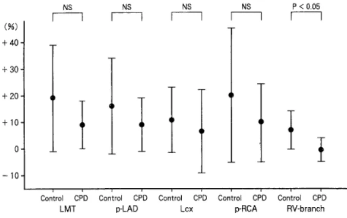
Table 3 showed the coronary artery diameters before and after the challenge. The coronary artery diameter was increased after hypoxic stimulation in both groups, but the increase was significantly greater in the control group at the main trunk of the left coronary artery and the right ventricular branch, and tended to be greater in the control group at the proximal right coronary artery (Fig. 5). The percent change in the coronary diameter [(diameter after hypoxia-diameter before hypoxia)/diameter before hy- poxial was also significantly greater in the control group than in the CPD group at the right ventricular branch (Fig. 6).
Fig. 5: Change in the coronary diameter. [diameter after hypoxia-diameter before hypoxial
DISCUSSION
Cor pulmonale is a serious cardiovascular disorder caused by respiratory diseases. The condition is defined as right ventricular dilation and/or hypertrophy eventually leading to right heart failure due to disturbances of the pulmonary circulation by organic or functional impair- ments of the pulmonary parenchyma or pulmonary vessels (1). The development of cor pulmonale exerts serious effects on the prognosis of CPD so its early diagnosis is imperative. However, it is difficult to evaluate the right ventricular load by routine 12-lead electrocardio-graphy (2). Pulmonary hypertension in CPD is considered to be induced by a combination of organic reduction in the vascular bed due to destruction of lung tissues, and functional reduction in the vascular bed due to hypoxic pulmonary vasoconstriction (HPV). This secondary pulmonary hypertension is considered to cause insuffi- ciency of right ventricular constriction and dilation due to
Fig. 6 : The percent change in the coronary diameter. [(di- ameter after hypoxia-diameter before hypoxia)/diameter before hypoxia I
relative ischemia of the right ventricle, following an increase in the afterload and myocardial hypertrophy (3).
Pulmonary hypertension is reported to increase the
coronary vascular resistance and reduce the coronary blood flow (4). However, Baroldi et al. (5) observed myocardial infarction during autopsy significantly less frequently in patients with pulmonary hypertension than in the control group. Also, Murphy (6) reported that right ventricular hypertrophy was not markedly correlated with the coro- nary artery diameter, and relative myocardial ischemia due to a reduction in the coronary vascular bed was unaccompanied by coronary stenosis. Both of these reports were based on autopsy findings, and few clinical studies have been reported. We therefore sought to elucidate the morphology of the coronary arteries in patients with CPD.
We used coronary angiography to investigate the coro- nary artery diameter in relation to the pulmonary circula- tion, pulmonary function and blood oxygen concentration.
Chronic pulmonary diseases were divided into restrictive disorders and obstructive disorders according to the pulmonary function. Pa02 and mean pulmonary artery pressure both revealed no significant difference.
The cardiac output is usually reduced in patients with restrictive disorders as compared with those with obstruc- tive disorders (7). In this study, however there was no reduction in the group with restrictive disorders, and it was in the normal range in both groups. According to the pulmonary artery pressure, the right coronary artery was dilated, and the main trunk of the left coronary artery also tended to be dilated in the group with pulmonary hypertension. According to the blood oxygen concentra- tion, the main trunk of the left coronary artery and the proximal anterior descending artery were dilated in patients showing hypoxemia. The correlation between the blood gas concentration and the pulmonary artery pressure is well known. In our study, PaO2 was significantly lower in the pulmonary hypertension group, leading us to hy- pothesize that the coronary artery may have dilated to compensate for the reduction in SVO2, therby increasing the cardiac output.
Many ischemic heart diseases are caused by narrowing or obstruction of the lumen of the coronary artery due to coronary sclerosis. Various factors including the sex and age are involved in the initiation and progression of coronary stenosis. Okada and Fukuda (8) showed that patients are at risk of ischemic heart disease when the stenosis index is 5 or above. Sugiura et al. (9) regarded an index of 10 or less to be physiologic in individuals aged 60 years or above, and in his study the index was 5 or less in 35 cases (12.6 %) of 275 autopsy cases. The present study showed a mean index of less than 1, which was lower than has been reported eleswhere. Hypolipidemia and hyper- HDL-cholesterolemia have been reported in patients with CPD (10). This low coronary risk associated with chronic respiratory insufficiency may have contributed the rarity of coronary stenosis among our patients. Our study also revealed that HDL cholesterol was significantly higher (p
< 0.001) in the CPD goup (53.5 ± 10.3 mg/dl) than in the
control goup (34.1±6.0 mg/dl) (11). Also in the hypoxia group, the significant coronary dilation may have sup- pressed the development of coronary sclerosis.
There have been a number of reports on changes in the cardiopulmonary hemodynamics during hypoxia (12, 13).
Additionally, Adachi et al. showed by the microsquare method that the blood flow of the heart is 5.2 % of the cardiac output at a normal PaO2, not significantly differ- ent (6.0 %) when Pa02 was 41.2 mmHg, but was signifi- cantly increased to 12.7 % when PaO2 was 24.3 mmHg (14).
The direct coronary vasodilating effect of hypoxemia is observed when PaO2 is reduced to 40 mmHg or less (15, 16).
However, the coronary artery is reported to be dilated by stimulation with nicotine and cyanide (17) so that the coronary blood flow increased by both direct and indirect effects of hypoxemia. The hemodynamics in patients with CPD during exercise have also been reported, but it is difficult to maintain the exercise load at a fixed level since the patients with CPD include many elderly individuals with reduced lower limb muscle strength. We employed a hypoxic challenge to equalize the degree of hypoxemia and to safely perform coronary angiography under the condi- tion of hypoxia. Coronary angiograms were obtained before and after the challenge. Various methods have been used to establish hypoxia. We induced hypoxia by having the subjects breathe 13 % oxygen for 15 minutes, following
the method of Weizenblum (13). No symptoms of hypoxia were noted in either the CPD group or the control goup.
After the challenge, the cardiac output was increased in both groups despite the increases in the pulmonary vascu- lar resistance, similarly to earlier reports, but no signifi- cant difference was observed in the percent changes of the cardiac output between the two groups.
The diameter of the coronary artery was noted to have increased after the challenge, but the dilation especially of the main trunk of the left coronary artery, right coronary artery, and the right ventricular branch was significantly greater in the control group than in the CPD group. In terms of the percent changes in the coronary artery
diameter between bf ore and after the hypoxic challenge, dilation of the right ventricular branch in the control group was significant. Hypoxia is known to cause hypoxic pulmonary vasoconstriction (HPV) in the pulmonary artery (18, 19). In contrast, the systemic vessels are
dilated during hypoxia. In this study, however, the aortic pressure increased after hypoxia in both groups, probably because the increase in the cardiac output exceeded the dilation of the systemic vassels. The degree of coronary vasodilation was particularly notable in the right ven- tricular branch in the CPD patients. The responsiveness to hypoxia was significantly reduced especially in the right ventricular branch in the patients as compared with normal controls. Chronic hypoxemia may thus reduce the responsiveness of the coronary arteries to hypoxia.
Acknowledgements
The authors greatefully acknowledge Professor Kohei Hara for for reading this manuscript and for his invalu- able comments, and to Associate Professor Toshiyuki Imamura for their critical comments and suggestion. This study was supported in part by the Second Department of Internal medicine Nagasaki University, japan.
REFERENCES
1) NVHO technical report : Chronic cor pulmonal. Circulation 27: 594, 1963 2) Murphy, M. L., Thenabadu, P. N., Soyza, N. D., et al_ Reevaluation of
electro cardiographic criteria for left, right and conbined cardiac
ventri-cular hypertrophy. Am J Cardio, 53: 1140, 1984
3) Morpurgo M, Denolin H. The heart in pulmpnary hypertension due to chronic lung disease in Pulmonary circulation : advances and controver-
sies. C. A. Wagenvoort, H. Denolin Elsevier Science Publishers, New
York, 1989, p. 166.
4) Morrison DA. Pulmonary hypertension in chronic obstructive pulmo- nary disase : the right ventricular hypothesis. Chest 92 : 387. 1987.
5) Baroldi G. The coronary arteries in chronic cor pulmonale. Acta Cardiol 26: 602, 1971
6) Baker BJ. Pathophysiological Aspects of Cor Pulmonale. In: Cor pulmonale in chronic bronchitis and emphysema. Murphy ML, Bone
RC. Mount Kisco : Futura, 1984, p. 65.
7) Imamura T, Hara K, et al. Hemodynamic Status in Patients with Chronic Respiratory Disease. Jpn thoracic dis 61. 3: 220, 1986.
(abstract in English)
8) Okada R, Fukuda K. A clinicopathological study on the coronary
narrowing in Japan. J Jpn Atheroscler Soc 7, 559, 1979. (abstract in English)
9) Sugiura M, Hirooka K, Ohkawa S. Severity of coronary sclerosis in the aged. Apathological study in 968 consecutive autopsy cases. Jpn Heart
J 17: 471, 1976. (abstract in English)
10) Tisi GM, Corrique A, Barret E, Grundy SM . Increased high density lipoprotein cholesterol in obstructive pulmonary disease (predominant
emphysematous type). Metabolism 30: 340, 1981.
11) Chyo Teiritu, Matsunaga K, Yamasa T , Imamura T, Hara K.
Increased Serum HDL-cholesterol in patients with CPD . in press (in
Japanese)
12) Cournand, A. Some aspects of the pulmonary circulation in normal man and in chronic cardiopulmonary disease . Circulation 2 : 641, 1950 13) Witzenblum E, Schrijin F, Mohan-Kumar T , Colas des Francs V,
Lockhart A. Variability of the Pulmonary Vascular Response to Acute Hypoxia in Chronic Bronchitis. Chest 94: 772, 1988.
14) Adachi H, Strauss HW, Ochi H, Wagner HN. The effect of hypoxia on the regional distribution of cardiac output in the dog. Circ Res 39 : 314,
1976.
15) Daugherty RM Jr, Scqott JB, Dabney JM, Hoddy FG. Local effects of 02 and C02 on limb, renal, and coronary vascular resistances . Am J
physiol 213: 1102, 1967.
16) Heistad DD, Abboud FM, Mark AL, et al. Effect of hypoxemia on responses to norepinephrine and angiotensin in coronary and muscular
vessels. J Pharmacol Expther 193: 941, 1975.
17) Hackett JB, Abboud FM, Mark AL, Schmid PD, Heistad DD . Coronary vascular responses to stimulation of chemoreceptors and
baroreceptors; evidence for reflex activation of vagal cholinergic
innervation. Circ Res 31 : 8, 1972.
18) Rendas A, branthwaite M, Lennox S, et al. Response of the pulmonary circulation to acute hypoxia in the growing pig. J Appl Physiol 52 : 811,
1981.
19) Damino KB, Borowec L, Alexander CM, et al. Infuluence of isoflurane on hypoxic pulmonary vasoconstriction.in dog. Anesthesiology 64 : 423,
1986.