Acta Med. Nagasaki 59: 41−45−
Introduction
The use of core needle biopsy (CNB) specimens collected under image guidance has been widely accepted for the di- agnosis of breast cancer. Additionally, CNB specimens can be used to assess the characteristics of tumors, including their lymphovascular invasion, nuclear grading, immuno- histochemical (IHC) findings (estrogen receptor (ER), pro- gesterone receptor (PR), human epidermal growth factor receptor 2 (HER-2), Ki-67), and so on
1, 2. Histological ob-
servations of CNB are sometimes the only information available to determine the therapeutic strategies, i.e., in cas- es with preoperative systemic therapy. Especially in pa- tients with ER+/HER2- tumors, the IHC findings are widely used as surrogate markers to classify luminal A and B tu- mors
3. The problem is whether a small amount of sample obtained by CNB reflects the overall histological features of the cancers that might have intrinsic heterogeneity. In order to evaluate the pathological reliability of CNB specimens for determining the therapeutic strategy for patients with
MS#AMN 07152
Is it practical to determine the therapeutic strategy for breast cancer by evaluating pathological findings in core needle biopsy specimens?
Kosho Y
amanouchi1, Shigeki M
inami1, Sayaka K
uba1, Fusako K
awakami1, Chika S
akimura1, Naoe K
inoshita2, Kuniko A
be2, Tomayoshi H
ayashi2, Tamotsu K
uroki1, Susumu E
guchi11 Department of Surgery, Nagasaki University Graduate School of Biomedical Sciences, Nagasaki, Japan
2 Department of Pathology, Nagasaki University Hospital
Background; Core needle biopsy (CNB) specimens have been widely used not only for the diagnosis of breast cancer, but also for assessing biomarkers, including lymphovascular invasion (ly and v), nuclear grading, the estrogen receptor (ER), proges- terone receptor (PR), human epidermal growth factor receptor 2 (HER-2) and Ki-67. We herein compared the pathological bio- markers of ER+/HER2- invasive breast cancers in CNB with those in the subsequent surgical specimens.
Methods; Patients with ER+/HER2- invasive breast cancer who presented to our department from August 2011 to July 2013 who had CNB and subsequent surgery were included. Lymphovascular invasion (ly, v) and nuclear grading were determined by hematoxylin and eosin staining, and the ER, PgR, HER-2, and Ki-67 status were evaluated by immunohistochemistry.
Results; The concordance rates between CNB and surgical specimens for the ly, v, nuclear grading, ER and PgR were 2.4%, 2.9%, 63.0%, 96.4% and 82.1%, respectively. Lymphovascular invasion and nuclear grading tended to be underestimated with CNB in discordant cases. The Ki-67 labeling index in CNB specimens was strongly correlated with that in surgical specimens (correlation coefficient 0.75, p<0.0001). Consequently, there was a reasonable level of agreement between CNB and surgical specimens for surrogate subtyping (82.1%).
Conclusions; CNB provided reliable information on the expression of hormone receptors, Ki-67 in ER+/HER2- invasive breast cancers. However, because of the substantial discordance between CNB and surgical specimens, the status of lymphovascular invasion and nuclear grading should not be concluded based on CNB specimens.
ACTA MEDICA NAGASAKIENSIA 59: 41−45, 2014 Key words: breast cancer, core needle biopsy, hormone receptor, Ki-67, lymphovascular invasion.
Address correspondence: Kosho Yamanouchi, Department of Surgery, Nagasaki University Graduate School of Biomedical Sciences, 1-7-1 Sakamoto, Nagasaki, 852-8501, Japan.
E-mail: [email protected], Phone: +81-95-819-7316, Fax: +81-95-849-7319 Received March 20, 2014; Accepted June 27, 2014
ER+/HER2- invasive breast cancer, we compared the tumor biomarker findings, including the lymphovascular invasion, nuclear grade, ER, PgR, and Ki67 status in CNB with those obtained in the subsequent surgical specimens.
Patients and methods
We reviewed the clinical and laboratory data of 29 pa- tients with ER+/HER2- primary invasive breast cancer de- termined by findings of surgical specimens, who underwent CNB and subsequent lumpectomy/mastectomy between August 2011 and July 2013 in our department. Patients were excluded if they were treated with systemic or local therapy before surgery. CNBs were performed under ultrasound guidance and were taken with an automated biopsy device fitted with 14-gauge needles (C. R. Bard, Inc., Covington, Georgia, U.S.A). The median number of CNBs was three (range; 2-3), and the median interval between CNB and sur- gery was 34 days (range; 20-61). CNB and surgical speci- mens were fixed in neutral buffered formalin and embedded in paraffin before the histopathological analysis. Fixative durations were 4 to 24 hours for the CNB samples and 24 to 48 hours for the surgical specimens.
Processing of samples
Five sections were prepared from each paraffin block.
One slide was stained for hematoxylin and eosin (HE), and the remaining slides were kept for IHC staining. In the IHC analysis, sections were incubated with antibodies against the ER (clone SP1, Ventana Medical Systems, Inc., AZ, USA.), PgR (clone 1E2, Ventana Medical Systems), and Ki- 67 (clone MIB1, DAKO, Tokyo, Japan). Blots were devel- oped by the labeled streptavidin biotinylated antibody method using automated staining system (BenchMark XT, Ventana Medical Systems).
Pathological evaluation
The histological findings of the surgical specimen were taken as the gold standard. Although the existence of lym- phovascular invasion (ly and v) in cancers was basically evaluated by HE staining, Elastica van Gieson staining or IHC analysis for anti-D2-40 was performed in the obscure cases with HE staining. The nuclear grading was based on the sum of the nuclear atypia score and the mitotic count score as follows
4: Grade 1: 2 or 3 points, Grade 2: 4 points, Grade 3: 5 or 6 points. The nuclear atypia score was graded
from 1 to 3 points according to the variation in the size and shape of nuclei and unevenness of chromatin. The mitotic counts score was graded from 1 to 3 points, increasing with the mitoses in the area in which mitoses were most abundant. The ER- and PgR-positive cell counts were determined by the J- score as follows
5: J-Score 0: not stained; J-Score 1: stained cells <1%; J-Score 2: stained cells ≥ 1% but <10%; J-Score 3a:
stained cells ≥10% but <50% and ;J-Score 3b: stained cells
≥50%. The Ki-67 labeling index (LI) was measured in ap- proximately 1,000 malignant invasive cells counted in hot spots in a high-power field (400 × )
6. The IHC-based surrogate sub- typing was determined using the following definitions adopted by the 2013 St. Gallen Consensus Panel
3: Luminal A-like; ER positive / PgR positive (≥ 20%) / HER2 negative / Ki-67<14%, Luminal B-like; ER positive / HER2 negative, and at least one of: Ki-67 ≥14%, PgR negative or low.
Statistical analysis
The agreement of the ly, v, nuclear grading and hormonal receptors was tested using the kappa test. The agreement of the Ki-67 LI between the results from CNB and those from surgical samples were statistically analyzed using Pearsonʼs correlation coefficient test. The results obtained were con- sidered to be significant at p < 0.05.
Results
Lymphovascular invasion
Pathological ly and v was identified in the CNB specimen in 21 of 29 cases, while the remaining eight CNB samples were too little to evaluate these parameters. The concor- dance rates for ly and v between CNB and surgical samples were only 23.8%, with a kappa value of 0.07 (Table 1) and 28.6%, with a kappa value of -0.18 (Table 2), respectively.
In 71.4% of cases, ly was underrated on CNB samples, and the v was underdiagnosed in 57.1%. Notably, ly and v were estimated to be absent in 90.5% and 76.2% of cases in CNB samples, although the final rates of absence in surgical sam- ples were only 28.6% and 38.1% respectively.
Nuclear grading
The nuclear grading was evaluated in 27 cases in both CNB and subsequent surgical samples, and there was 63.0%
(17 out of 27 cases) agreement, with a kappa of 0.41 (Table
3). The grading was underrated in 33.3% of the cases, while
it was overrated in only 3.7% of the cases.
43 Kosho Yamanouchi et al.: Diagnostic value of core needle biopsy
Estrogen and progesterone receptor status
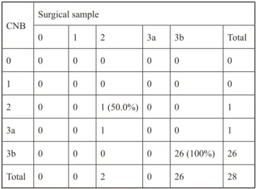
Based on the surgical samples, the ER was expressed in 100% of the cases (Table 4), while PgR was expressed in 85.7% (24/28) (Table 5). The concordance rate of the J-score was 96.4% (27/28), with a kappa of 0.74, for the ER and was 82.1% (23/28) for the PgR, with a kappa of 0.68.
Ki-67 labeling index
The correlation coefficient of the Ki-67 LI between the CNB and surgical samples was 0.75 (p<0.001, 95% CI 0.61 – 1.28) (Figure 1).
CNB Surgical sample
0 1 2 3 Total
0 5 (83.3%) 8 4 2 19
1 1 0 (0%) 0 0 1
2 0 0 0 (0%) 1 1
3 0 0 0 0 (0%) 0
Total 6 8 4 3 21
Table 1. Lymphatic invasion
Percentages in parentheses indicate the degree of concordance.
CNB Surgical sample
0 1 2 3 Total
0 5 (62.5%) 8 3 0 16
1 3 1 (11.1%) 1 0 5
2 0 0 0 (0%) 0 0
3 0 0 0 0 (0%) 0
Total 8 9 4 0 21
Table 2. Vessel invasion
Percentages in parentheses indicate the degree of concordance.
CNB Surgical sample
1 2 3 Total
1 12 (92.3%) 0 5 17
2 1 2 (100%) 4 7
3 0 0 3 (25.0%) 3
Total 13 2 12 27
Table 3. Nuclear grading
Percentages in parentheses indicate the degree of concordance.
CNB Surgical sample
0 1 2 3a 3b Total
0 0 0 0 0 0 0
1 0 0 0 0 0 0
2 0 0 1 (50.0%) 0 0 1
3a 0 0 1 0 0 1
3b 0 0 0 0 26 (100%) 26
Total 0 0 2 0 26 28
Table 4. Degree of estrogen receptor expression by J-Score
Percentages in parentheses indicate the degree of concordance.
CNB Surgical sample
0 1 2 3a 3b Total
0 4 (100%) 0 0 0 0 4
1 0 0 1 0 0 1
2 0 0 0 (0%) 0 0 0
3a 0 0 1 3 (100%) 3 7
3b 0 0 0 0 16 (84.2%) 16
Total 4 0 2 3 19 28
Table 5. Degree of progesteron receptor expression by J-Score
Percentages in parentheses indicate the degree of concordance.
44 Kosho Yamanouchi et al.: Diagnostic value of core needle biopsy
Surrogate subtyping
The concordance rate of the surrogate subtyping based on the IHC findings was 82.1% (23 out of 28 cases), with a kappa of 0.38 (Table 6). All five of the remaining cases were evaluated as Luminal A-like based on the CNB and Luminal B-like based on the corresponding surgical sam- ples, four of which showed discordance in the Ki-67 LI and one in the PgR expression.
Discussion
Breast cancer is regarded as a systemic disease, and most patients require individualized systemic therapy before or after surgery, based on the biology of the tumor
3. Addition- ally, the most favorable treatment varies depending on the progression of the disease. CNB specimens have been wide- ly used for the diagnosis of breast lesions that can be seen under image guidance. It is sometimes necessary to predict the prognosis or determine the therapeutic strategies of the patients with primary invasive breast cancer by evaluating
only CNB samples, for instance, in cases where preopera- tive systemic therapy has been given, or in cases with ac- companying metastatic disease.
Lymphovascular invasion is a prognostic factor
7or a pre- dictive factor for loco-regional recurrence in node-negative breast cancer
8and its initial precise evaluation is desirable.
In this study, ly and v were accurately diagnosed in only 23.8% and 28.6% of CNB samples, respectively. Lymphatic vessel invasion in invasive breast cancer is seen only in the peripheral areas, not in the central areas of tumors in re- sected specimens
9, and this heterogeneous distribution might be one of the reasons for the low accuracy by CNB.
In early breast cancer patients, a high nuclear grade is as- sociated with occult metastases and isolated tumor cells in non-sentinel lymph nodes
10. Our concordance rate for nu- clear grading was 63.0%, and this value was comparable to that reported by Badoual et al. and Harris et al
11, 12. Lym- phovascular invasion and nuclear grading tended to be un- derestimated with CNB, as reported previously
13, 14. In terms of the number of CNB, the accuracy of the histologi- cal grading or hormone receptor status increased with the number of CNB passes
15, 16. This implies that the portions sampled and number of passes of CNB are relevant for the precise evaluation of the cancer characteristics.
One of the purposes of preoperative systemic therapy is to permit breast-conserving surgery for patients who would otherwise require total mastectomy. Additionally, preop- erative systemic therapy can provide information on the drug sensitivity, which could be beneficial during the subse- quent treatment. Rapidly proliferating tumor subtypes, such as the nonluminal HER2 type and triple negative dis- eases, could subsequently turn into a pCR with systemic cytotoxic chemotherapy
3. Preoperative hormone therapy is, in general, carried out for the patients with hormone re- ceptor-positive breast cancer, for whom chemotherapy is unsuitable
17. ER+/HER2- invasive breast cancer consists of heterogeneous entities, and the tumor subtypes vary ac- cording to the pathological biomarkers, including the de- gree of ER and PgR expression, nuclear grading and mark- ers of proliferation such as Ki-67 LI. In randomized trials of preoperative hormone therapy in postmenopausal pa- tients with ER+ breast cancer, higher ER levels significantly correlated with a higher response
18. On the other hand, the degree of expression of the ER and PgR was inversely cor- related with the response to neoadjuvant chemotherapy
19. These reports suggest that the proportion of hormone recep- tor expression might be more useful to predict the therapeu- tic efficacy and to determine the therapeutic strategy than a dichotomous evaluation of their expression.
Figure 1 (Yamanouchi, K)
0 20 40 60 80
0 10 20 30 40 50 60 70
CNB
Surgical samples
(%) (%)
Figure 1. Comparison of the Ki-67 labeling index between CNB and surgical specimens (Pearsonʼs correlation coefficient test, cor- relation coefficient 0.75, p<0.0001).
CNB Surgical sample
Luminal-A like Luminal-B like Total
Luminal-A like 2 (100%) 5 7
Luminal-B like 0 21 (80.8%) 21
Total 2 26 28
Table 6. Surrogated subtyping
Percentages in parentheses indicate the degree of concordance.
45 Kosho Yamanouchi et al.: Diagnostic value of core needle biopsy
In our study, the proportion of ER and PgR expression showed good concordance rates (96.4 and 82.1%, respec- tively) between CNB and surgical samples, comparable to the findings of previous reports. With respect to prolifera- tion, hormone therapy and chemotherapy showed similar clinical response rates in the patients with a low Ki-67 LI, but patients with a high Ki-67 LI had a better response with chemotherapy
20. The 2013 St. Gallen Consensus Panel strongly endorsed preoperative hormone therapy for post- menopausal patients with highly positive expression of hor- mone receptors and disease with a low proliferation rate
3. We demonstrated that the Ki-67 LI in CNB was positively correlated with that in surgical samples (correlation coeffi- cient 0.75, p<0.0001).
In addition to the ER and Ki-67 LI, the PgR expression has been considered as the factor that distinguishes Luminal A- like and Luminal B-like/HER2-negative disease, based on the 2013 St. Gallen Consensus Panel. The concordance rate of the surrogate subtyping based on the IHC findings was 82.1% (23 out of 28 cases) in our study. The remaining five cases showed discordance due to an underestimation of the Ki-67 LI or PgR. The problem with the Ki-67 LI and PgR measurement is that it is not standardized among laborato- ries, and the former is lacking a consensus regarding the threshold for the use of adjuvant cytotoxic chemotherapy for patients with ER+/HER2- disease
3. That is why the ready availability of multigene molecular assays is expected to help provide accurate and reproducible prognostic information and prediction of the response to chemotherapy.
In conclusion, since the ER, PgR and Ki-67 LI, all of which are factors defining surrogate subtyping, were well correlated between CNB and surgical samples, the appro- priate systemic therapy could be determined based on the findings of CNB specimens. On the other hand, since the lymphovascular invasion and nuclear grading were fre- quently discordant, their status should be determined based on the surgical samples whenever possible.
References
1. Tamaki K, Sasano H, Ishida T, et al. Comparison of core needle biopsy (CNB) and surgical specimens for accurate preoperative evaluation of ER, PgR and HER2 status of breast cancer patients. Cancer Sci 101:
2074-2079, 2010
2. Rakha EA, Ellis IO. An overview of assessment of prognostic and predictive factors in breast cancer needle core biopsy specimens. J Clin Pathol 60: 1300-1306, 2007
3. Goldhirsch A, Winer EP, Coates AS, et al. Personalizing the treatment of women with early breast cancer: highlights of the St Gallen Interna- tional Expert Consensus on the Primary Therapy of Early Breast Can- cer 2013. Ann Oncol 24: 2206-2223, 2013
4. Sakamoto G, Inaji H, Akiyama F, et al. General rules for clinical and pathological recording of breast cancer 2005. Breast cancer12 Suppl:
S1-27, 2005
5. Kurosumi M. Immunohistochemical assessment of hormone receptor status using a new scoring system (J-Score) in breast cancer. Breast cancer 14: 189-193, 2007
6. Dowsett M, Nielsen TO, AʼHern R, et al. Assessment of Ki67 in breast cancer: recommendations from the International Ki67 in Breast Can- cer working group. J Natl Cancer Inst103: 1656-1664, 2011
7. Lee AH, Pinder SE, Macmillan RD, et al. Prognostic value of lympho- vascular invasion in women with lymph node negative invasive breast carcinoma. Eur J Cancer 42: 357-362, 2006
8. Sundquist M, Thorstenson S, Klintenberg C, Brudin L, Nordenskjold B. Indicators of loco-regional recurrence in breast cancer. The South East Swedish Breast Cancer Group. Eur J Surg Oncol 26: 357-362, 9. Ito M, Moriya T, Ishida T, et al. Significance of pathological evaluation 2000 for lymphatic vessel invasion in invasive breast cancer. Breast cancer 14: 381-387, 2007
10. Takeshita T, Tsuda H, Moriya T, et al. Clinical implications of occult metastases and isolated tumor cells in sentinel and non-sentinel lymph nodes in early breast cancer patients: serial step section analysis with long-term follow-up. Ann Surg Oncol 19: 1160-1166, 2012
11. Badoual C, Maruani A, Ghorra C, Lebas P, Avigdor S, Michenet P.
Pathological prognostic factors of invasive breast carcinoma in ultra- sound-guided large core biopsies-correlation with subsequent surgical excisions. Breast 14: 22-27, 2005
12. Harris GC, Denley HE, Pinder SE, et al. Correlation of histologic prognostic factors in core biopsies and therapeutic excisions of inva- sive breast carcinoma. Am J Surg Pathol 27: 11-15, 2003
13. Park SY, Kim KS, Lee TG, et al. The accuracy of preoperative core biopsy in determining histologic grade, hormone receptors, and hu- man epidermal growth factor receptor 2 status in invasive breast can- cer. Am J Surg 197: 266-269, 2009
14. Sharifi S, Peterson MK, Baum JK, Raza S, Schnitt SJ. Assessment of pathologic prognostic factors in breast core needle biopsies. Modern pathology 12: 941-945, 1999
15. McIlhenny C, Doughty JC, George WD, Mallon EA. Optimum num- ber of core biopsies for accurate assessment of histological grade in breast cancer. Br J Surg 89: 84-85, 2002
16. Greer LT, Rosman M, Mylander WC, et al. Does breast tumor hetero- geneity necessitate further immunohistochemical staining on surgical specimens? J Am Coll Surg 216: 239-251, 2013
17. Colleoni M, Montagna E. Neoadjuvant therapy for ER-positive breast cancers. Ann Oncol 23 Suppl 10: x243-x248, 2012
18. Smith IE, Dowsett M, Ebbs SR, et al. Neoadjuvant treatment of post- menopausal breast cancer with anastrozole, tamoxifen, or both in combination: the Immediate Preoperative Anastrozole, Tamoxifen, or Combined with Tamoxifen (IMPACT) multicenter double-blind ran- domized trial. J Clin Oncol 23: 5108-5116, 2005
19. Colleoni M, Bagnardi V, Rotmensz N, et al. Increasing steroid hor- mone receptors expression defines breast cancer subtypes non respon- sive to preoperative chemotherapy. Breast Cancer Res Treat 116: 359- 369, 2009
20. Alba E, Calvo L, Albanell J, et al. Chemotherapy (CT) and hormono- therapy (HT) as neoadjuvant treatment in luminal breast cancer pa- tients: results from the GEICAM/2006-03, a multicenter, randomized, phase-II study. Ann Oncol 23: 3069-3074, 2012