Ⅰ.Introduction
The polychaetes of Siboglinidae in Annelida are divided into the following three groups: Frenulata (beard worm), Vestimentifera (tube worm), and Monilifera (Sclerolinum). Although they have neither a mouth nor a digestive tract, they harbor chemosynthetic bacteria in their body (Bright and Giere, 2005; Southward et al., 2005). The symbiosis of the siboglinid polychaetes with bacteria was confirmed by enzyme histochemistry for the first time in tube worms that inhabit hydrothermal vents. It was suggested that the bacteria were sulfur-oxidizing ones (Cavanaugh et al., 1981). In the trophosome of tube worms, the existence of sulfur has been investigated in detail. In Lamellibrachia sp.
inhabiting the sea bottom, where hydrogen sulfide and
methane seep, there is variation in the morphology of symbiotic bacteria (Naganuma et al., 1997). Using an X-ray microanalyzer, the sulfur content has been shown to vary among bacteria of different morphology. In addition, Pflugfelder et al. (2005) noted variation in the color of the trophosome in Riftia pachyptila. Using an electronic energy-loss spectroscopy, they found that the sulfur content of synthetic bacteria was different in individuals of different color.
Also in Frenulata, symbiotic bacteria are regarded as sulfur-oxidizing (Southward et al., 1986; Schulze and Halanych, 2003). In this group, however, there is no direct evidence of which symbiotic bacteria are sulfur-oxidizing. In this study, we tried to detect the existence of sulfur from the trophosome of Oligobrachia mashikoi using an X-ray analytical microscope.
1Division of Biodiversity, Noto Marine Laboratory, Institute of Nature and Environmental Technology, Kanazawa University, Kakuma-machi, Kanazawa, 920-1192 Japan
2Department of Life Science, Gradulate School of Natural Science and Technology, Kanazawa University, Kakuma-machi, Kanazawa, 920-1192 Japan
3Kanazawa University Health Center, Kakuma-machi, Kanazawa, 920-1192 Japan
4Division of Biological Measurement and Applications, Institute of Nature and Environmental Technology, Kanazawa University, Kakuma-machi, Kanazawa, 920-1192 Japan
日本海域研究,第40号,13-18ページ,2009 Nihon-Kaiiki Kenkyu, vol. 40, p. 13-18, 2009
Detection of Sulfur Using an X-ray Analytical Microscope from the Trophosome of a Beard Worm, Oligobrachia mashikoi, Frenulate, Siboglinidae
Yuichi SASAYAMA1, Yoshihiro FUKUMORI2, Hajime NAKABAYASHI3 and Nobuaki SHIMIZU4
2008年9月5日受付,Recceived 5 September 2008 2008年11月18日受理,Accepted 18 November 2008
Abstract
In one species of Frenulata which has neither a mouth nor a digestive tract, element analysis of the part including the trophosome, in which chemosynthetic bacteria are harbored, and of a non-trophosome not including the bacteria was conducted using an X-ray analytical microscope. As a result, signals of sulfur were detected predominantly from the part including the trophosome.
Key Words: Siboglinidae polychaetes, Oligobrachi maoshikoi, X-ray analytical microscope, sulfur, chemosynthetic bacteria, trophosome
Ⅱ.Materials and Methods
O. mashikoi was collected from the sea bottom at a 25 m depth in Tsukumo Bay, Noto Peninsula, Ishikawa Prefecture, using a previously described method (Sasayama et al., 2003). They were preserved in 70%
ethanol after fixation with a 4% paraformaldehyde PBS solution for about 12 hours.
The samples were taken to the Horiba, Ltd., Technical Plaza (Tokyo), where an attempt was made to detect sulfur from the trophosome using an X-ray analytical microscope (XGT-5000, Horiba, Ltd., Kyoto), which is an advanced type of X-ray fluorescence spectrometer. The principle of the X-ray analytical microscope is to detect the fluorescence X-rays excited from materials irradiated with X-rays, to identify elements by the specific wavelength of the fluorescence X-ray of each element, and to quantify the element by its wave strength. Every element from sodium to uranium can be detected at the same time.
Each sample, both with and without the trophosome, was about 1.5cm long. The samples were supported with a mylar membrane to prevent evaporation of the 70% alcohol and placed on a sample stage. We observed the samples through a microscope display and
decided which parts should be irradiated by X-rays.
The diameter of the sample containing a trophosome was 0.66 mm. Five spots upon which X-rays were perpendicularly irradiated were identified (Fig.1a). The diameter of the sample not containing the trophosome was 0.73 mm, and seven spots were identified (Fig.1b).
In this study, the diameter of the X-ray guide tube was set at 10 µm in order to examine the smallest spots possible. The electric current and the voltage were set to 1 mA and 50kV, respectively. The irradiation time on any one spot was set to 100 seconds. The pulse processing time was adjusted for the P3 level to efficiently detect only major elements. These conditions can detect only masses of sulfur element, not sulfur included in an amino acid. The quantity of elements was calculated using the fundamental parameter method.
After observation with this microscope, the samples were remade into paraffin sections of 10 µm in thickness by a routine method and stained with hematoxylin and eosin. This procedure is necessary to identify the tissue inside of the X-ray-irradiated spot, as shown in Fig.1.
Ⅲ.Results
1) Part containing the trophosome
In Figure 2, cross section of a specimen observed under an X-ray analytical microscope and X-ray irragiated parts are shown; it corresponds to spots 1 to 5 in Figure 1a.
Fig.1a Part of the whole body containing the trophosome. Bar = 0.3mm. 1b Part of the whole body not containing the trophosome.
Bar = 0.3mm. The spots and numbers correspond to the parts irradiated with X-rays.
Fig.2 X-ray-irradiated parts (1 to 5) in the specimen containing the trophosome. DV:
dorsal vessel; BC: bacteriocytes. Bar = 0.1 mm.
BC
a
b
Figure 3 shows element spectrum from irradiation spots 1 to 5.
As is evident in Figure 3, only sulfur was detected, and the other elements were equal to or less than the detection limit. The broad peak observed always on the right side of the sulfur peak is rhodium, which is a constituent element of an X-ray gun used for the generation of X-rays. Therefore, it is not a true ingredient of the specimen.
At spot 1, when the X-rays irradiated the upper end of the dorsal blood vessel, no sulfur was detected.
From spot 2, however, in which the X-rays irradiated through the dorsal blood vessel, sulfur was detected.
The value of the X-ray intensity was 6.90 cps/mA. In spot 3, the value was 11.90 cps/mA. Furthermore, a higher level of sulfur was detected from spot 4, which is the central part of the trophosome. The value was 13.84 cps/mA. Even at spot 5, which is the basal part of the trophosome, the sulfur density still showed a high value, 11.30 cps/mA.
2) Part not including the trophosome
In Figure 4, the part inside of the specimen that was X-ray-irradiated is shown; it corresponds to spots 1 to 7 in Figure 1b.
Fig.4 X-ray-irradiated parts in a specimen not including the trophosome. Bar = 0.1 mm. SB:
sperm bundles; SD: sperm duct SB
SD SD
Fig.3 Element distribution patterns obtained in spots 1 to 5.
Spot 1
Spot 2
Spot 3
Spot 4
Spot 5
Figure 5 shows element detection patterns from irradiation spots 1 to 7.
The non-trophosome part included a testis. In spots 3, 4, and 5, sulfur was detected. Their X-ray intensity levels were, however, rather low, i.e., 4.16, 7.41, and 7.91 cps/mA, respectively.
Ⅳ.Discussion
Recently, we analyzed the mud from the sea bottom of the bay inhabited by O. mashikoi and detected
hydrogen sulfide, although the concentration was not high (Sasayama et al., 2007). On the other hand, we succeeded in the analysis of the three-dimensional structure of the extra-cellular hemoglobin of this species (Numoto et al., 2005). In this molecule, there was a non-paired cysteine residue, and it was suggested that the sulfur of the hydrogen sulfide was bound by this residue In the part containing the trophosome, a peak of the sulfur was at first detected in spot 2, where X-rays irradiated the central part of the dorsal vessel. This fact appears to reflect the existence of large quantities of Fig.5 Element distribution patterns obtained in the part not containing the trophosome in spots 1 to 7.
Spot 6
Spot 7 Spot 5
Spot 4 Spot 3 Spot 2 Spot 1
hemoglobin bound with sulfur. In addition, in the part not containing the trophosome, sulfur was detected at spots 3, 4, and 5. In these regions, no large blood vessels were found. However, considering that the peak height is similar to that in spot 2 of the dorsal vessel in the trophosome, hemoglobin bound with sulfur might be present in the capillary blood vessels. Furthermore, in the part containing the trophosome, high peaks of sulfur were obtained at spots 3 and 4, where X-rays irradiated the trophosome. The peak of sulfur was still high at spot 5 of the basal part of the trophosome.
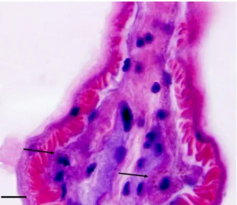
Although the existence of symbiotic bacteria is not clear in the resolution of the light microscope used in this study, it is known that, in so-called bacteriocytes, which harbor chemosynthetic bacteria, there are specific lysosomal granules (Southward, 1993; Deguchi et al., 2007). Therefore, the regions in which such granules can be recognized must be bacteriocytes (Fig.6).
Consequently, it is probable that the chemosynthetic bacteria living in O. mashikoi are sulfur-oxidizing bacteria.
Acknhowledgments: We thank Mr. Kenichi Watanabe (Horiba, Ltd., Nagoya, the chief manager at chemical machinery team), who kindly arranged for us to use the X-ray analytical microscope. We also thank Ms.
Tomoko Nishikawa (an application engineer with Horiba Ltd., Technical Plaza, Application Center), who operated
the microscope and provided a thorough explanation of the procedure.
References
Bright, M. and Giere, O., 2005: Microbial symbiosis in Annelida. Symbiosis, 38, 1-45.
Cavanaugh, C.M., Gardiner, S.L., Jones, M.L., Jannasch, H.W.
and Watervury, J.B., 1981: Prokaryotic cells in the hydrothermal vent tube worm Riftia pachyptila Jones:
Possible chemoautotrophic symbionts. Science, 213, 340-342.
Deguchi, M., Kubota, N., Matsuno, A., Kanemori, M., Fukumori, Y., and Sasayama, Y., 2007: Actual distribution of bacteriocytes in the trophosome of a beard worm (Oligobrachia mashikoi, Siboglinidae, Annelida):
Clarification using whole-mount in situ hybridization. Acta Zool., 88, 129-135.
Naganuma, T., Naka, J., Okayama, Y., Minami, A., and Horikoshi, K., 1997: Morphological diversity of the microbial population in a vestimentiferan tubeworm. Mar.
Biotechnol., 5, 119-123.
Numoto, N., Nakagawa, T., Kita, A., Sasayama, Y., Fukumori, Y., and Miki, K., 2005: Structure of an extracellular giant hemoglobin of the gutless beard worm Oligobrachia mashikoi. Proc. Nat. Acad. Sci. USA, 102, 14521-14526.
Pflugfelder, B., Fisher, C.R. and Bright, M., 2005: The color of the trophosome: elemental sulfur distribution in the endosymbionts of Riftia pachyptila (Vestimentifera;
Siboglinidae). Mar. Biol., 146, 895-901.
Sasayama, Y., Matada, M., Fukumori, Y., Umebayashi, M., Matsuno, A., Nakagawa, T. and Imajima, M., 2003: External morphology of the posterior end, the “opisthosoma”, of the beard worm Oligobrachia mashikoi (Pogonophora). Zool.
Sci., 20, 1411-1416.
Sasayama, Y., Higashide, Y., Sakai, M., Matada, M., and Fukumori, Y., 2007: Relationship between the lifestyle of a Siboglinidae polychaete, Oligobrachia mashikoi (also known as Pogonophora), and the total sulfide/nitrogen levels in its habitat. Zool. Sci., 24, 131-136.
Schulze, A., and Halanych, K.M., 2003: Siboglinid evolution shaped by habitat preference and sulfide tolerance.
Hydrobiologia, 496, 199-205.
Southward, E.C., 1993: Pogonophora In “Microscopic Anatomy of Invertebrates” ed. by FW Harrison and ME Rice.
Vol.12, pp.327-369. Wiley-Liss, New York.
Southward, E.C., Schulze, A., and Gardiner, S.L., 2005:
Fig. 6 A photograph showing the presence of bacteriocytes (arrows) in the trophosome, which include lysosomes (red-colored small granules) around a nucleus (dark-bluish large granule). Bar = 0.02mm.
Pogonophora (Annelida): form and function. Hydrobiologia, 535/536, 227-251.
Southward, A.J., Southward, E.C., Dando, P.R., Barrett, R.B.,
and Ling, R.L., 1986: Chemoautotrophic function of bacterial symbionts in small Pogonophora. J. Mar. Biol. Ass.
U. K., 66, 415-437.
Siboglinidae 科, Frenulate 類のマシコヒゲムシ,Oligobrachia mashikioiの 栄養体から,X線分析顕微鏡を用いてイオウ元素の検出
笹山雄一1・福森義宏2・中林 肇3・清水宣明4
要 旨
ヒゲムシ類は口も消化管も無い。そのかわり,体内に化学合成細菌を共生させ,それが生産 する炭水化物をもらい生きている。一方,共生細菌は,これまで 16SrDNA の塩基配列や生息 環境に硫化水素が存在することなどより,酸化エネルギー基質としてイオウを要求するイオウ 酸化細菌であると考えられてきた。本研究ではマシコヒゲムシを用いて,細菌を共生させてい る栄養体を含む部分と,栄養体を含まない部分をX線分析顕微鏡で元素分析を行った。その結 果,共生細菌が含まれる部分からイオウ元素が検出された。このことは,宿主が環境にある硫 化水素を栄養体に棲む共生細菌まで血流によって運び,細菌にそれをわたしている可能性を示 唆している。また直接,イオウが検出されたことにより,この細菌はイオウ酸化細菌であるこ とをさらに補強するものである。
1金沢大学環日本海域環境研究センター生物多様性研究部門(能登臨海実験施設) 〒920-1192 石川県金沢市角間町
2金沢大学大学院自然科学研究科生命科学専攻 〒920-1192 石川県金沢市角間町
3金沢大学保健管理センター 〒920-1192 石川県金沢市角間町
4金沢大学環日本海域環境研究センター生体機能計測部門 〒920-1192 石川県金沢市角間町