Anticancer Activity of Atractylodes lancea (Thunb.) DC. in Hamster Model and Application of PET-CT for Early Detection
Asian Pac J Cancer Prev, 16 (15), 6279-6284
Introduction
Cholangiocarcinoma (CCA) or bile duct cancer is a malignant cancer of the biliary duct system that originates in the liver and extrahepatic bile ducts which terminate at the ampulla of Vater (Mosconi et al., 2009). Recent statistics showed an increasing worldwide incidence and mortality rate of 3% of all gastrointestinal cancers, and is the second usual primary hepatic tumor (Rizvi and Gores, 2013). Most CCA cases occur in populations aged over 60 years with yearly incidence of approximately 5.5 and 7.3 cases per 100,000 populations in Japan and Israel, and 1-2 cases per 100,000 populations in the UK and the USA, respectively (Singh and Facciuto, 2012).
The highest incidence rate is reported in the northeastern region of Thailand particularly in Khon Kaen province, with age standardized rate of 89.2 per 100,000 in men, and 37.4 per 100,000 in women (range: 93.8 to 317.6 per 100,000) (Sriamporn et al., 2004). The major cause of CCA in Thailand is consumption of improperly cooked and fermented fresh water cyprinoid fish called ‘Pla-ra’,
1Center of Excellence in Pharmacology and Molecular Biology of Malaria and Cholangiocarcinoma, Chulabhorn International College of Medicine, Bangkok, Thailand, 2Division of Radiation Biology and Protection, Radioisotope Center, 3Department of Clinical Product Development, 4Department of Immunogenetics, Institute of Tropical Medicine (NEKKEN), Nagasaki University, Nagasaki, Japan *For correspondence: [email protected]
Abstract
Opisthorchis viverrini (OV)-induced cholangiocarcinoma (CCA) is an important cancer in the Great Mekong
region, particularly in Thailand. Limitations of treatment options and the lack of an effective diagnostic tool for early detection of CCA are major concerns for the control of this type of cancer. The aim of the study was to investigate anti-CCA activity of the ethanolic extract of Atractylodes lancea (Thunb.) DC., and the applicability of positron emission tomography-computed tomography (PET-CT) as a tool for detection and monitoring the progression of CCA in Opisthorchis viverrini (OV)/dimethylnitrosamine (DMN)-induced CCA hamsters. Male Syrian hamsters were used for toxicity tests and anti-CCA activity evaluation. Development of CCA was induced by initial feeding of 50 metacercariae of OV, followed by drinking water containing 12.5 ppm of DMN in hamsters.
The ethanolic extract of A. lancea (Thunb.) DC. was administered orally for 30 days. PET-CT was performed every 4 weeks after initiation of CCA using 18F-fluorodeoxyglucose (
18F-FDG). Results from the present study suggest that the ethanolic extract of A. lancea (Thunb.) DC. rhizome exhibited promising anti-CCA activity and safety profile in the OV/DMN-induced hamster model. To successfully apply PET-CT as a tool for early detection of tumor development and progression, modification of radiolabeling approach is required to improve its specificity for CCA cells.
Keywords: Cholangiocarcinoma - anticancer - PET-CT - Atractylodes lancea (Thunb.) DC
RESEARCH ARTICLE
Anticancer Activity of Atractylodes lancea (Thunb.) DC in a Hamster Model and Application of PET-CT for Early Detection and Monitoring Progression of Cholangiocarcinoma
Tullayakorn Plengsuriyakarn
1, Naoki Matsuda
2, Juntra Karbwang
3, Vithoon Viyanant
1, Kenji Hirayama
4, Kesara Na-Bangchang
1*
‘Pla-som’, or ‘Koi-pla’ which contains the liver fluke Opisthorchis viverrini (OV) and the pre-carcinogen nitrosamine (Sithithaworn et al., 2012). The risk factors in western countries are infection with hepatic C virus and primary sclerosing cholangitis (PSC) (Khan et al., 2008).
The dried rhizomes of Atractylodes lancea (Thunb.) DC. (Khod-Kha-Mao or Cang Zhu), family Compositae, is a common medicinal plants used in traditional Chinese medicine and Japanese campo, as well as in Thai traditional medicine in drug formulation for treatment of fever, colds, flu, sore throat, rheumatic diseases, digestive disorders, night blindness, and influenza (Chayamarit, 1995; Xio, 2002). The major constituents are atractylodin, β-eudesmol, atractylon, and hinesol (Ouyang et al., 2007).
β-eudesmol has been reported to be the main and active
component of A. lancea (Thunb.) DC (Ouyang et al.,
2007; Chen et al., 2007). These compounds including
the rhizome extract have been shown to possess several
pharmacological activities including anti-inflammatory
(Resch et al., 2001), anti-angiogenic (Tsuneki et al.,
2005), antimicrobial (Wang et al., 2009; Chen et al.,
2012), antihypertensive (Plengsuriyakarn et al., 2012), anti-ulcer and inhibitory activities on gastric secretion (Kubo et al., 1983; Nogami et al., 1986), as well as activity on nervous system (Nojima et al., 1992). Results of our previous studies demonstrated promising anti-CCA activity of the ethanolic extract of A. lancea (Thunb.) DC. (rhizome) in various in vitro and in vivo models.
The cytotoxic activity against the CCA cell line CL-6 was shown with IC
50(concentration that inhibits cell growth by 50%) of 24.09 µg/ml. In addition, the extract also produced inhibitory activity on tube formation, cell invasion (Mahavorasirikul et al., 2010; Plengsuriyakarn et al., 2012), and anti-CCA activity in CCA-xenografted nude mice at all dose levels (1,000, 3,000, and 5,000 mg/
kg body weight) (Plengsuriyakarn et al., 2012).
Besides the limitation of effective chemotherapeutics, one of the major problems for the treatment and control of CCA is the lack of effective diagnostic tool for early detection of the cancer. The imaging technology Positron Emission Tomography with Computed Tomography (PET- CT) with
18F-FDG has been shown to be a promising approach for screening of various types of human cancer, i.e., lung cancer (Lardinois et al., 2003), colorectal cancer (Cohade et al., 2003), and lymphoma (Tatsumi et al., 2005), including regional and distal metastases of CCA (Breitenstein et al., 2008; Sacks et al., 2011). The aim of the present study was to investigate anti-CCA activity of the ethanolic extract of Atractylodes lancea (Thunb.) DC. In addition, the applicability of Positron Emission Tomography-Computed Tomography (PET-CT) imaging system as a tool for detection and monitoring the progression of CCA was also investigated in Opisthorchis viverrini (OV)/dimethylnitrosamine (DMN)-induced CCA hamsters.
Materials and Methods
Chemicals and reagents
5-fluorouracil (5-FU), dimethylnitrosamine (DMN), Tween-80, and ethanol were purchased from Wako Pure Chemical Industries, Ltd. (Osaka, JPN).
18F-fluorodeoxyglucose (
18F-FDG) was purchased from Japan Radioisotope Association (JRIA).
Animals
Male and female Syrian hamsters (6 weeks of age, weighting 100-120 g) were purchased from Japan SLC Inc. (Japan). They were housed under standard conditions and fed with a stock diet and water ad libitum. Approval of the study protocol was obtained from the Ethics Committee of Nagasaki University, Japan (Approval number 1303011046) and Thammasat University, Thailand (Approval number 013/2556).
Preparation of plant extract
The rhizomes of A. lancea (Thunb.) DC. (voucher number SKP 051011201) were rinsed thoroughly with tap water and cut into small pieces, oven-dried at 50°C until stability of the dry weight was observed, and then ground into powder with an electric-grinder (NBMT 21;
Chennai, India). Extraction was carried out by macerating
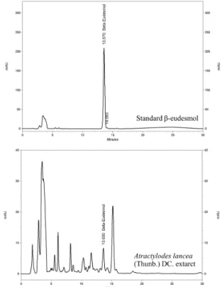
the powdered A. lancea (Thunb.) DC. (100 g) in a stopper flask containing 500 ml of 95% ethanol at room temperature (25-30°C) for 7 days. The extracted solvent was separated and filtered through Whatman no. 1 filter paper (GE Healthcare, Maidstone, UK). After filtration, the extract was evaporated under reduced pressure by rotary evaporation. The dried powder were weighed and stored at -20°C until use. The extract was standardized using high-performance liquid chromatography (HPLC) to determine the amount of β-eudesmol. Chromatographic separation condition used was as follow: Thermo Hypersil Gold C18 column (Thermo Scientific, Rockford, IL, USA), and mobile phase consisting of a mixture of water and acetonitrile with isocratic elution of 40:60 (v:v) at the flow rate of 1 ml/min and run time of 30 min. The wavelength was set at 203 nm.
In vivo model for evaluation of toxicity
Acute and repeated doses toxicity tests were performed in accordance with the OECD guideline for testing of the chemicals (OECD 2004; OECD 2008). For acute toxicity, healthy hamsters (5 males and 5 females) were fed (via intragastric gavage) with a single dose of 5,000 mg/kg body weight of A. lancea (Thunb.) DC. (resuspended in a mixture of distilled water and tween-80 at the ratio of 4:1, v:v). For repeated doses toxicity, three dose levels of A. lancea (Thunb.) DC., i.e., 1,000, 3,000, and 5,000 mg/kg body weight were administered to the healthy hamsters (5 males and 5 females for each group) daily for 28 days. The control hamsters were fed with the mixture of distilled water and Tween-80. Animals were observed for awareness, status of mood, motor activity, CNS excitation, posture, muscle tone, reflexes, and autonomic signs during the first 30 minutes, periodically during the first 24 hours, and then daily for 14 days (acute toxicity) or 28 days (repeated doses toxicity). At the end of the observational period, all animals were sacrificed under ether anesthesia and vital organs were removed from all animals.
In vivo model for evaluation of activity against cholangiocarcinoma
The metacercariae of OV were collected as the naturally infected cyprinoids fish captured from an endemic area of Khon Kaen, northeast Thailand. The parasite species were confirmed under light microscope and the metacercariae were minced and digested with pepsin-hydrichloric acid, then filtrated and washed with normal saline. Development of CCA was induced by initial feeding of male hamsters (by gastric gavage) with 50 metacercariae of OV, followed four weeks later by drinking water containing 12.5 ppm of DMN for eight weeks (Thamavit et al., 1987). The first five groups were treated as OV-infected groups and the last group served as normal control of healthy hamsters. The occurrence and development of CCA was detected and confirmed by ultrasonography throughout the investigation period and divided into six groups (6 males for each group) as follows:
Group 1 (5-FU treated control): CCA hamsters treated with 5-FU (40 μg/ml) daily for 14 doses.
Group 2 (untreated control): CCA hamsters treated
Anticancer Activity of Atractylodes lancea (Thunb.) DC. in Hamster Model and Application of PET-CT for Early Detection
with vehicle (a mixture of distilled water and tween-80)
daily for 30 days.
Group 3 (high dose): CCA hamsters treated with 5000 mg/kg body weight of A. lancea (Thunb.) DC. daily for 30 days.
Group 4 (medium dose): CCA hamsters treated with 3000 mg/kg body weight of A. lancea (Thunb.) DC. daily for 30 days.
Group 5 (low dose): CCA hamsters treated with 1000 mg/kg body weight of A. lancea (Thunb.) DC. daily for 30 days.
Group 6 (normal control): Healthy hamsters treated with vehicle (a mixture of distilled water and tween-80) daily for 30 days.
Body weight, food and water consumption were recorded daily for 30 days. Survival time and survival rate were the primary endpoint parameters for evaluation of the anticancer activity of the crude ethanolic extract of A. lancea (Thunb.) DC.
PET-CT for assessment of activity against cholangiocarcinoma
PET-CT imaging was applied for detection and monitoring the progression of CCA using a small animal PET scanner, FLEX
TMTriumph
TM(Gamma Medica-Ideas, CA, USA). Before experiment, all hamsters were fasted overnight and injected with 10-15 MBq of
18F-FDG via the cephalic vein. After a 30 minutes period for the accumulation of
18F-FDG inside the internal organs, animals were anesthetized with isoflurane and started perform Triumph XO CT system and following by acquisition of PET image using LabPET system for 1h.
The images were reconstructed by the ordered subsets expectation maximization algorithm and analyzed using
VIVIDTM program (Gamma Medica-Ideas, CA, USA).
The standardized UV uptake (SUV) was calculated using the formula:
SUV =
Tumor FDG concentration (Bq/ml) Injected dose (Bq)/Body weight (g)Autopsy and histopathology
All organs were removed at autopsy and observed macroscopically. Samples were fixed with 10% buffer formalin solution. Specimens were washed in phosphate buffer three times, dehydrated in an ascending series of ethanol for 15 min each, embedded in paraffin, followed by sectioning and staining with hematoxylin and eosin.
Statistical analysis
All quantitative variables are presented as medians with 95% CI (confidence interval). Comparison of the quantitative variables between unrelated groups was performed using ANOVA and Mann-Whitney U test. For the quantitative two related groups, Wilcoxon Signed Rank test was applied. Statistical significance level was set at α= 0.05 for all tests.
Results
Standardization of A. lancea (Thunb.) DC. extract The yield of A. lancea (Thunb.) DC. following extraction of the dried rhizomes with 95% ethanolic was 16.89%. The amount of β-eudesmol analyzed by HPLC was 6.73%. The chromatogram of separation was shown in Figure. 1.
Toxicity test
No animal died during the observation period. Only mild signs of acute toxicity occurred in all hamsters receiving all dose levels of A. lancea (Thunb.) DC. as well as the control vehicle. These included stomach irritation, and general CNS depressant signs (reduced alertness and locomotion, and diminished response to touch and balance) was observed within 1 hour after feeding with highest dose (5,000 mg/kg) of the A. lancea (Thunb.) DC.
No histopathological abnormality was observed in any vital organ at autopsy.
Figure 1. Standardization of the Ethanolic Extract of
A. lancea (Thunb.) DC. Using High-PerformanceLiquid Chromatography Figure 2. Change in the Body Weights (Median with
95% CI) of Hamsters in the 6 Group
Anticholangiocarcinoma activity
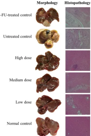
The average body weights of the OV/DMN-induced CCA hamsters in groups 1-5 were significantly reduced after DMN withdrawal compared with normal control hamsters (Figure 2). The morphological examination of OV/DMN-induced hamsters (group 1-5) but normal hamsters (group 6) showed identical features of bile duct epithelium and liver parenchymal cells such as the enlargement of bile duct, pus and shape change of the liver (Figure 3). Histopathological examination of all OV/DMN-induced CCA hamsters (group 1-5), but not normal control group (group 6), confirmed the occurrence of CCA with proliferation and cystic formation of bile duct and infiltration of inflammatory cells (lymphocytes, plasma cells, and macrophages) around portal areas. For the untreated control group, the OV parasite was found in bile duct with severe degeneration and necrosis of hepatocytes (Figure 3). The histopathological lesion of the bile ducts was markedly less pronuced in A. lancea
(Thunb.) DC. treated hamsters, with only mild diffuse proliferation, cystic formation and fibrosis of bile ducts (Figure 3). No remarkable lesion was found in normal control group (group 6).
The median survival rate and survival time were significantly prolonged (about 2 times) in animals treated with the extract at all dose levels (group 3-5) compared with 5-FU treated (group 1), and untreated control (group 2) groups during the 4-6 months observation period (p<0.01, Table 1). At week 36, all animals except those treated with the extract at the highest dose level (5,000 mg/kg: group 3); only one animal died (80% survival rate). The untreated control animals started to die as early as 14 weeks.
PET-CT imaging
PET-CT imaging system was used for first time evaluation and detection of CCA development in OV/
DMN-induced CCA hamster with
18F-FDG. PET images were fused with CT images to improve interpretation accuracy to compare between CCA-bearing hamster and normal group in Figure 4. Coronal views of PET-CT studies showed
18F-FDG accumulation inside the livers, and kidneys to be similar in both the CCA-bearing and control hamsters. PET-CT images of cancerous hamster exhibited
18F-FDG uptake throughout the liver at 8 to Table 1. Survival Time (Days: Median with 95% CI) of OV/DMN-induced CCA-hamsters following Treatment with A. Lancea (Thunb.) DC. in Comparison with Untreated Control
Control hamsters Treated hamsters
Untreated a 5-FU-treated b High dose Medium dose Low dose
(Group 1) (Group 2) (Group 3) (Group 4) (Group 5)
15.5 (14.3-16.7) 20.7 (19.0-22.4) 37.3 (36.1-38.5) 34.2 (32.8-35.6) 27.9 (26.1-29.7)
aStatistically significant difference with Group 3 (p<0.01), Group 4 (p<0.01), and Group 5 (p<0.01); bStatistically significant difference with Group 3 (p<0.01), Group 4 (p<0.01), and Group 5 (p<0.01)
Figure 3. Morphology and Histopathology of Livers in OV/DMN-induced CCA-Hamsters Following Treatment with A. lancea (Thunb.) DC. in Comparison with Untreated Control, and 5-FU (Reference Control)
Figure 5. Tumor
18F-FDG Uptake Presented as Percentage (Median with 95% CI) of SUV Baseline for Cancerous and Normal Hamsters
Figure 4. Representative PET-CT Images of CCA in
OV/DMN-Induced Hamster Model
Anticancer Activity of Atractylodes lancea (Thunb.) DC. in Hamster Model and Application of PET-CT for Early Detection
16 weeks after infection with OV. The median (95% CI)
tumor
18F-FDG uptake (percentage of baseline) in CCA- baring and normal control hamsters at week 4, 8, 12, and 16 after OV-infection were comparable [90 (86.7-93.3) vs 100 (97.7-102.3), 130 (119.4-140.6) vs 140 (131.7-148.3), 170 (156.5-183.5) vs 175 (162.4-187.6), and 200 (184.2- 215.8) vs 205 (188.8-221.2) at week 4, 8, 12, and 16 after OV-infection, respectively] (Figure 5).
Discussion
The ethanolic extract of A. lancea (Thunb.) DC. was shown to exhibit a promising anti-CCA activity and safety profile in OV/DMN-induced CCA hamster model. Its anti-CCA activity, particularly at the highest dose level of 5,000 mg/kg body weight, was clearly demonstrated with a marked inhibition of tumor growth and prolongation of survival time compared with the standard drug 5-FU and untreated control. These observations confirm the previous reports for the potent activity of the plant extract against CCA in xenograft mouse model where significant reduction in tumor size (by 97.3%), prolongation of survival time (by 208.5%), and inhibition of lung metastasis (by 95% of total lung mass) were found, compared with the untreated control (Plengsuriyakarn et al., 2012). Its anti-CCA activity together with other complementary pharmacological activities on gastrointestinal system and inflammatory process support further development of this medicinal plant as a promising candidate for treatment of CCA. In previous studies in rats, the extract was shown to increase the levels of gastric hormone motilin and gastrin, while decreasing the level of somatostatin and corticotrophin-releasing factor, which result in improving the gastric emptying condition (Nakai et al., 2003; Zhang et al., 2008). Furthermore, it also showed potent anti-inflammatory activity through inhibition of 5-lipoxygenase (5-LOX) and cyclooxygenase-1 (COX- 1) (Resch et al., 1998) as well as antimicrobial activity against Rhodotorula glutinis and Saprolegnia (Wang et al., 2009).
Several animal models were used to investigate the anti-CCA activities of candidate compounds or medicinal plant extracts. These include xenograft and orthotopic models, carcinogen-induced CCA model, and genetically engineered mouse model (Thamavit et al., 1987; O’Dell et al., 2012; Zhang et al., 2012). CCA-xenograft mouse model in conjunction with PET-CT is widely applied in research on targeted drug therapy in cancers. PET-CT was also shown to be successfully applied in thioacetamide (TAA)- induced CCA rat model for detection and monitoring the progression of CCA (Laverman et al., 2007; Yeh et al., 2008). The present study was the first study which applied PET-CT as a tool (for detection and monitoring the progression of cancer) for investigation of anti-CCA activity of a candidate medicinal plant in OV/DMN- induced CCA hamster model. The imaging approach is quantitative and noninvasive without sacrificing animals (Cherry and Gambhir, 2007). The carcinogen-induced CCA model by OV infection followed by induction with the carcinogen DMN in hamsters mimics the pathogenicity of human CCA. The development and progress of CCA
following treatment could be periodically monitored using the radio tracer
18F-FDG throughout the observation period. Unfortunately the accumulation of this marker compound is non-specific.
18F-FDG is accumulated in all tissues with high glucose uptake. It is thus, intensely uptaken into all living cells with high metabolic rates, which include not only the cancer cells but also other vital organs especially liver and kidney cells. This problem has been overcome by labeling the radio tracers with ligands such as monoclonal antibodies that are specific to the target tumors. Benvacizumab labeling with
64Cu was successfully developed for imaging intratumoral vascular endothelial growth factor (VEGF) content in renal carcinoma xenograft mouse model (Chang et al., 2013). The elimination half-life of the
64Cu-monoclonal antibody label is relatively longer (~ 12 hours) compared with 18F-mono clonal antibody label (~ 2 hours).
In conclusion, results from the present study support the potential role of A. lancea (Thunb.) DC. rhizome for treatment of CCA. To successfully apply PET-CT as a tool for early detection of tumor development and progression, modification of radiolabeling approach is required to improve its specificity to CCA cells
Acknowledgements
The study was supported by the National Research University Project of Thailand (NRU), Office of Higher Education Commission of Thailand and Thammasat University (Center of Excellence in Pharmacology and Molecular Biology of Malaria and Cholangiocarcinoma).
Tullayakorn Plengsuriyakarn receives financial support from the Thailand Research Fund (Grant No.
TRG5680040).
References
Breitenstein S, Apestegui C, Clavien PA (2008). Positron emission tomography (PET) for cholangiocarcinoma. HPB (Oxford), 10, 120-1.
Chang AJ, Sohn R, Lu ZH, Arbeit JM, Lapi SE (2013). Detection of rapalog-mediated therapeutic response in renal cancer xenografts using 64Cu-bevacizumab immunoPET. PLoS One, 8, 58949.
Chayamarit K (1995). Thai Medicinal Plants, 5th edn. Department of Forestry, Bangkok.
Chen YM, Chou GX, Wang ZT (2007). Determination of beta- eudesmol in rhizome of Atractylodes lancea by RP-HPLC.
Zhongguo Zhong Yao Za Zhi, 32, 2265-7
Chen Y, Wu Y, Wang H, Gao K (2012). A new 9-noratractylodin from Atractylodes lancea and the antibacterial activity of the atractylodin derivatives. Fitoterapia, 83, 199-203.
Cherry S, Gambhir S (2007). Use of positron emission tomography in animal research. ILAR J, 42, 219-32.
Cohade C, Osman M, Leal J, Wahl RL (2003). Direct comparison of 18
F-FDG
PET and PET/CT in patients with colorectal carcinoma. J Nucl Med, 44, 1797-803.Khan SA, Toledano MB, Taylor-Robinson SD (2008).
Epidemiology, risk factors, and pathogenesis of cholangiocarcinoma. HPB (Oxford), 10, 77-82.
Kubo M, Nogami M, Nishimura M, Moriura T, Arichi S (1983). Origins, processing, and qualities of crude drugs (1). Preventive effects of a Chinese crude drug, Zhu,
evaluation. Yakugaku Zasshi, 103, 442-8.
Lardinois D, Weder W, Hany TF, et al (2003). Staging of non- small-cell lung cancer with integrated positron-emission tomography and computed tomography. N Engl J Med, 348, 2500-7.
Laverman P, Blokx WA, Te Morsche RH, et al (2007). [(18)F]
FDG accumulation in an experimental model of multistage progression of cholangiocarcinoma. Hepatol Res, 37, 127- Mahavorasirikul W, Viyanant V, Chaijaroenkul W, Itharat A, Na-32.
Bangchang K (2010). Cytotoxic activity of Thai medicinal plants against human cholangiocarcinoma, laryngeal and hepatocarcinoma cells in vitro. BMC Complement Altern Med, 10, 55.
Mosconi S, Beretta GD, Labianca R, et al (2009).
Cholangiocarcinoma. Crit Rev Oncol Hematol, 69, 259-70.
Nakai Y, Kido T, Hashimoto K, et al (2003). Effect of the rhizomes of Atractylodes lancea and its constituents on the delay of gastric emptying. J Ethnopharmacol, 84, 51-5.
Nogami M, Moriura T, Kubo M, Tani T (1986). Studies on the origin, processing and quality of crude drugs. II.
Pharmacological evaluation of the Chinese crude drug
“zhu” in experimental stomach ulcer. (2). Inhibitory effect of extract of Atractylodes lancea on gastric secretion. Chem Pharm Bull (Tokyo), 34, 3854-60.
Nojima H, Kimura I, Kimura M (1992). Blocking action of succinylcholine with beta-eudesmol on acetylcholine- activated channel activity at endplates of single muscle cells of adult mice. Brain Res, 575, 337-40.
O’Dell MR, Huang JL, Whitney-Miller CL, et al (2012).
Kras(G12D) and p53 mutation cause primary intrahepatic cholangiocarcinoma. Cancer Res, 72, 1557-67.
OECD (2004). Acute Oral Toxicity, Guideline 420, the OECD guideline for testing of chemical.
OECD (2008). Repeated dose 28-day oral toxicity study in rodents, guideline 407, the OECD guideline for testing of chemical.
Ouyang Z, Yang L, Su SL, Feng X, Wang M (2007). Fingerprint of volatile oil of Atractylodes lancea by GC-MS. Acta Pharmaceutica Sinica, 42, 968-72.
Plengsuriyakarn T, Viyanant V, Eursitthichai V, et al (2012).
Anticancer activities against cholangiocarcinoma, toxicity and pharmacological activities of Thai medicinal plants in animal models. BMC Complement Altern Med, 12, 23.
Plengsuriyakarn T, Viyanant V, Eursitthichai V, Na-Bangchang K (2012). In vitro investigations on the potential roles of Thai medicinal plants in treatment of cholangiocarcinoma.
Int Res Pharm Pharmacol, 2, 52-63.
Resch M, Steigel A, Chen Z, Bauer R (1998). 5-Lipoxygenase and cyclooxygenase-1 inhibitory active compounds from Atractylodes lancea. J Nat Prod, 61, 347-50.
Resch M, Heilmann J, Steigel A, Bauer R (2001). Further phenols and polyacetylenes from the rhizomes of Atractylodes lancea and their anti-inflammatory activity. Planta Med, 67, 437-42.
Rizvi S, Gores GJ (2013). Pathogenesis, diagnosis, and management of cholangiocarcinoma. Gastroenterol, 145, 1215-29.
Sacks A, Peller PJ, Surasi DS, et al (2011). Value of PET/CT in the management of primary hepatobiliary tumors, part 2.
AJR, 197, 260-65.
Singh MK, Facciuto ME (2012). Current management of cholangiocarcinoma. Mt Sinai J Med, 79, 232-45.
Sithithaworn P, Andrews RH, Nguyen VD, et al (2012). The current status of opisthorchiasis and clonorchiasis in the Mekong Basin. Parasitol Int, 61, 10-6.
Sriamporn S, Pisani P, Pipitgool V, Suwanrungruang K (2004).
of cholangiocarcinoma in Khon Kaen, Northeast Thailand.
Trop Med Int Health, 9, 588-94.
Tatsumi M, Cohade C, Nakamoto Y, Fishman EK, Wahl RL (2005). Direct comparison of FDG PET and CT findings in patients with lymphoma: initial experience. Radiology, 237, 1038-45.
Thamavit W, Kongkanuntn R, Tiwawech D, Moore MA (1987).
Level of opisthorchis infestation and carcinogen dose- dependence of cholangiocarcinoma induction in Syrian golden hamsters. Virchows Arch B Cell Pathol Incl Mol Pathol, 54, 52-8.
Tsuneki H, Ma EL, Kobayashi S, et al (2005). Antiangiogenic activity of beta-eudesmol in vitro and in vivo. Eur J Pharmacol, 512, 105-15.
Wang Y, Dai CC, Chen Y (2009). Antimicrobial activity of volatile oil from Atractylodes lancea against three species of endophytic fungi and seven species of exogenous fungi.
Ying Yong Sheng Tai Xue Bao, 20, 2778-84.
Xiao PG, Li DP, Yang SL (2002). Modern Chinese materia medica. chemical industry press, Beijing.
Yeh CN, Lin KJ, Hsiao IT, et al (2008). Animal PET for thioacetamide-induced rat cholangiocarcinoma: a novel and reliable platform. Mol Imaging Biol, 10, 209-16.
Zhang H, Han T, Sun LN, et al (2008). Regulative effects of essential oil from Atractylodes lancea on delayed gastric emptying in stress-induced rats. Phytomed, 15, 602-11.
Zhang J, Han C, Wu T (2012). MicroRNA-26a promotes cholangiocarcinoma growth by activating β-catenin.
Gastroenterol, 143, 246-56.