Mem. Natl. Mus. Nat. Sci., Tokyo, (47): 339–365, April 15, 2011
Introduction
The galatheoid fauna of the oceanic Ogasawara Islands, located at about 1000 km south of Tokyo, central Japan, is little known, although some pub- lications have been published (Stimpson, 1858;
Balss, 1913; Melin, 1939; Miyake and Baba, 1965; Osawa and Okuno, 2002). With regard to the genus Munida Leach, 1820, only a few spe- cies have been reported from the area. Melin (1939) recorded
Munida japonica Stimpson,1858, and M. heteracantha Ortmann, 1892 (as
Munida japonica var. heteracantha) from Chichi-jima Island, but the identification of his record of
M. japonica still needs to be verified (Baba et al.,2008). Takeda and Kurata (1976) mentioned an unidentified specimen obtained from gut contents of fish as Munida sp. Osawa and Okuno (2002) reported the occurrence of M. olivarae Macpher- son, 1994 in Otouto-jima Island. The genus is currently represented by 246 species worldwide (De Grave et al., 2009), and recent studies have shown the great species diversity of the genus in the tropical and subtropical waters in the West
Pacific (e.g., Baba, 1988; 1994; 2005; Baba et al., 2009; Macpherson, 1993; 1994; 1996a; 1996b;
1997; 1999a; 1999b; 2000; 2004; 2006a; 2006b;
2009; Macpherson and de Saint Laurent, 1991;
Macpherson and Baba, 1993; Macpherson and Machordom, 2005; Machordom and Macpherson, 2004; Ahyong and Poore, 2004; Ahyong, 2007).
There is no doubt that many species await discov- ery in the Ogasawara Islands.
During a five-year project carried out by the National Museum of Nature and Science, Tokyo,
“Studies on the Origin of Biodiversity in the Saga- mi Sea, Fossa Magna Element and the Izu-Ogasa- wara Arc” (2006–2010), dredgings and/or trap samplings using research vessels were made in the shallow to upper bathyal zone in waters around the Ogasawara Islands. Numerous deca- pod crustaceans were collected. The present study reports on species of Munida represented by six species, including four new to science and one new to the Japanese fauna. Supplemental collec- tions housed in the Natural History Museum and Institute, Chiba, were also examined.
The material examined is deposited in the Na-
Squat Lobsters of the Genus Munida (Crustacea: Decapoda: Anomura: Munididae)
from the Ogasawara Islands, with Descriptions of Four New Species
Tomoyuki Komai
Natural History Museum and Institute, Chiba, 955–2 Aoba-cho, Chuo-ku, Chiba-shi, Chiba 260–8682, Japan
E-mail: [email protected]
Abstract. The present study reports on the squat lobster genus Munida Leach, 1820 (Anomura:
Munididae) collected in the Ogasawara Islands during the Project “Studies on the Origin of Bio- diversity in the Sagami Sea Fossa Magna Element and the Izu-Ogasawara (Bonin) Arc” in 2006–2010, carried out by the National Museum and Nature and Science. Six species were iden- tified, including four new species: M. disiunctus sp. nov., M. honshuensis Benedict, 1902, M.
koyo sp. nov., M. longinquus sp. nov., M. munin sp. nov., and M pectinata Macpherson and Ma- chordom, 2005. The two previously described species are newly recorded from the area, of them M. pectinata is first recorded from waters outside New Caledonia. Affinities of the four new spe- cies are discussed.
Key words: Crustacea, Munididae, Munida, new species, Ogasawara Islands
tional Museum of Nature and Science (NSMT) and Natural History Museum and Institute, Chiba (CBM). The carapace length (cl), as the indica- tion of specimen size, was measured from the level of the sinus between rostrum and supraocu- lar spines to the midpoint of the posterodorsal margin in the midline. The lengths of segments of cheliped are measured along the dorsomesial margin and those of ambulatory legs are along the dorsal or extensor margin. The higher classifica- tion follows Ahyong et al. (2010).
Taxonomic Account
Superfamily Galatheoidea
Family Munididae Genus Munida Leach, 1820
Munida disiunctus
sp. nov. [New Japanese:
Hanare-chū-koshiori-ebi] (Figs. 1–2)
Materials examined. Holotype: R/V
Koyo,2010 cruise, stn 17, W of Mago-jima Island, 27°12.78′N, 142°05.14′ E, 160 m, 7 July 2010, male (cl 6.9 mm), NSMT-Cr S 853.
Paratype: R/V Koyo, 2008 cruise, stn 23, W of Chichi-jima Island, 27°12.04′ N, 142°04.28′E, 202–199 m, 30 October 2008, 1 male (cl 5.2 mm), NSMT-Cr S 854.
Description. Carapace (excluding rostrum) (Fig. 1A) about 1.1 times longer than wide. Dor- sal surface gently convex transversely; main transverse ridges mostly interrupted; only few secondary transverse striae present between main ridges; most ridges and striae with dense short, non-iridescent setae. Gastric region slightly ele- vated, with 6 pairs of epigastric spines and medi- an spine or tubercle. Cervical groove distinct.
Parahepatic, hepatic, anterobranchial spines pres- ent, these spines small; postcervical spines absent.
Anterior part of branchial region between cervical groove and transverse groove without granules;
lateral part of posterior branchial region with 8 or 9 transverse ridges. Intestinal region with 1 rather broad scale. Frontal margins nearly transverse.
Lateral margins feebly convex in dorsal view. An- terolateral spines each located at anterolateral an-
gle, relatively long, reaching sinus between ros- trum and supraocular spines. Second marginal spine anterior to cervical groove less than 0.2 length of anterolateral spine. Branchial margins each with 5 small spines, anteriormost spine larg- er than other spines.
Rostrum (Fig. 1A) spiniform, about 0.5 times as long as carapace, nearly horizontal in lateral view. Supraocular spines moderately long and slender, parallel in dorsal view and very slightly ascending in lateral view, about 0.4 length of ros- trum.
Pterygostomial flap with sharp spine anteriorly, lateral face rugose with irregular transverse or obliquely transverse ridges.
Third thoracic sternite (Fig. 1B) about 2.9 times wider than long, almost as wide as anterior margin of fourth sternite; anterior margin faintly granulate, with distinct median notch. Fourth ster- nite with few transverse striae. Fifth to seventh sternites nearly smooth. Transverse ridges nearly smooth, with row of short setae.
Second abdominal somite (Fig. 1A) with un- armed anterior ridge and 2 or 3 transverse striae on tergum. Third somite (Fig. 1A) with unarmed anterior ridge and 1–3 transverse striae. Fourth somite with unarmed anterior ridge and few striae interrupted medially. Sixth somite (Fig. 1C) with 2 transverse striae interrupted laterally. Telson (Fig. 1C) distinctly wider than long, with numer- ous squamiform ridges on proximal half.
Eyes (Fig. 1A) large. Cornea strongly dilated and somewhat flattened dorsoventrally, corneal width much greater than sinus between rostrum and supraocular spine and 0.25–0.30 of carapace length. Eyestalk slightly narrowed proximally, with 1 stria on dorsal surface; eyelash short, not covering corneal surface.
Basal segment of antennular peduncle (Fig.
1D) moderately stout, reaching distal corneal
margin, length excluding distal spines about 2.0
of width; distal spines moderately long and slen-
der, unequal with distomesial spine somewhat
shorter than distolateral spine; 2 lateral spines
present,
first spine far overreaching distal spines,located somewhat proximal to base of distolateral
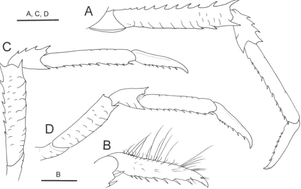
Fig. 1. Munida disiunctus sp. nov., holotype, male (cl 6.9 mm), NSMT-Cr S 853. A, carapace, first to third abdominal somites, and cephalic appendages, dorsal view (distal two segments of antennular peduncles omitted); B, thoracic sternum, ventral view; C, sixth abdominal somite and telson, external view; D, basal segment of left antennular pe- duncle, ventral view; E, left antennal peduncle, ventral view; F, endopod of left third maxilliped, lateral view; G, right cheliped, dorsal view; H, same, merus, ventral view; I, same, carpus ventral view. Setae omitted. Scale bars: 2 mm for A, C, G–I; 1 mm for B, D–F.
spine, second spine short, located slightly proxi- mal to base of first spine; statocyst lobe not par- ticularly inflated.
Antennal peduncle (Fig. 1E) moderately stout, not reaching distal corneal margins. First segment with moderately long distomesial spine not reach- ing distal margin of third segment; distolateral angle unarmed. Second segment with distomesial spine overreaching distal margin of fourth seg- ment, distolateral spine reaching slightly beyond third segment, mesial spine tiny. Third and fourth segments unarmed.
Third maxilliped (Fig. 1F) moderately slender.
Ischium distinctly longer than merus, with strong ventrodistal spine, produced dorsodistal angle ter- minating in sharp spine. Merus with 2 greatly un- equal spines on ventral margin, distal spine small, proximal spine arising at about midlength very strong; dorsodistal margin unarmed. Carpus smooth on extensor surface. Propodus subequal in length to carpus, not expanded. Dactylus short- er than propodus.
Chelipeds (Fig. 1G–H) strongly squamous, similar, subequal in length, 3.4 times as long as carapace at most, equally broad on merus, carpus and palm; mesial face of merus to palm with mix- ture of short plumose setae and stiff iridescent se- tae. Merus with row of 4–6 spines on dorsal sur- face laterally (spines increasing in size distally) and 3–5 spines mesially (spine at distomesial an- gle strong and diverging); lateral face with small spine at ventrodistal angle, no other spines; mesi- al face with row of 3 spines adjacent to dorsal margin and row of 3 spines adjacent to ventral margin, ventromesial distal angle with spine. Car- pus shorter than palm; dorsolateral margin with 5 small spines (distalmost spine strongest), dor- somesial margin also with 6 spines; ventrodistal angle with small spine. Palm slightly widened distally, 2.6–3.2 times longer than wide; dorsal surface with row of median row of 4 or 5 small spines and 1 spine at articulation to dactylus, dor- solateral margin with 2 or 3 small spines, dor- somesial margin with 7 small spines; mesial face with row of 3 small spines adjacent to dorsal mar- gin. Fixed finger nearly straight, terminating in
sharp claw, with 1 or 2 small subdistal spines;
cutting edge weakly denticulate; surfaces with scattered short setae dorsally, sparse long stiff se- tae laterally and mesially. Dactylus 0.8–1.0 times as long as palm, unarmed or armed with some small granules proximally on dorsal surface, ter- minating in sharp, curved claw crossing tip of fixed finger; lateral margin with proximal and subterminal spines (two additional spines present on lateral margin in holotype, otherwise unarmed in paratype), with several long stiff setae; cutting edge with denticulate over entire length; narrow proximal hiatus between dactylus and fixed finger in holotype, no hiatus in paratype.
Ambulatory legs (second to fourth pereopods) moderately long and slender, decreasing in length posteriorly; setal row on dorsal or extensor mar- gin of merus to propodus consisting of thin plu- mose and thick iridescent setae. Second pereopod (Fig. 2A, B) about 2.5 times as long as carapace;
merus 0.7–0.8 times as long as carapace, about 6.4 times longer than high, dorsal margin with row of 6–8 spines, distal spine strongest, ventral margin with 1 strong distal spine followed by 2–4 smaller spines and short transverse ridges, lateral face with few short striae or squamiform ridges;
carpus about 0.3 length of propodus, with promi- nent extensor distal spine and 2 or 3 small spines or spinules on extensor margin, flexor distal mar- gin produced in spine; propodus unarmed on ex- tensor margin, lateral face almost smooth,
flexormargin with row of 10 movable spines; dactylus (Fig. 2B) about 0.7 times as long as propodus and about 6.0 times as long as high, slightly curved sinuously, bearing sparse short to long stiff setae, flexor margin slightly sinuous, with 8 corneous spines in proximal 0.7–0.8, notably decreasing in length proximally, distal 0.2–0.3 only with sub- terminal spinule closely appressed to unguis.
Third pereopod (Fig. 2C) with merus bearing row
of 6 spines increasing in size distally on dorsal
margin and row of 2–4 spines and short trans-
verse ridges on ventral margin; carpus with 1
prominent extensor distal spine and 2 or 3 addi-
tional smaller spines on extensor margin; propo-
dus with 10 or 11 movable spines on
flexor mar-gin; dactylus with 8 corneous spines on flexor margin in proximal 0.7–0.8 and subterminal spinule. Fourth pereopod (Fig. 2D) reaching to lateral end of cervical groove of carapace by me- ro-carpal articulation; merus about 0.6 length of that of second pereopod, bearing dorsodistal spine, dorsal margin otherwise unarmed, ventral margin with prominent distal spine followed by 1 or 2 small spines and short transverse ridges; car- pus with prominent dorsodistal and ventrodistal spines; propodus with 9–11 movable spines on
flexor margin; dactylus with 8 corneous spines on flexor margin in proximal 0.7–0.8 and subtermi-nal spinule.
Fifth pereopod without distinctive features.
Uropodal protopod with 1 small spine posteri- orly on outer side.
Color in life. Not known.
Distribution. At present known only from Oga- sawara Islands, 160–202 m.
Remarks. The two male specimens of the pres-
ent new species seem to attain maturity because
the gonopods are fully developed. Munida disi-
unctus sp. nov. closely resembles M. notataMacpherson, 1994 from the Southwest Pacific in
sharing five spines on the branchial carapace mar-
gins, nearly transverse frontal margin, absence of
a postcervical spine on the carapace, smooth tho-
racic sternites, unarmed anterior ridge of the sec-
ond abdominal somite, distomesial spine of the
basal segment of the antennular peduncle slightly
longer than distolateral spine, distomesial spine
of the first segment of the antennal peduncle
reaching the distal margin of the third segment,
distomesial spine of the second segment of the
antennal peduncle overreaching the fourth seg-
Fig. 2. Munida disiunctus sp. nov., holotype, male (cl 6.9 mm), NSMT-Cr S 853. Right ambulatory legs, lateral view, setae omitted except for B. A, second pereopod; B, same, distal part of propodus and dactylus, setation shown; C, third pereopod; D, fourth pereopod. Scale bars: 1 mm.ment, and dilated corneas. However, the new spe- cies is distinguishable from M. notata by several morphological characters (Macpherson, 1994).
The carapace has fewer transverse ridges in the new species than in M. notata. For example, there are eight transverse ridges on the lateral part of the posterior branchial region in
M. disiunctus,whereas
M. notata has 13 ridges there. Similarly,the abdominal tergites have fewer transverse stri- ae in M. disiunctus than in M. notata. The number of the striae on the carapace and abdomen may increase with increase of the body size, but this character still works when specimens in similar developmental stages are compared. The cornea is larger in M. disiunctus than in M. notata (the corneal width 0.25–0.30 in the new species versus less than 0.20 in
M. notata). The distomesialspine of the first segment of the antennal peduncle is less elongate in the new species than in M. no-
tata (reaching the distal margin of the third seg-ment in the former versus nearly reaching the fourth segment in the latter). The merus of the third maxilliped is unarmed on the dorsodistal margin in the new species, rather than armed with a small dorsodistal spine in M. notata. The dacty- lus of the second pereopod is unarmed in the dis- tal 0.25 in M. disiunctus, rather than 0.30 or more in M. notata.
Except for the character of the distal spines of the basal segment of the antennular peduncle, the new species also resembles M. leagora Macpher- son, 1994 from the Southwest Pacific and M. pse-
liphora Macpherson, 1994 from New Caledonia,in sharing the characters mentioned above (Macpherson, 1994). The latter two species differ from
M. disiunctus in the subequal distal spinesof the first segment of the antennular peduncle, although this character is rather delicate.
Munida leagora further differs from the new species inhaving more numerous transverse ridges or striae on the carapace and abdomen, the elongate disto- lateral spine of the second segment of the anten- nal peduncle that overreaches the fourth segment, and the proportionally shorter dactylus of the sec- ond pereopod (less than 0.5 times as long as the propodus versus about 0.7 times as long). Munida
pseliophora is distinguished from M. disiunctus
by having more elongate and slender anterolateral spines of the carapace (clearly overreaching the sinus between the supraocular spine and the ros- trum versus just reaching it), the smaller cornea (the corneal width 0.20 or less in M. pseliophora versus 0.25–0.30 in M. disiunctus) and three, in- stead of two, ventral spines on the third maxil- liped merus.
Etymology. From the Latin “disiunctus” (= re- mote), alluding to the locality of the new species remote from the Japanese mainland.
Munida honshuensis Benedict, 1902 [New Jn.:
Suji-chū-koshiori-ebi] (Fig. 11A)
Munida honshuensis Benedict, 1902: 261, fig. 11
(type locality: off Honshu, Japan, 110–128 m);
Macpherson and Baba, 1993: 396, fig. 7; Baba, 2005: 264.
Munida japonica: Ortmann, 1892: 254, pl. 11, fig.
11, 11i, 11k. Not
Munida japonica Stimpson,1858.
Materials examined. FR/V Koyo, 2009 cruise, stn 10, N of Haha-jima Island, 26°50.25′ N, 142°07.09′E, 145–150 m, 13 July 2009, dredge, 1 juv. (cl 4.7 mm), CBM-ZC 10102; stn 26, NE of Mago-jima Island, 27°14.36′N, 142°16.04′E, 230–250 m, 15 July 2009, dredge, 1 male (cl 7.5 mm), CBM-ZC 10103.
R/V Tansei-maru, KT09-2 cruise, stn TW1-1, W o f C h i c h i - j i m a I s l a n d , 2 7 ° 0 1 . 4 0
′N , 142°07.41
′E, 145–138 m, 19 March 2009, dredge, 1 male (cl 6.9 mm), CBM-ZC 10104; stn TW2-3, W of Chichi-jima Island, 27°03.03′N, 142°05.29′E, 166 m, 19 March 2009, 1 male (cl 6.0 mm), CBM-ZC 10105.
Coloration. Fig. 11A. Ground color of cara- pace, abdomen, and appendages orange, trans- verse ridges or squamiform ridges or tubercles darker. Cornea brown. Chelipeds with reddish tinge on distal parts of merus and carpus and dis- tal part of palm at articulation to dactylus; distal part of fingers whitish; dorsal spines on palm also whitish. Ambulatory legs each with white band at articulation between dactylus and propodus.
Distribution. Previously known from the Pacif-
ic coast of the Japanese mainland, 110–300 m.
The present specimens extend the geographical range of the species to the Ogasawara Islands.
Remarks. The present specimens agree well with the redescription of
Munida honshuensisgiven by Macpherson and Baba (1993), particu- larly in the following diagnostic characters: cara- pace with five branchial spines; fourth to seventh thoracic sternites with arcuate striae; lateral parts of seventh thoracic sternite with fine granules;
second abdominal somite with 9–10 spines dis- tributed over entire length of anterior ridge; cor- nea large, strongly dilated; basal segment of an- tennular peduncle with distomesial spine longer than distolateral spine; distomesial spine of sec- ond segment of antennal peduncle overreaching fourth segment; merus of third maxilliped with small spine on dorsodistal margin. Therefore, the specimens are identified with
M. honshuensiswith little hesitation.
Melin (1939) reported Munida japonica from the Ogasawara Islands, but Baba et al. (2008) questioned the identity. It is likely that Melin’s specimens are actually referable to M. honshuen-
sis, because the two species are superficially verysimilar (Macpherson and Baba, 1993).
Munida koyo sp. nov. [New Jn: Koyo-chū-koshi-
ori-ebi] (Figs. 3–5)
Materials examined. Holotype: FR/V Koyo, 2008 fishery ground survey, S of Chichi-jima Is- land, Ogasawara Islands, about 1000 m deep, 14 October 2008, male (cl 17.4 mm), CBM-ZC 10121.
Paratypes: same data as holotype, 4 males (cl 16.0–19.2 mm), 1 ovig. female (cl 17.2 mm), CBM-ZC 10120; same data, 2 males (cl 17.0.
18.9 mm), NSMT-Cr S 855.
Description. Carapace (excluding rostrum) (Fig. 3A) about 1.1–1.2 times longer than wide.
Dorsal surface gently convex transversely; main transverse ridges mostly interrupted; numerous secondary transverse striae or transverse rows of small scale-like ridges between main ridges; most ridges and striae with dense short, non-iridescent setae. Gastric region with 10–12 epigastric spines
and median tubercle. Cervical groove distinct.
Parahepatic, anterobranchial and postcervical spines usually present, these spines small. Anteri- or part of branchial region between cervical groove and transverse groove with small scale- like ridges, but non-granulate; lateral part of pos- terior branchial region with 12 or 13 transverse ridges. Intestinal region with one or few short transverse striae. Frontal margins very slightly oblique. Lateral margins very slightly convex in dorsal view. Anterolateral spines each located at anterolateral angle of carapace, moderately long, falling short of sinus between rostrum and su- praocular spines, slightly diverging. Anterolateral margin with 1 or 2 marginal spines posterior to anterolateral spine, these spines distinctly shorter than anterolateral spine. Branchial margins each with 5 small spines, anteriormost spine strongest.
Rostrum (Figs. 3A, 4A) spiniform, about 0.5 times as long as carapace, directed forward, slightly sinuously curved in lateral view; distal part of lateral margins faintly denticulate. Su- praocular spines moderately long and slender, parallel or slightly diverging in dorsal view and slightly ascending in lateral view, about 0.4 length of rostrum.
Pterygostomial flap (Fig. 4A) unarmed anteri- orly, lateral face rugose with irregular transverse or obliquely transverse ridges.
Third thoracic sternite (Fig. 3B) about 2.9 times wider than long, much wider than anterior margin of fourth sternite; anterior margin faintly granulate, with shallow median notch. Fourth sternite with faint stria medially. Fifth to seventh sternites smooth. Transverse ridges nearly smooth, with row of short setae.
Second abdominal somite (Fig. 3A) with 10
spines in 5 pairs on anterior ridge and 2 trans-
verse striae on tergum and several small scales on
each pleuron. Third somite (Fig. 3A) with un-
armed anterior ridge and 2 transverse striae (ante-
rior stria interrupted medially and laterally, poste-
rior stria complete); pleura each with short striae
or scales. Fourth somite (Fig. 3A) with unarmed
anterior ridge and 2 interrupted striae. Sixth
somite (Fig. 4B) with 2 main transverse striae in-
Fig. 3. Munida koyo sp. nov., holotype, male (cl 17.4 mm), CBM-ZC 10121. A, carapace, first to third abdominal somites, and cephalic appendages, dorsal view (distal two segments of antennular peduncles omitted); B, thoracic sternum, ventral view; C, right cheliped, dorsal view; D, left cheliped, dorsal view. Setae omitted (A, B) or denuded (C, D). Scale bars: 5 mm.
terrupted medially and few short striae laterally.
Telson (Fig. 4B) distinctly wider than long, with numerous squamiform ridges except for smooth broad oblique sulci.
Eyes (Figs. 3A, 4A, C) large. Cornea strongly dilated, somewhat flattened dorsoventrally, corne- al width much greater than sinus between rostrum and supraocular spine and about 0.25 of carapace length. Eyestalk slightly narrowed proximally, with 1 stria on dorsal surface; eyelashes short, not covering corneal surface.
Basal segment of antennular peduncle (Fig.
4C) moderately stout, reaching distal corneal margin, length excluding distal spines about 2.0 of width; distal spines relatively long and slender, distolateral spine subequal in length to slightly longer than distomesial spine; 2 lateral spines present, first spine very slender, not reaching dis- tal spines, faintly sinuously curved in ventral view, slightly ascending in lateral view, arising somewhat proximal to base of distolateral spine, second spine small, arising at midlength of seg- ment; statocyst lobe not particularly inflated; ven- tral surface with scattered squamiform ridges.
Antennal peduncle (Fig. 4C) moderately stout, not reaching distal corneal margins. First segment with short, subacute distomesial spine reaching only midlength of second segment; distolateral angle unarmed. Second segment with distomesial spine reaching distal margin of third segment, distolateral spine not reaching distal margin of third segment; mesial margin with small spine at midlength. Third and fourth segments unarmed.
Third maxilliped (Fig. 4D) moderately slender.
Ischium longer than merus, with small ventrodis- tal spine, distolateral angle terminating in small spine; lateral face with minute scale-like ridges.
Merus with 2 unequal spines on ventral margin, proximal spine arising at about midlength stron- ger than distal spine; dorsodistal margin unarmed;
lateral face also with minute scale-like ridges.
Carpus smooth on extensor surface. Propodus subequal in length to carpus, not expanded. Dac- tylus slender, shorter than propodus.
Chelipeds (Figs. 3C, D, 4E–H) relatively stout, 3.5 times longer than carapace at most, equally
broad on merus, carpus and palm, usually similar and subequal, but in males occasionally slightly dissimilar and unequal; entire surfaces with dense covering of soft plumose setae, scattered stiff iri- descent setae also present on ventral surface of merus, and mesial faces of carpus and chela.
Merus having row of 9–10 spines on dorsal sur- face laterally (spines noticeably increasing in size distally) and 3 prominent spines mesially (stron- gest spine at distomesial angle reaching proximal 0.2 of carpus); lateral face with scattered scale- like tubercles becoming larger ventrally, ventro- lateral distal angle with small spine; mesial face with sparse granules, 1 or 2 strong spines on mid- line, and 2 strong spines adjacent to ventral mar- gin, ventromesial distal angle with strong spine;
ventral surface with numerous scale-like ridges and tubercles. Carpus about 0.7 length of palm;
dorsal surface with small scale-like tubercles, dorsolateral and dorsomesial margins each with 5 small spines including distal marginal spine; lat- eral face covered with squamiform ridges and 2 small spines adjacent to dorsal margin; mesial face with scattered spinules or spinulose tubercles and 3 strong spines along midline; ventral surface with scattered scale-like tubercles and with 1 middle spine. Palm 2.1–2.4 times longer than wide; dorsal surface with scattered small tuber- cles, median row of 3–6 small spines and 1 spine at articulation to dactylus, dorsolateral margin with 5–6 small spines, dorsomesial margin 2–4 small spines; mesial face with row of 3 small spines adjacent to dorsal margin; ventral surface with few short transverse ridges and small spine at articulation to dactylus. Fixed finger nearly straight to arched, terminating in sharp claw, bearing small tubercles on surfaces and 1–4 spines on mesial margin (including 1 or 2 subter- minal spines), cutting edge minutely denticulate;
surfaces with scattered short setae dorsally, sparse
long stiff setae laterally and mesially. Dactylus
subequal in length to palm, terminating in sharp,
curved claw crossing tip of fixed
finger; surfacescovered with numerous small tubercles or gran-
ules, dorsomesial margin with only 1 proximal
spine; cutting edge denticulate over entire length,
occasionally with small rectangular tooth proxi- mally; narrow proximal hiatus between dactylus and fixed finger in males, no hiatus in females.
Ambulatory legs (second to fourth pereopods)
squamous, moderately long and relatively slen- der, decreasing in length posteriorly. Second pe- reopod (Fig. 5A) about 2.1 times longer carapace;
merus about 0.9 times as long as carapace, about Fig. 4. Munida koyo sp. nov., holotype, male (cl 17.4 mm), CBM-ZC 10121. A, anterior part of carapace and left ce-
phalic appendages, lateral view; B, sixth abdominal somite, telson and left uropod, external view; C, left eye, basal segment of antennular peduncle and antennal peduncle, ventral view; D, endopod of left third maxilliped, lateral view; E, merus of right cheliped, lateral view; F, same, mesial view; G, carpus of right cheliped, lateral view; H, same, dorsomesial view. Setae omitted (A–D) or denuded (E–H).
7.9 times longer than high, dorsal margin with numerous plumose setae and row of 12–13 spines, distal spine strongest, ventrolateral margin with 1 strong distal spine followed by 1 or 2 spines and squamiform ridges, ventromesial mar- gin with 1 distal spine, lateral face with numerous scale-like tubercles or squamiform ridges stron- gest adjacent to ventral margin; carpus about 0.3 length of propodus, with prominent extensor dis- tal spine and 1 or 2 spines and few spinules on extensor margin, flexor distal margin produced in spine; propodus unarmed on extensor margin, lat- eral face almost with scattered small scale-like tubercles, flexor margin with row of 9–10 mov- able spines; dactylus (Fig. 5B) about 0.6 times as long as propodus and 4.6–5.0 times as long as high, nearly straight, bearing numerous short to long stiff setae, flexor margin slightly sinuous,
with 9–11 corneous spinules over entire length, including subterminal spinule closely appressed to unguis. Third pereopod (Fig. 5C) generally similar to second pereopod; merus bearing row of 12 spines increasing in size distally on dorsal margin, ventral margin with 1 prominent ventro- distal spine followed by 1 small spine and squam- iform ridges; carpus with 1 prominent extensor distal spine and 1 or 2 additional spines on exten- sor margin; propodus with 9 movable spines on
flexor margin; dactylus with 8 corneous spinuleson flexor margin. Fourth pereopod (Fig. 5D, E) not reaching to lateral end of cervical groove of carapace by mero-carpal articulation; merus about half length of that of second pereopod, bearing small dorsodistal spine, dorsal margin otherwise unarmed, ventral margin with prominent distolat- eral spine and squamiform ridges, no distomesial
Fig. 5. Munida koyo sp. nov., holotype, male (cl 17.4 mm), CBM-ZC 10121. Right ambulatory legs, setae omitted ex- cept for B. A, second pereopod, lateral view; B, same, distal part of propodus and dactylus, lateral view, setation shown; C, third pereopod, lateral view; D, fourth pereopod, lateral view; E, merus of fourth pereopod, mesial view.
Scale bars: 5 mm for A, C–E; 2 mm for B.
spine; some tiny spines on mesial face adjacent to ventral margin; carpus with prominent extensor distal and flexor distal spines, otherwise unarmed;
propodus with 9 movable spines on flexor mar- gin; dactylus with 8 corneous spinules on flexor margin. Fifth pereopod without distinctive fea- tures.
Uropodal protopod (Fig. 4B) with 1 small spine posteriorly.
Color in life. Unknown. In preservative in for- malin, carapace, anterior three somites of abdo- men and pereopods pinkish, basal parts of su- praocular spines white.
Distribution. Known only from off Chichi-jima Island, Ogasawara Islands, about 1000 m.
Remarks. Munida koyo sp. nov. appears closest to M. militaris Henderson, 1885 in sharing five branchial spines on the carapace, the third thorac- ic sternite being much wider than the anterior margin of the fourth sternite, one or two trans- verse striae on the second and third abdominal tergites, absence of granular patches on the lateral parts of the seventh thoracic sternite, a row of spines along the anterior ridge of the second ab- dominal somite, eye large, subequal or slightly unequal distal spines on the basal segment of the antennular peduncle (the distolateral spine is slightly longer than the distomesial spine, when unequal) and moderately elongate chelipeds.
However, the new species is readily distinguished from M. militaris in the presence of many second- ary striae or transverse rows of small scale-like ridges on the carapace, which are very few in M.
militaris (cf. Baba and Macpherson, 1991; Baba et al., 2009). Furthermore, in the new species, the
anterolateral spine on the carapace does not reach the level of the sinus between the rostrum and su- praocular spine, instead of reaching or slightly overreaching it in
M. militaris; the distomesialspine on the second segment of the antennal pe- duncle only reaches the distal margin of the third segment in M. koyo, rather than overreaching it in
M. militaris; the merus of the second pereopodhas only one distinct spine on the ventrolateral margin proximal to the distal spine in M. koyo, but there are three distinct spines in M. militaris.
Munida howensis Ahyong, 2007 is also similar
to
M. koyo sp. nov. in the ornamentation of thecarapace and the characters above mentioned, but the former is readily distinguished from the new species by the more strongly unequal distal spines on the basal segment of the antennular peduncle, the more elongate distomesial spine on the second segment of the antennal peduncle, which reaches the distal margin of the fourth segment, and the possession of three, rather than two, ventral spines on the merus of the third maxilliped.
Baba and Macpherson (1991) clarified that the type series of M. militaris contained three species,
M. militaris, M. japonica Stimpson, 1858, and M.inornata Henderson, 1885. One of the four speci-
mens from “Challenger” station 173, off Matuku Fiji Islands, was selected as a lectotype by the au- thors. Later, it has been shown that the specimens referred to
M. japonica actually contained twospecies, M. japonica and M. agave Macpherson and Baba, 1993 (Macpherson and Baba, 1993;
Baba et al., 2008). The type specimens referred to
M. militaris now include the four specimens from“Challenger” station 173 and one specimen from Ambon, Indonesia. There is little doubt that the latter specimen is specifically distinct from the former lot containing the lectotype, because it has numerous secondary striae on the carapace and the more elongate distomesial spine on the second segment of the antennal peduncle. The latter char- acter provides evidence that this specimen from Ambon is also specifically distinct from
M. koyosp. nov.
Etymology. Named for the FR/V Koyo of the Ogasawara Fisheries Center, from which many specimens of Munida studied here were collected.
Noun in apposition.
Munida longinquus sp. nov. [New Jn.: Adeyaka-
chū-koshiori-ebi:] (Figs. 6–7, 11B)
Materials examined. Holotype: R/V
Koyo,2008 cruise, stn 19, E of Chichi-jima Island, 27°06.07′N, 142°18.56′E, 175–176 m, ovig. fe- male (cl 4.4 mm), NSMT-Cr S 856.
Paratypes: R/V Koyo, 2008 cruise, stn 4, W of
Chichi-jima Island, 27°03.60′N, 142°04.22′ E,
211–214 m, 1 male (cl 3.4 mm), 1 ovig. female (cl 3.9 mm), NSMT-Cr S 857.
Description. Carapace (excluding rostrum) (Fig. 6A) about 1.2 times longer than wide. Dor- sal surface gently convex transversely; main transverse ridges mostly interrupted; no second- ary transverse striae between main ridges; most ridges with dense short, non-iridescent setae.
Gastric region slightly elevated, with 9 (5 on left, 4 on right) epigastric spines and median scale.
Cervical groove distinct. Parahepatic and postcer- vical spines on each side, these spines small; an- terobranchial spine absent. Anterior part of bran- chial region between cervical groove and transverse groove without granules; lateral part of posterior branchial region with 6 or 7 transverse ridges. Intestinal region without scale. Frontal margins somewhat oblique. Lateral margins fee- bly convex in dorsal view. Anterolateral spines each located at anterolateral angle, relatively short, not reaching sinus between rostrum and su- praocular spines. Second marginal spine anterior to cervical groove less than half-length of antero- lateral spine. Branchial margins each with 4 small spines, decreasing in size posteriorly.
Rostrum (Fig. 6A) spiniform, about 0.4 times as long as carapace, nearly horizontal in lateral view. Supraocular spines moderately long and slender, parallel in dorsal view and very slightly ascending in lateral view, about 0.4 length of ros- trum.
Pterygostomial flap with sharp spine anteriorly, lateral face rugose with irregular transverse or obliquely transverse ridges.
Third thoracic sternite (Fig. 6B) about 4.8 times wider than long, almost as wide as anterior margin of fourth sternite; anterior margin faintly granulate, with very shallow median notch.
Fourth sternite with 2 submedian short transverse striae on either side of midline. Fifth and sixth sternites nearly smooth. Seventh sternite with scattered granules on lateral parts. Transverse ridges nearly smooth, with row of short setae.
Second abdominal somite (Fig. 6A) with 2 pairs of minute lateral spines on anterior ridge and 1 transverse stria on tergum. Third somite
(Fig. 6A) with unarmed anterior ridge and 1 transverse stria. Fourth somite (Fig. 6A) with un- armed anterior ridge, no stria on tergum. Sixth somite (Fig. 6C) with 1 medially interrupted stria.
Telson (Fig. 6C) distinctly wider than long, with several squamiform or short transverse ridges.
Eyes (Fig. 6A, D) moderately large. Cornea di- lated, corneal width much greater than sinus be- tween rostrum and supraocular spine and 0.22–
0.23 of carapace length. Eyestalk not narrowed proximally, without striae on dorsal surface; eye- lash sparse, very short.
Basal segment of antennular peduncle (Fig.
6D) moderately stout, overreaching distal corneal margin, length excluding distal spines about 2.5 of width; distal spines relatively short and stout, distinctly unequal with distolateral spine longer than distomesial spine; 2 lateral spines present,
first spine reaching only midlength of distolateralspine, arising slightly distal to midlength of seg- ment,, second spine short, arising at midlength of segment; statocyst lobe not particularly inflated.
Antennal peduncle (Fig. 6D) relatively short and stout, far falling short of distal corneal mar- gins. First segment with short distomesial spine reaching distal margin of second segment; disto- lateral angle unarmed. Second segment with dis- tomesial spine reaching distal margin of third segment, distolateral spine subequal to distomesi- al spine, reaching slightly beyond third segment, mesial spine absent. Third and fourth segments unarmed.
Third maxilliped moderately slender. Ischium distinctly longer than merus, with strong ventro- distal spine, distolateral angle terminating in sharp spine (Fig. 6E). Merus with 2 greatly un- equal spines on ventral margin, distal spine small, proximal spine arising at about midlength very strong; dorsodistal margin unarmed (Fig. 6E).
Carpus smooth on extensor surface. Propodus subequal in length to carpus, not expanded.
Propodus not expanded. Dactylus shorter than propodus.
Chelipeds (Fig. 6F–H) not squamous, similar,
subequal in length, about 2.5 times longer than
carapace, equally broad on merus, carpus and
Fig. 6. Munida longinquus sp. nov., holotype, ovig. female (cl 4.4 mm), NSMT-Cr S 856. A, carapace, first to third ab- dominal somites, and cephalic appendages, dorsal view (distal two segments of antennular peduncles omitted); B, thoracic sternum, ventral view; C, sixth abdominal somite and telson, external view; D, right eye, basal segment of antennular peduncle and antennal peduncle, ventral view; E, distal part of ischium and merus of right third maxil- liped, lateral view; F, left cheliped, dorsal view; G, same, merus, ventral view; H, same, carpus ventral view. Setae omitted. Scale bars: 1 mm for A–D, F–I; 0.5 mm for E.
palm; mesial face of merus and palm with numer- ous long iridescent setae, mesial face of carpus with mixture of numerous plumose setae and few iridescent setae. Merus with row of 6 spines on dorsal margin (spines increasing in size distally) and 3 spines mesially (spine at distomesial angle strong and diverging); lateral face with short transverse ridges and strong ventrodistal spine;
ventromesial margin with 3 spines including strong ventrodistal spine; ventral surface with scattered short transverse ridges. Carpus subequal in length to palm; dorsolateral margin with 4 small spines (distalmost spine small, marginal), dorsomesial margin also with 5 strong spines; lat- eral face with short transverse, occasionally den- ticulate ridges; ventral face with 2 small strong spines, ventrodistal angle with spine. Palm not widened distally, about 2.0 times longer than wide; dorsal surface with longitudinal row of 6 small spines along midline, dorsolateral margin with 4 small spines, dorsomesial margin 3 small spines; mesial face with longitudinal row of 3 spines; ventral surface with scattered low tuber- cles and 1 spine located as base of dactylus; fixed
finger nearly straight, terminating in sharp claw,with 6 spines on lateral margin, cutting edge weakly denticulate; surfaces with scattered short setae dorsally, sparse long stiff setae laterally and mesially. Dactylus about 1.5 times as long as palm, unarmed proximally on dorsal surface, ter- minating in sharp, curved claw crossing tip of
fixed finger; mesial margin with 5 spines includ-ing 1 proximal and 2 subterminal spines, and with several long stiff setae; cutting edge with minute- ly denticulate over entire length; no proximal hia- tus between dactylus and fixed finger.
Ambulatory legs (second to fourth pereopods) moderately long and slender, decreasing in length posteriorly. Second pereopod (Fig. 7A) about 2.0 times as long as carapace; merus about 0.8 times as long as carapace, about 6.0 times longer than high, dorsal margin with row of 9–10 spines de- creasing in size proximally and row of plumose and iridescent setae (tuft of long iridescent setae at base of strong dorsodistal spine), ventral mar- gin with 1 strong distal spine followed by 1–2
small spines and short transverse ridges, lateral face with few short striae; carpus about 0.3 length of propodus, with prominent extensor distal spine and 1 small spines on extensor margin, flexor dis- tal margin produced in strong spine; propodus unarmed on extensor margin, lateral face almost smooth, flexor margin with row of 9–11 movable spines; dactylus (Fig. 7B) about 0.8 times as long as propodus and about 5.8 times as long as high, nearly straight, bearing sparse short to long stiff setae, flexor margin faintly sinuous, bearing 9–10 slender corneous spines over entire length, in- cluding subterminal spinule closely appressed to unguis. Third pereopod (Fig. 7C) similar to sec- ond: merus bearing row of 4–7 small spines on dorsal margin, ventral margin with prominent ventrodistal spine followed by 1–2 small spines and short transverse ridges; carpus with 1 promi- nent extensor distal spine and 2 additional small spines on extensor margin; propodus with 6–8 movable spines on flexor margin; dactylus with 7–8 corneous spines on flexor margin. Fourth pe- reopod (Fig. 7D) reaching to lateral end of cervi- cal groove of carapace by mero-carpal articula- tion; merus about 0.6 length of that of second pereopod, unarmed on dorsal margin, ventral margin with prominent distal spine followed by short transverse ridges; carpus with small dorso- distal and strong ventrodistal spines; propodus with 6–9 movable spines on flexor margin; dacty- lus with 7–8 corneous spines on flexor margin.
Fifth pereopod without distinctive features.
Uropodal protopod with 1 small spine posteri- orly on outer side.
Color in life. Fig. 11B. Carapace yellow, with pale purple bands posterior to epigastric spines, along cervical groove and across cardiac region;
rostrum and frontal margin red; dorsal surface with pair of distinct red spots on branchial region just posterior to cervical groove. Second abdomi- nal somite pale purple, with red line along anteri- or ridge and yellow transverse band on middle potion of tergum; remaining abdominal somites brownish, with distinct white marking on third to fourth somites. Eye with reddish brown cornea;
eyestalk red. Antennular and antennal peduncles
translucent, with tint of red. Cheliped generally pale red, fingers and spines dark red. Ambulatory legs with white and reddish bands.
Distribution. Known only from the Ogasawara Islands, at depths of 175–214 m.
Remarks. Munida longinquus sp. nov. resem- bles
M. gordoae Macpherson, 1994 from theSouthwest Pacific and
M. rogeri Macpherson,1994 from the Southwest Pacific and Western Australia in sharing four branchial marginal spines on the carapace, second abdominal somite bearing spines on the anterior ridge, granular patches on the lateral parts of the seventh thoracic sternite, somewhat elongate basal segment of the antennular peduncle, and distolateral spine of the
basal segment of the antennular peduncle being
longer than the distomesial spine (Macpherson,
1994). The new species differs from these latter
two species in the lack of submedian spines on
the anterior ridge of the second abdominal
somites, which are present in the latter two spe-
cies, arranged in two pairs. In
M. longinquus,there are only two minute spines at each lateral
angle of the anterior ridge. Furthermore, the cor-
nea is relatively larger in the new species than in
M. gordoae and M. rogeri (the corneal width ismore than 0.20 of the carapace length in M. long-
inquus, rather than less than 0.20 in M. gordonaeand M. rogeri. The basal segment of the antennu-
lar peduncle is less elongate in the new species
Fig. 7. Munida longinquus sp. nov., holotype, ovig. female (cl 4.4 mm), NSMT-Cr S 856. Left ambulatory legs, lateral view, setae omitted except for B. A, second pereopod; B, same, distal part of propodus and dactylus, setation shown; C, third pereopod; D, fourth pereopod. Scale bars: 1 mm for A, C, D; 0.5 mm for B.than in the latter two species (about 2.5 times lon- ger than wide except for distal spines in M. long-
inquus versus more than 3.0 times in the lattertwo species).
Munida rogeri further differs fromthe new species in the granular patches extending onto the lateral parts of the sixth thoracic sternite and the stronger distomesial spine of the first seg- ment of the antennal peduncle that reaches the distal margin of the third segment, rather than only reaching to the distal margin of the second segment in M. longinquus.
Munida pasithea Macpherson and de Saint
Laurent, 1991 from French Polynesia is also very similar to M. longinquus, but it is distinguished from the new species by the subequal distal spines of the basal segment of the antennular pe- duncle and the presence of two pairs of submedi- an spines on the anterior ridge of the second ab- dominal somite (Macpherson and de Saint Laurent, 1991).
Etymology. From the Latin longinquus, mean- ing distant, in reference to the type locality of the new species, which is far distant from the Japa- nese mainland.
Munida munin sp. nov. [New Jn: Munin-chū-ko-
shiori-ebi] (Figs. 8–9, Fig. 11C)
Materials examined. Holotype: R/V
Koyo,2009 cruise, stn 30, E of Nishi-jima Island, 27°07.22′N, 142°10.60′ E, 50–52 m, 16 July 2009, dredge, male (cl 8.0 mm), CBM-ZC 10106.
Paratypes: R/V
Koyo, 2008 cruise, stn 20, 29October 2008, 1 ovig. female (cl 5.7 mm), NSMT-Cr S 858; stn 26, 30 October 2008, 1 male (cl 7.2 mm), 1 female (cl 8.0 mm), 1 ovig. female (cl 7.2 mm), 1 juv. (cl 3.7 mm), NSMT-Cr. 2009 cruise, stn 4, W of Minami-jima Island, 26°58.64′N, 142°04.64′E, 470 m, 13 July 2009, 1 female (cl 4.9 mm), NSMT-Cr S 860; stn 14, S of Haha-jima Island, 26°34.03′N, 142°10.80′E, 92–
93 m, 14 July 2009, dredge, 1 male (cl 6.4 mm), CBM-ZC 10108; stn 29, S of Nishi-jima Island, 27°06.59′ N, 142°10.25′E, 60–61 m, 16 July 2009, dredge, 1 female (cl 5.1 mm), 2 ovig. fe- males (cl 6.4, 6.6 mm), CBM-ZC 10109. 2010 cruise, stn 3, N of Haha-jima Island, 26°45.32′N,
142°05.99′E, 97–100 m, 5 July 2010, dredge, 1 ovig. female (cl 6.2 mm), NSMT-Cr S 861; stn 27, S of Nishi-jima Island, 27°06.65′ N, 142°10.42′E, 59–60 m, 9 July 2010, dredge, 2 ovig. females (cl 7.2, 7.6 mm), NSMT-Cr S 862.
Non-type. R/V Koyo, 2008 cruise, stn 6, W of Chichi-jima Island, 27°04.64′N, 142°08.52′E, 88 m, 24 October 2008, dredge, 1 male (cl 9.9 mm, damaged), NSMT-Cr S 863. 2009 cruise, stn 8, N of Haha-jima Island, 26°45.20′N, 142°06.44′ E, 98–102 m, 13 July 2009, dredge, 1 juv. (cl 3.6 mm), CBM-ZC 10110; stn 30, same data as holo- type, 1 juv. (cl 4.7 mm), CBM-ZC 10107. 2010 cruise, stn 21, E of Higashi-jima Island, 27°06.20′N, 142°18.82′E, 176–178 m, 21 Octo- ber 2010, dredge, 1 juv. (cl 4.4 mm), NSMT-Cr S 864; stn 31, Chichi-jima Island, W of Futami Port, 27°05.18′N, 142°08.48′E, 96–97 m, 9 July 2010, dredge, 1 male (cl 5.0 mm), 2 juv. (cl 3.8, 4.2 mm), NSMT-Cr S 865.
Description. Carapace (excluding rostrum) (Fig. 8A) about 1.2 times longer than wide. Dor- sal surface gently convex transversely; main transverse ridges mostly interrupted; only few secondary transverse striae between main ridges;
most ridges and striae with dense short, non-iri- descent setae. Gastric region with 5 or 6 pairs of epigastric spines and median spine or tubercle.
Cervical groove distinct. Parahepatic, anterobran- chial and postcervical spines on each side, these spines small. Anterior part of branchial region be- tween cervical groove and transverse groove without granules; lateral part of posterior branchi- al region with 9 or 10 transverse ridges. Intestinal region without transverse ridge. Frontal margins strongly oblique. Lateral margins feebly convex in dorsal view. Anterolateral spines each located at anterolateral angle of carapace, relatively short, far falling short of sinus between rostrum and su- praocular spines. Anterolateral margin with 1 or 2 marginal spines posterior to anterolateral spine, these spines distinctly smaller than anterolateral spine. Branchial margins each with 5 small spines, posteriormost spine smaller than other subequal spines.
Rostrum (Fig. 8A) spiniform, about 0.5 times
Fig. 8. Munida munin sp. nov., holotype, male (cl 8.0 mm), CBM-ZC 10106. A, carapace, first to third abdominal somites, and cephalic appendages, dorsal view (distal two segments of antennular peduncles omitted); B, thoracic sternum, ventral view; C, sixth abdominal somite and telson, external view; D, left eye, basal segment of left anten- nular peduncle and antennal peduncle, ventral view; E, endopod of left third maxilliped, lateral view; F, right che- liped, dorsal view; G, same, merus, ventral view; H, same, carpus ventral view. Setae omitted. Scale bars: 2 mm for A, F–H; 1 mm for B–E.
as long as carapace, nearly horizontal in lateral view; distal part of lateral margins minutely den- ticulate. Supraocular spines relatively short and slender, parallel in dorsal view and very slightly ascending in lateral view, 0.25 length of rostrum.
Pterygostomial flap unarmed anteriorly, lateral face rugose with irregular transverse or obliquely transverse ridges.
Third thoracic sternite (Fig. 8B) about 2.9 times wider than long, almost as wide as anterior margin of fourth sternite; anterior margin faintly granulate, with wide V-shaped median notch.
Fourth sternite with 2 transverse striae. Fifth to seventh sternites nearly smooth. Transverse ridges nearly smooth, with row of short setae.
Second abdominal somite (Fig. 8A) with un- armed anterior ridge and 3 or 4 transverse striae on tergum. Third somite (Fig. 8A) with unarmed anterior ridge and 3 or 4 transverse striae. Fourth somite (Fig. 6A) with unarmed anterior ridge and 3 or 4 striae. Sixth somite (Fig. 8C) with 2 striae (posterior one interrupted laterally). Telson (Fig.
8C) distinctly wider than long, with several squamiform ridges.
Eyes (Fig. 8A) moderately large. Cornea dilat- ed, not flattened dorsoventrally, corneal width much greater than sinus between rostrum and su- praocular spine and about 0.20 of carapace length.
Eyestalk slightly narrowed proximally, with 1 stria on dorsal surface; eyelash short, not cover- ing corneal surface.
Basal segment of antennular peduncle (Fig.
8D) moderately stout, reaching distal corneal margin, length excluding distal spines about 2.0 of width; distal spines relatively long and slender, subequal in length; 2 lateral spines present, first spine elongate, far overreaching distal spines, sin- uously curved, arising somewhat proximal to base of distolateral spine, second spine short, arising anterior to midlength of segment; statocyst lobe not particularly inflated; ventral surface with scat- tered squamiform ridges.
Antennal peduncle (Fig. 8D) moderately stout, not reaching distal corneal margins. First segment with moderately long distomesial spine reaching or slightly overreaching distal margin of second
segment; distolateral angle unarmed. Second seg- ment with distomesial spine reaching or slightly falling short of distal margin of fourth segment, distolateral spine reaching third segment; mesial margin unarmed. Third segment unarmed or armed with small distolateral spine; fourth seg- ments unarmed.
Third maxilliped (Fig. 8E) moderately slender.
Ischium longer than merus, with strong ventrodis- tal spine, distolateral angle terminating in small spine. Merus with 3 unequal spines on dorsal margin, proximalmost spine arising at about mid- length stronger than 2 distal spines; dorsodistal margin with small spine. Carpus smooth on ex- tensor surface. Propodus subequal in length to carpus, not expanded. Dactylus shorter than propodus.
Chelipeds (Fig. 8F–H) squamous, similar, sub- equal in length, 3.4 times longer carapace at most, equally broad on merus, carpus and palm; mesial face of merus to chela with short to very short plumose setae and rather few longish iridescent setae. Merus with row of 9–10 spines on dorsal surface laterally (spines increasing in size distal- ly) and 3–4 spines mesially (strongest spine at distomesial distal angle relatively short); ventro- lateral distal angle with small spine; mesial face with row of 2–3 spines adjacent to dorsal margin and row of 3–5 spines (including ventrodistal spine) adjacent to ventral margin. Carpus about half length of palm; dorsolateral margin with 3 small spines (distalmost marginal spine smallest), dorsomesial margin also with 4 spines; ventrolat- eral distal angle produced in prominent, rounded lobe bearing small spine; lateral face with few spinules or granules and 1 spine located at about midlength; mesial face with row of 4 spines adja- cent to ventral margin (distalmost spine stron- gest), ventromesial distal angle with small spine;
ventral surface with 1 small spine at middle. Palm
slightly widened distally, 2.8 times longer than
wide; dorsal surface with median row of 5 small
spines and 1 spine at articulation to dactylus, dor-
solateral margin with 2–4 small spines, dorsome-
sial margin with 6 small spines; mesial face with
row of 3–4 small spines. Fixed finger nearly
straight, terminating in sharp claw, with 4–5 spines including 1 proximal and 1–2 subdistal spines; cutting edge denticulate; surfaces with scattered short setae dorsally, sparse long stiff se- tae laterally and mesially. Dactylus 0.8 times as long as palm, bearing few tiny spines proximally on dorsal surface, terminating in sharp, curved claw crossing tip of fixed finger; dorsomesial margin with 5–6 small spines (including 1 proxi- mal and 2 subdistal spines) and with several long stiff setae; cutting edge with denticulate over en- tire length, occasionally with small rectangular tooth proximally; in large males (including holo- type), narrow proximal hiatus between dactylus and fixed finger present.
Ambulatory legs (second to fourth pereopods) moderately long and slender, decreasing in length posteriorly; setal row on dorsal or extensor mar- gin of merus to propodus consisting of thin plu- mose and thick iridescent setae. Second pereopod (Fig. 9A) about 2.3 times as long as carapace;
merus about 0.8 times as long as carapace, about 5.6 times longer than high, dorsal margin armed with 8–10 spines, distal spine strongest, ventral margin with 1 strong distal spine followed by 2 small spines and short transverse ridges, lateral face with few short striae or squamiform ridges;
carpus about 0.3 length of propodus, with exten- sor distal spine and 3 spines or spinules on exten- sor margin, flexor distal margin produced in spine; propodus unarmed on extensor margin, lat- eral face almost smooth, flexor margin with row of 10–11 movable spines; dactylus (Fig. 9B) 0.6–
0.7 times as long as propodus and 5.0–6.0 times as long as high, nearly straight, bearing sparse short to long stiff setae, flexor margin slightly sinuous, with 7–9 corneous spines over entire length, including subterminal one closely ap- pressed to unguis. Third pereopod (Fig. 9C) with merus bearing row of 6–8 spines increasing in size distally on dorsal margin and row of 2–4 spines, ventral margin with 1 prominent ventro-
Fig. 9. Munida munin sp. nov., holotype, male (cl 8.0 mm), CBM-ZC 10106. Right ambulatory legs, lateral view, setae omitted except for B. A, second pereopod; B, same, distal part of propodus and dactylus, setation shown; C, third pereopod; D, fourth pereopod. Scale bars: 2 mm for A, C, D; 1 mm for B.
distal spine followed by short transverse ridges;
carpus with 1 extensor distal spine and 2 or 3 ad- ditional spines on extensor margin; propodus with 10–11 movable spines on
flexor margin; dactyluswith 7–8 corneous spines on flexor margin, distal fourth unarmed. Fourth pereopod (Fig. 9D) reach- ing to lateral end of cervical groove of carapace by mero-carpal articulation; merus about 0.6 length of that of second pereopod, bearing small dorsodistal spine, dorsal margin otherwise un- armed, ventral margin with prominent distal spine and short transverse ridges; carpus with promi- nent extensor distal and flexor distal spines, oth- erwise unarmed; propodus with 10 movable spines on flexor margin; dactylus with 7 corneous spines on flexor margin.
Fifth pereopod without distinctive features.
Uropodal protopod with 1 small spine posteri- orly.
Color in life. Fig. 11C. Ground color of cara- pace and abdomen orange, abdominal somites with white spots or markings. Third maxilliped orangish, with distinct dark red spots at dorsodis- tal part of merus. Chelipeds with obscurely de- limited transverse whitish and reddish bands.
Ambulatory legs also having whitish and reddish or brownish bands.
Distribution. Known only from Ogasawara Is- lands, at depths of 50–178 m.
Remarks. Munida munin sp. nov. is morpho- logically very similar to M. clinata Macpherson, 1994 from New Caledonia and
M. llenasiMacpherson, 2006 from the Austral Archipelago in having the following features: strongly oblique frontal margins of the carapace, five spines on the branchial margin of the carapace posterior to the cervical groove, smooth thoracic sternites, un- armed abdominal somites, merus of the third maxilliped having a dorsodistal spine, and fingers of the cheliped bearing rows of spines on lateral and mesial margins, respectively (Macpherson, 1994; 2006). From M. clinata, the new species can be distinguished by the following minor, but constant characters: the distomesial spine of the second segment of the antennal peduncle is rela- tively short, slightly falling short of or just reach-
ing the distal margin of the fourth segment in M.
munin, rather than clearly overreaching it in M.
clinata; and the supraocular spines are less elon-
gate in the new species, only reaching the level of the midlength of the eye, rather than reaching the distal corneal margins in M. clinata. From M. lle-
nasi, the new species can be distinguished bysubequal distal spines on the basal segment of the antennular peduncle, the relatively short distome- sial spines on the first segment of the antennal pe- duncle and the more elongate palm of the che- liped (2.8 times longer than wide versus about 2.0 times). In M. llenasi, the distal spines on the basal segment of the antennular peduncle are unequal with the distomesial spine being longer than dis- tolateral spine.
Following the key of Baba (2005), this new species keys out in couplets with
M. roshaneiTirmizi, 1966 from the western Indian Ocean.
Macpherson (1994), who examined the types and additional material of M. roshanei, clarified that this species is characterized by the possession of numerous striae on the thoracic sternites and the
fingers of the chelipeds lacking rows of spines onthe lateral and mesial margins, respectively. These characters immediately separate M. roshanei from the new species and the above-mentioned allies.
Etymology. “Munin” is an old name of the Ogasawara Islands, meaning “no residents”. Used as a noun in apposition.
Munida pectinata Macpherson and Machordom,
2005 [New Jn: Kushinoha-chū-koshiori-ebi] (Fig.
10, 11D)
Munida pectinata Macpherson and Machordom,
2005: 828, fig. 3 [type locality: New Caledo- nia, 190–212 m]; Baba et al., 2008: 112.
Materials examined. R/V
Koyo, 2008 cruise,stn 25, 27°07.31′N, 142°07.70′ E, 129 m, 30 Oc-
tober 2008, dredge, 1 male (cl 5.5 mm), NSMT-
Cr S 866; 1 male (cl 5.5 mm), NSMT-Cr. 2009
cruise, stn 7, W of Minami-jima Island,
27°01.72′N, 142°07.39′ E, 136–138 m, 10 July
2009, dredge, 2 males (cl 4.8, 5.0 mm), CBM-ZC
10111; stn 9, N of Haha-jima Island, 26°45.64′N,
142°05.75′E, 102–118 m, 13 July 2009, dredge, 1
male (cl 8.2 mm), 2 ovig. females (cl 5.6, 8.4 mm), CBM-ZC 10112; stn 12, W of Haha-jima Island, 26°42.24′N, 142°05.80′E, 96 m, 1 ovig.
female (cl 5.2 mm), NSMT-Cr S 867; stn 14, S of Haha-jima Island, 26°34.03′N, 142°10.80′E, 92–
93 m, 14 July 2009, dredge, 1 male (cl 4.9 mm), CBM-ZC 10113; stn 15, S of Haha-jima Island, 26°24.80′N, 142°10.92′ E, 106–109 m, 14 July 2009, dredge, 1 male (cl 5.4 mm), CBM-ZC 10114. 2010 cruise, stn 21, E of Higashi-jima Is- land, 27°06.20′N, 142°18.82′E, 175–178 m, 8 July 2010, dredge, 2 males (cl 4.3, 4.8 mm), 1 juv. (cl 3.2 mm), NSMT-Cr S 868; stn 23, E of Higashi-jima Island, 27°06.23′N, 142°18.82′E, 178–179 m, 8 July 2010, dredge, 1 female (cl 5.2 mm), NSMT-Cr S 869; stn 31, W of Futami Port, Chichi-jima Island, 27°05.18′N, 142°08.48′E, 96–97 m, 9 July 2010, dredge, 2 males (cl 6.7, 7.3 mm), 3 females (cl 4.4–5.3 mm), NSMT-Cr S 870.
TR/V Shin’yo-maru, 1997 cruise, stn 13, W of Otouto-jima Island, 27°11.21′N, 142°05.32′E, 154–151 m, 16 October 1997, dredge, 1 male (cl 4.5 mm), 2 females (cl 6.0, 6.1 mm), CBM-ZC 9669; stn 14, similar locality, 27°10.91′ N, 142°07.99′E, 151 m, coral rock and sand, 16 Oc- tober 1997, dredge, 3 juv. (cl 3.4–3.6 mm), CBM- ZC 9683. 2009 cruise, stn 4, E of Muko-jima Is- lands, 27°44.99′N, 142°10.52′E, 159–152 m, 16 November 2009, 1 ovig. female (cl 6.6 mm), 1 juv. (cl 3.6 mm), NSMT-Cr S 871; stn 5, E of Muko-jima Islands, 27°45.10′N, 142°11.05′E, 193–172 m, 16 November 2009, 2 males (cl 6.8, 7.6 mm), 1 female (cl 4.2 mm), 5 ovig. females (cl 5.2–6.5 mm), 2 juv. (cl 2.7, 3.0 mm), NSMT- Cr S 872.
R/V
Tansei-maru, KT09-02 cruise, stn KK-1-2 ( 1 ) , K a i k a t a S e a m o u n t , 2 6 ° 4 0 . 0 0′ N , 140°55.54′E, 165–172 m, 16 March 2009, chain bag dredge, 3 males (cl 5.0–9.8 mm), CBM-ZC 10119; stn TW-1-1, W of Chichi-jima Island, 27°01.40′N, 142°07.41′E, 138–145 m, 19 March 2009, dredge, 3 males (cl 5.1–6.8 mm), 1 ovig.
female (cl 6.0 mm), 2 juv. (cl 4.1, 4.3 mm), CBM- ZC 10115; stn TW-2-1, W of Chichi-jima Island, 27°03.01′N, 142°04.84′E, 190–191 m, 19 March
2009, dredge, 1 male (cl 10.1 mm), CBM-ZC 10116; stn TW-2-3, W of Chichi-jima Island, 27°03.03′N, 142°05.29′E, 165–166 m, 19 March 2009, dredge, 3 males (cl 4.6–5.3 mm), 3 juv. (cl 4.3–5.3 mm), CBM-ZC 10117; stn TW-02-04, W of Chichi-jima Island, 27°02.94′N, 142°07.17′E, 194–221 m, 19 March 2009, dredge, 2 males (cl 4.6, 5.2 mm), 2 juv. (cl 4.2, 5.3 mm), CBM-ZC 10118.
Abbreviated description based on newly col- lected specimens. Carapace (Fig. 10A) about 1.2 times longer than wide; transverse ridges on dor- sal surface mostly interrupted; few scales or sec- ondary striae between main ridges, no scale on intestinal region; gastric region with 5 or 6 pairs of epigastric spines; parahepatic, anterobranchial and postcervical spines usually present on each side; frontal margins somewhat oblique; lateral margins slightly convex; anterolateral spine mod- erately strong, located at anterolateral angle of carapace, not reaching level of sinus between ros- trum and supraocular spines; 2 or 3 marginal spines anterior to cervical groove; branchial mar- gin with 5 moderately small spines. Rostrum (Fig.
10A) spiniform, 0.6–0.7 times as long as cara- pace, horizontal with distal part slightly upturned.
Supraocular spines slender, nearly parallel, slight- ly falling short of midlength of rostrum or distal corneal margins.
Second abdominal somite (Fig. 10A) unarmed on anterior ridge, with 3 or 4 striae on tergum.
Third and fourth abdominal somites (Fig. 10A) unarmed on anterior ridges, each with 2 or 3 stri- ae on tergum.
Eyes (Fig. 10A, C) moderately large, corneal width about 0.2 of carapace length, eye lashes very short.
Basal segment of antennular peduncle (Fig.
10C) with distomesial spine slightly longer than distolateral spine; lateral margin with 2 spines, laterodistal spine elongate, overreaching distal spines, lateroproximal spine small.
Antennal peduncle (Fig. 10C) with first seg-
ment bearing long distomesial spine, reaching
distal margin of third segment. Second segment
with moderately long distomesial spine slightly
Fig. 10. Munida pectinata Macpherson and Machordom, 2005, male (cl 9.8 mm), CBM-ZC 10119. A, carapace, first to third abdominal somites, and cephalic appendages, dorsal view (distal two segments of antennular peduncles omit- ted); B, thoracic sternum, ventral view; C, right eye, basal segment of antennular peduncle and antennal peduncle, ventral view; D, endopod of left third maxilliped, lateral view; E, left cheliped, dorsal view; F, left second pereo- pod, lateral view; G, same, distal part of propodus and dactylus, lateral view, setation shown. Setae omitted except for G. Scale bars: 2 mm for A–F; 1 mm for G.
overreaching distal margin of fourth segment;
distolateral spine somewhat shorter than distome- sial spine, reaching or slightly overreaching distal margin of third segment; mesial margin with 1 spinule. Third segment unarmed.
Third maxilliped (Fig. 10D) with ischium bear- ing strong ventrodistal spine. Merus deep, with 3 spines on ventral margin in distal half, proximal- most spine strongest, dorsodistal margin with
small spine.
Chelipeds (Fig. 10E) moderately squamous, 3.5 times longer than carapace at most. Merus armed with some spines, strongest spine on distal mar- gin moderately short to moderately long, reaching proximal 0.1–0.2 of carpus. Carpus with row of spines on each dorsolateral and dorsomesial mar- gin. Palm 2.7 times longer than wide at most, with small spines arranged in longitudinal rows.
Fig. 11. Coloration in life. A, Munida honshuensis Benedict, 1902, juvenile (cl 4.7 mm), CBM-ZC 10102; B, Munida longinquus sp. nov., paratype, (cl 3.4 mm), NSMT-Cr S 857, photo H. Komatsu; C, Munida munin sp. nov., para- type, ovig. female (cl 6.2 mm), NSMT-Cr S 861, photo H. Komatsu; D, Munida pectinata Macpherson and Ma- chordom, 2005, ovig. female (cl 8.4 mm), CBM-ZC 10112.