Title: Filamentous structures in skeletal muscle: anchors for the subsarcolemmal 1
space 2
3
Authors: Astrid Feinisa Khairani1, 2, Yuki Tajika1, Maiko Takahashi1, Hitoshi Ueno1, 4
Tohru Murakami1, Arifin Soenggono2, Hiroshi Yorifuji1. 5
Academic affiliation: 1: Department of Anatomy, Gunma University Graduate 6
School of Medicine, Gunma, Japan, 2: Department of Anatomy, Faculty of Medicine, 7
Universitas Padjadjaran, Bandung, West Java, Indonesia 8
9
Corresponding author: Hiroshi Yorifuji, MD, Ph.D 10
Department of Anatomy, Gunma University Graduate School of Medicine, 3-39-22 11
Showa-machi, Maebashi, Gunma 371-8511, Japan, TEL: +81-27-220-7912, FAX:
12
+81-27-220-7916, E-mail: yorifuji@gunma-‐u.ac.jp 13
Proofs to: Astrid Feinisa Khairani, astrid.khairani@gmail.com 14
15
Keywords: cytoskeleton, filamentous anchoring structure, subsarcolemmal space, 16
costameres, actin filament, intermediate filament, transmission electron microscopy 17
18 19 20 21 22 23 24 25
Abstract 1
2
In skeletal muscle fibers, intermediate filaments and actin filaments, provide 3
structural support to the myofibrils and the sarcolemma. For many years, it was 4
poorly understood from ultrastructural observations how these filamentous structures 5
were kept anchored. The present study was conducted to determine the architecture of 6
filamentous anchoring structures in the subsarcolemmal space and the intermyofibrils.
7
The diaphragms (Dp) of adult wild type and mdx mice (mdx is a model for Duchenne 8
muscular dystrophy (DMD)) were subjected to tension applied perpendicular to the 9
long axis of the muscle fibers, with or without treatment with 1% Triton X-100 or 10
0.03% saponin. These experiments were conducted to confirm the presence and 11
integrity of the filamentous anchoring structures. Transmission electron microscopy 12
(TEM) revealed that these structures provide firm transverse connections between the 13
sarcolemma and peripheral myofibrils. Most of the filamentous structures appeared to 14
be inserted into subsarcolemmal densities, forming anchoring connections between 15
the sarcolemma and peripheral myofibrils. In some cases, actin filaments were found 16
to run longitudinally in the subsarcolemmal space to connect to the sarcolemma or in 17
some cases to connect to the intermyofibrils as elongated thin filaments. These 18
filamentous anchoring structures were less common in the mdx Dp. Our data suggest 19
that the transverse and longitudinal filamentous structures form an anchoring system 20
in the subsarcolemmal space and the intermyofibrils.
21 22 23 24 25
Introduction 1
2
The cytoskeletons of muscle fibers, except those of myofibrils, contain several types 3
of filamentous structures. The three major types of filamentous structures in the 4
cytoskeleton are actin filaments, microtubules and intermediate filaments. Proper 5
organization of these filamentous structures is critical for establishing the internal 6
architecture of muscle fibers, as well as for maintaining the mechanical integration 7
and stability of the myofibrils and the plasma membrane (sarcolemma) [1-4].
8
The myofibrils, which are key components of skeletal muscle, are composed 9
of sarcomeres. Sarcomeres are repeating units of interdigitating actin (thin filaments) 10
and myosin (thick filaments) [4, 5]. The sarcomeres are laterally attached to the 11
sarcolemma at the costameres [4-6].
12
Recently, research on the cytoskeletons of skeletal muscle fibers has expanded 13
the definition of costameres. In the earliest report, costameres were recognized as rib- 14
like structures (costa is Latin for “rib”) overlying the Z lines of nearby myofibrils [6].
15
Costameres are currently described as having three distinct domains. Two domains 16
run transversely from the peripheral myofibrils to the sarcolemma; Z-domains are 17
linked to the Z disks of peripheral myofibrils, and M-domains are aligned with the M 18
lines of peripheral sarcomeres. L-domains, the third type, run longitudinally [2, 7, 8].
19
The correlation of L-domains with the internal parts of myofibrils remains unknown 20
[2, 6-15]. The functions of costameres have been reported to include maintaining the 21
internal framework that links peripheral myofibrils to the sarcolemma [1-4, 16-18].
22
Other functions include the transmission of force and the stabilization of the 23
sarcolemma during the contractile cycle [2, 4-7, 9, 10, 12, 19]. Even though 24
costameres have such important functions, the ultrastructural characteristics of their 25
components remain poorly understood.
1
Filamentous structures in the subsarcolemmal and intermyofibrillar spaces have 2
been studied by electron microscopy, and they have been described as anchoring 3
structures [16-21]. Some researchers have also proposed that actin and intermediate 4
filaments could be components of the filamentous anchoring structures. Detailed 5
studies have focused on Z-domains, whereas descriptions of M-domains and L- 6
domains have been limited. Some filamentous structures have also reportedly been 7
observed at subsarcolemmal densities connected to the Z disks of peripheral 8
myofibrils [16, 22, 23]. Subsarcolemmal densities are distinctive electron-dense 9
plaques that occur on the cytoplasmic side of the sarcolemma [16, 22].
10
Subsarcolemmal densities have been proposed to be associated with costameres, 11
though this association is still under discussion [2, 4-7, 9, 10, 12, 19, 22]. Several 12
other studies have shown that the sarcolemmal regions between the costameres can 13
bulge outwards during muscle contraction, whereas the costameres remain tightly 14
connected to the Z disks of the peripheral myofibrils [1, 2, 16, 19, 22]. These bulges, 15
or “festoons” [4, 5, 19], are indicative of the presence of very firm connections 16
between the sarcolemma and the peripheral myofibrils [4-7, 19].
17
Recently, some groups have studied the characteristics of costameric molecules 18
[3, 4, 9], but these studies have mainly relied on observations using light microscopy 19
[9]. Immunofluorescence studies have suggested that vinculin, gamma-actin, spectrin 20
and intermediate filament proteins are structural components of costameres and 21
intermyofibrils at the Z disk level [6, 10, 11, 18, 23, 24]. Ultrastructural studies have 22
also been conducted to study the characteristics of costameres. Immunoelectron 23
microscopic studies have indicated that desmin intermediate filaments serve as 24
physical links between myofibrils, especially at Z-domains. These filaments have also 25
been shown to connect the Z disks of peripheral myofibrils to the sarcolemma through 1
their associations with plectin 1, dystrophin, vinculin, β-synemin, α-dystrobrevin, 2
actin and subsarcolemmal densities [18, 23, 25]. Molecules in the other costameric 3
domains, such as the M-domains and the longitudinal domains, have not yet been 4
identified.
5
For the filamentous anchoring structures, the complete picture has yet to be 6
elucidated. To uncover the precise spatial relationships of the components of the 7
anchoring structures that connect the sarcolemma to the peripheral myofibrils or 8
intermyofibrils, we conducted ultrastructural analyses of the filamentous anchoring 9
structures in the subsarcolemmal space and the intermyofibrils.
10 11 12
Materials and methods 13
14
Animals 15
C57BL/10ScN mice and mdx mice were obtained from the Central Institute for 16
Experimental Animals (Kawasaki, Japan). The mice (2-4 months old) were deeply 17
anesthetized by inhalation of diethyl ether and intraperitoneal injection of 18
pentobarbital. The protocol used in this study was approved by the Animal Care and 19
Experimentation Committee of Gunma University (#10-061).
20 21
Muscle preparation 22
Diaphragm (Dp) muscles from adult wild-type (WT) mice were used as samples 23
without saponin or detergent treatment. After removal, muscles were directly washed 24
in calcium-free rat Ringer’s solution (156 mM NaCl, 5.4 mM KCl, 5 mM HEPES (pH 25
7.4), 10 mM glucose, 5 mM EGTA, 6 mM MgCl2, 0.5 mM NaH2PO4) at 4 °C. They 1
were then pinned down for transverse tension treatment [16, 17] and fixed by 2
immersion in 2% paraformaldehyde (PFA), 2.5% glutaraldehyde (GA) and 0.1 M 3
sodium cacodylate buffer (pH 7.3) containing 0.2% tannic acid [2, 6, 7, 9, 10, 12, 23]
4
at 4 °C for 30 minutes to one hour. Samples were trimmed into small blocks and fixed 5
in the same solution at 4 °C overnight. The following day, samples were post-fixed in 6
1% OsO4 in the same buffer, and they were block-stained with 1% aqueous uranyl 7
acetate. The samples were embedded in Epon 812. For samples treated with saponin, 8
Dp muscles from WT mice were used. After being pinned down, they were treated 9
with 0.03% saponin in calcium-free rat Ringer’s solution containing a protease 10
inhibitor cocktail (1:100; Nacalai Tesque code no. 25955-11, Kyoto, Japan) for 30 11
minutes at room temperature, then fixed in 2.5% glutaraldehyde (GA) and 0.2%
12
tannic acid in 0.1 M sodium cacodylate buffer (pH 7.3) [23] at 4 °C overnight. For 13
samples treated with detergent, Dp muscles from WT and mdx mice were used. These 14
samples were treated with 1% Triton X-100 in calcium-free rat Ringer’s solution 15
containing the protease inhibitor cocktail, then fixed in 2.5% glutaraldehyde in 0.1 M 16
sodium cacodylate buffer (pH 7.3) containing 0.2% tannic acid. The remaining steps 17
of the procedure were performed as described above.
18 19
Transmission electron microscopy (TEM) 20
Ultrathin sections were observed using a Hitachi H-800 or JEOL JEM-1010 21
transmission electron microscope. Filament diameter was measured on printed images 22
at ×50,000 or ×60,000 using a ×7 micrometer eyepiece [26]. Only straight filaments 23
with a distinct margin and little overlying debris were identified and measured.
24 25
1
Results 2
3
To reveal the filamentous architecture as transverse and longitudinal anchoring 4
structures in the subsarcolemmal space and the intermyofibrils, we used several 5
treatments of the TEM samples. Transverse tension treatment, with or without 1%
6
Triton X-100 or 0.03% saponin, was used to confirm the existence and integrity of the 7
filamentous structure. Dp was used because it is easier to stretch transversely than 8
other skeletal muscles [16]. Moreover, in mdx mice, Dp shows the typical features of 9
muscular dystrophy, including degeneration, fibrosis and severe functional deficit.
10
mdx mice are dystrophin-deficient mice that serve as an animal model for Duchenne 11
muscular dystrophy (DMD) [27, 28]. We also used mdx mice for comparisons with 12
WT animals.
13 14
Ultrastructure of the filaments in the subsarcolemmal space and the intermyofibrils of 15
WT Dp 16
17
We found that tension treatment, with or without detergent or saponin, made it 18
possible to observe filamentous anchoring structures in the subsarcolemmal space and 19
the intermyofibrils. Dp samples without transverse tension or detergent (Fig. 1a) 20
failed to show filamentous structures providing anchorage between the sarcolemma 21
and peripheral myofibrils. The subsarcolemmal space was not identifiable because the 22
sarcolemma was closely attached to the peripheral myofibrils. When tension was 23
applied without detergent treatment (Fig. 1b), the subsarcolemmal space was 24
identified between the sarcolemma and peripheral myofibrils. Some filamentous 25
structures were observed within the subsarcolemmal space. Many membrane 1
organelles of unknown origin remained, most likely because no detergent treatment 2
was applied. Dp samples subjected to 1% Triton X-100 without tension treatment 3
(Fig. 1c) also failed to show filaments. Dp samples with both 1% Triton X-100 and 4
transverse tension treatment (Fig. 1d) yielded better results for the observation of 5
filamentous anchoring structures.
6
Three independent Dp samples (Figs. 2a-c) were stretched transversely and 7
then fixed without detergent. In these samples, the subsarcolemmal space could be 8
visualized, but organelles of unknown origin obscured the underlying structures and 9
interfered with observation in some locations. The filamentous anchoring structures 10
appeared to survive the transverse tension treatment. Filaments from the same group 11
(Figs. 2a-c) that had less overlying debris and more distinct margins were measured.
12
Higher magnification figures from the boxed areas that illustrated with two white 13
arrows showing a 10-nm space between them are shown to clarify the measuring 14
process of filaments diameter (Figs. 2d-f). The diameter of thin filaments were within 15
the space of two white arrows, while the diameter of intermediate filaments were 16
exceeded. Length between the filaments margins were measured and then calculated 17
to have the value of filament diameter. From the samples (Figs. 2a-c), the diameters 18
of the thin filaments were 8.20 ± 1.16 nm (n = 12) and the 10-nm-filaments were 19
11.65 ± 1.63 nm (n = 4). Based on the measured diameters, it is highly possible that 20
these thin filaments (8.20 ± 1.16 nm) represent actin filaments. The 10-nm filaments 21
(11.65 ± 1.63 nm) appeared to be intermediate filaments [29]. This interpretation is 22
consistent with previous reports that the costameric cytoskeleton mainly comprises 23
intermediate filaments and actin filaments [3, 4]. Some elongation of the thin 24
filaments was apparent in both the subsarcolemmal space and the intermyofibrils. As 25
reported previously, subsarcolemmal densities can sometimes be observed at the 1
membrane level at attachment sites of the filamentous systems from the Z lines or M 2
lines of peripheral myofibrils [16, 22]. Most actin filaments were found to be inserted 3
into these subsarcolemmal densities. Transverse sections of Dp samples treated in the 4
same manner (Figs. 3a, b) confirmed the appearance of the filamentous anchoring 5
structures that connect the sarcolemma to peripheral myofibrils. Thin filaments (8.29 6
± 0.55 nm; n = 11) were clearly visible originating from the A-band, and some 7
filaments stretched from the I-band to the sarcolemma. As seen in longitudinal 8
sections (Figs. 2a-c), these thin filaments may represent actin filaments. They appear 9
to make direct contact with the subsarcolemmal densities.
10
To ensure the presence and integrity of the filamentous structures, three 11
independent samples of Dp (Figs. 4a-c) were treated with transverse tension and 1%
12
Triton X-100. The subsarcolemmal space was clearly exposed. Higher magnification 13
figure showed that the diameter of thin filament was less than the 10-nm space of the 14
two white arrows, while the diameter of intermediate filament was exactly filled the 15
space (Figs. 4d). The filamentous structures, including actin filaments (8.23 ± 0.44 16
nm; n = 9) and intermediate filaments (10.38 ± 0.52 nm; n = 8), formed firm 17
transverse connections between the sarcolemma and the Z disks and M lines of 18
peripheral myofibrils. Most of the longitudinal structures were elongated thin 19
filaments. Some subsarcolemmal densities were observed, especially above the Z disk 20
and M line areas. Membrane organelles of unknown origin were less common but still 21
present. The persistent appearance of firm anchoring structures, despite treatment with 22
1% Triton X-100 and the application of transverse tension, made it clear that the 23
filamentous structures are transverse and longitudinal anchoring structures that 24
connect the sarcolemma to peripheral myofibrils.
25
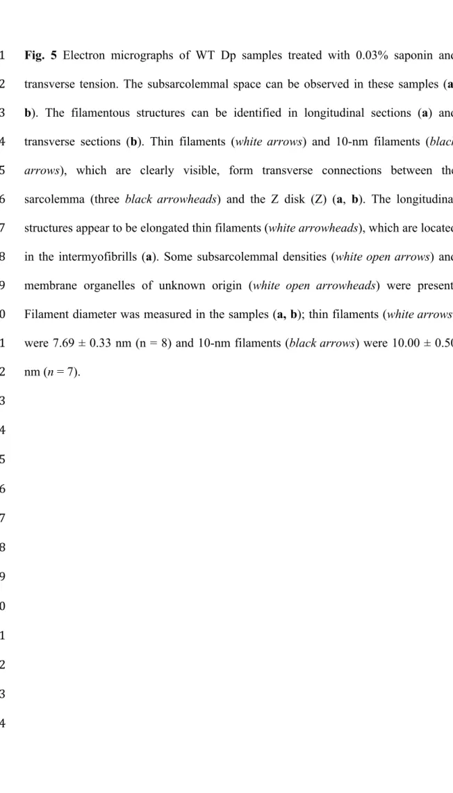
Transverse and longitudinal filamentous structures were also present in Dp 1
samples treated with 0.03% saponin and transverse tension (Figs. 5a, b). Those 2
filamentous structures could be identified in longitudinal sections (Fig. 5a) and in 3
transverse sections (Fig. 5b). Actin filaments (7.69 ± 0.33 nm; n = 8) and intermediate 4
filaments (10.00 ± 0.50 nm; n = 7) were clearly visible in the sections, and they 5
formed transverse connections between the sarcolemma and the Z disks. The 6
longitudinal structures appeared as elongated thin filaments that were mainly located 7
in the intermyofibrils. The insertion of some actin filaments into subsarcolemmal 8
densities was also observed.
9 10
Loss of filamentous architecture in the M line domain in mdx Dp 11
12
We demonstrated that treatment with transverse tension and 1% Triton X-100 or 13
0.03% saponin allows observation of the filamentous anchoring structures in the 14
subsarcolemmal space and intermyofibrils (Figs. 4 and 5). In the next set of 15
experiments, we used mdx mice to determine whether particular ultrastructural 16
features of the anchoring system were affected by these treatments.
17
Samples of mdx Dp [Fig. 6a, b (insert)] were treated with transverse tension 18
and 1% Triton X-100. As with the WT Dp samples that received the same treatment, 19
the subsarcolemmal space was clearly exposed. However, there were differences in 20
the appearance of the filamentous anchoring structures. There were fewer filamentous 21
structures in the mdx Dp. Connections between the M lines and the sarcolemma were 22
barely observed. Some 10-nm filaments were present above the Z disk areas.
23 24 25
Discussion 1
2
In this study, we provide morphological evidence of filamentous anchoring structures 3
in the subsarcolemmal space and intermyofibrils. The filamentous anchoring 4
structures discussed here are applied to the transverse and longitudinal filamentous 5
structures that appear in the three costameric domains and the intermyofibrils. These 6
filamentous structures can survive treatment with transverse tension and 1% Triton X- 7
100 or 0.03% saponin. Moreover, these structures are able to connect the peripheral 8
myofibrils to the sarcolemma or intermyofibrils (Figs. 4 and 5). Functionally, these 9
lateral linkages would help individual muscle fibers avoid disruptive contraction and 10
would aid in the generation of force [7, 18, 19].
11
Because our ultrastructural evidence for filamentous anchoring structures in 12
the subsarcolemmal space and intermyofibrils was obtained by electron microscopy of 13
intact muscle treated with Triton X-100 or saponin, we must consider the possibility 14
that these procedures may introduce artifacts, such as filament redistribution or 15
superimposition onto the subsarcolemmal space, Z disks or M lines. A classic 16
experiment by Pierobon-Bormioli demonstrated that muscle framework is difficult to 17
preserve [16]. However, our results, which were obtained from several independent 18
mice and several different treatments, suggest that the observed filamentous 19
anchoring structure distribution was not artifactual. First, Dp samples with no 20
transverse tension or detergent treatment (Fig. 1a) failed to show filamentous 21
structures because the subsarcolemmal space could not be visualized. Second, when 22
tension was applied without detergent treatment (Fig. 1b), some filamentous 23
structures could be observed within the subsarcolemmal space, but the presence of 24
many membrane organelles interfered with observation. Third, Dp samples subjected 25
to 1% Triton X-100 treatment but not tension treatment (Fig. 1c) also failed to show 1
filaments. Fourth, Dp samples treated with both 1% Triton X-100 and transverse 2
tension (Fig. 1d) yielded better results for the observation of filamentous anchoring 3
structures. Treatment with only transverse tension or transverse tension treatment 4
combined with 1% Triton X-100 or 0.03% saponin (Figs. 2, 4, 5) enabled observation 5
of the filamentous anchoring structures in the subsarcolemmal space and 6
intermyofibrils. These findings are in agreement with previous studies [6, 10, 16-24], 7
and we have obtained new evidence that provides a more comprehensive 8
understanding of the filamentous anchoring structures.
9
Based on the present and previous data, filamentous anchoring structures in 10
the subsarcolemmal space may be depicted, as shown in Fig.7. Transverse anchoring 11
filamentous structures that laterally interlink the Z disks and M lines of peripheral 12
myofibrils to the sarcolemma were clearly visible, and they were composed of actin 13
and intermediate filaments (Figs. 2-5). By measuring the filament diameters, we 14
confirmed that actin filaments and intermediate filaments are the most likely 15
candidates for linking peripheral myofibrils to the cytoplasmic surface of the 16
sarcolemma [1, 3, 4, 7, 9]. The possibilities that the filamentous anchoring structures 17
were also composed of membrane skeleton protein, such as dystrophin and/or spectrin 18
are low because these molecules are thinner than actin filament [30, 31]. At the Z- 19
domains of costameres, actin and intermediate filaments appeared to cooperate to 20
attach the Z disks of peripheral myofibrils to the sarcolemma (Figs. 4a, 4c, 5a). Actin 21
filaments in particular seem to take the form of longitudinal anchoring structures or 22
elongated filaments, not only in the subsarcolemmal space (Figs. 2-5) but also in the 23
intermyofibrils (Figs. 2 and 5). This result supports the findings of previous 24
immunofluorescence studies, which indicated that costameric actin filaments and 25
intermediate filaments serve as structural components of both costameres and 1
intermyofibrils at the level of Z lines [10, 18, 23, 24]. Different from the Z-domains, 2
our findings at the M-domains of costameres suggest that only intermediate filaments 3
form the connections between the M lines of peripheral myofibrils and the 4
sarcolemma (Figs. 2b, 4a, 4b). This result is in accordance with previous reports [2, 3, 5
19, 32]. In the later researches, not only desmin [2, 18, 23, 25] but also keratin 6
filaments [2, 3, 8, 12], are the types of intermediate filaments that are being proposed 7
as anchoring structures between sarcolemma and peripheral myofibril. Desmin 8
enriched at the Z-domains of costameres [2, 3], but was not present in significant 9
amounts at M-domains or L-domains of costameres [7]. The other type is composed 10
of keratin filaments containing keratin 8 (K8) and keratin 19 (K19) [3, 8]. Although 11
keratins are present in smaller amounts than desmin, K8 and K19 are found at both 12
the Z-domains and M-domains of costameres [8, 12]. Our ultrastructural findings 13
ascertained the previous reports by clearly showing that the intermediate filaments are 14
component of filamentous anchoring structures between sarcolemma and peripheral 15
myofibrils.
16
We were able to visualize the elongation of thin filaments in the 17
subsarcolemmal spaces and in the intermyofibrils (Figs. 2-5). The path of elongation 18
of thin filaments from the peripheral myofibrils to the sarcolemma was clearly 19
demonstrated by our data. This information was lacking in previous studies by Bard 20
and Franzini-Armstrong [17]. Those authors suggested that peripheral filaments are 21
composed of actin and are anchored to Z lines [17], which is consistent with our 22
results. In the subsarcolemmal space, the elongated thin filaments obliquely arose 23
from the peripheral myofibrils and then longitudinally extended before finally 24
inserting into the subsarcolemmal densities (Figs 2-5). Based on this evidence, it is 25
possible that these elongated thin filaments extending from the peripheral myofibrils 1
to the sarcolemma are actually the ultrastructural L-domains of the costameres.
2
In Figures 2-5, we also observed the appearance of electron-dense plaques on 3
the cytoplasmic side of the sarcolemma. These plaques are most likely 4
subsarcolemmal densities, as reported previously by Pierobon-Bormioli [16] and 5
Shear [22]. Membrane skeleton proteins such as vinculin [6, 10, 22], dystrophin and 6
β-spectrin [11] are considered to be components of subsarcolemmal densities. From 7
our results, the subsarcolemmal densities appeared in all three domains of costameres.
8
We clearly visualized the association of subsarcolemmal densities with the 9
filamentous anchoring structures, particularly the elongated thin filaments coming 10
from the peripheral myofibrils (Figs. 2, 3, 5). These results remind us of a previous 11
study that mentioned the relationship of subsarcolemmal densities with extracellular 12
structures [22]. In 1985, Shear observed that the densities were associated with 13
extracellular thin filaments that extend from the sarcolemma through the basal lamina 14
[22]. Our observations complete the picture by revealing that the subsarcolemmal 15
densities are also associated with filamentous anchoring structures from the peripheral 16
myofibrils. This association might play a role in anchoring the sarcolemma to the 17
peripheral myofibrils. Thus, we propose that subsarcolemmal densities and their 18
associated filamentous anchoring structures constitute the ultrastructural 19
representation of costameres.
20
Previous experiments suggest that costameres may serve to laterally transmit 21
contractile forces from the sarcomeres across the sarcolemma to the extracellular 22
matrix, ultimately transmitting the force to neighboring muscle cells [9, 10, 19].
23
Dystrophin and its associated proteins are found at the sarcolemma in association with 24
the Z-domains of costameres [33, 34]. Confocal immunofluorescence analysis showed 25
that dystrophin forms a strong mechanical attachment to the sarcolemma [24, 33].
1
Immunoelectron microscopy revealed that dystrophin distributed close to the 2
cytoplasmic surface of the plasma membrane [35, 36]. In muscle fibers skinned with 3
Triton X-100, immunoelectron microscopy labeled dystrophin at outer-side surface of 4
subsarcolemmal densities [23]. Freeze-fracture replica immunoelectron microscopy 5
showed that labeling of spectrin and dystrophin were at the cytoplasmic surface of the 6
plasma membrane [37]. Dystrophin in the subsarcolemmal densities is associated with 7
integral membrane proteins such as β-dystroglycans [3, 24, 38, 39]. These proteins 8
interaction are associated subsequently with suprasarcolemmal α-dystroglycans, 9
forming a structural links in the sarcolemma and with the basal lamina by binding 10
laminin-2 [23, 39, 40]. On the other hand, dystrophin do not make filaments between 11
sarcolemma and sarcomeres. Dystrophin links to the sarcomeres through other 12
proteins interaction, such as gamma-actin filaments [3, 24]. Our results showed that 13
the subsarcolemmal densities still remained after tension treatment. Thus, dystrophin, 14
spectrin, and other associated proteins might still retained in the densities. DMD is 15
caused by mutations in the gene encoding dystrophin [41-43]. The mdx mouse, which 16
is an animal model for DMD, carries a mutation in the dystrophin gene and lacks the 17
native protein [28, 44]. When dystrophin is absent, the link between the costamere 18
and sarcolemma is disrupted, resulting in compromised sarcolemmal integrity [9].
19
This study provides the first ultrastructural evidence showing the differences between 20
the filamentous anchoring structures of WT and mdx Dp samples subjected to the 21
same treatment (Figs. 4 and 6). There were fewer filamentous anchoring structures in 22
the mdx Dp samples. Especially connections between the M lines and the sarcolemma 23
were barely observed in the mdx samples. Our data support previous studies that 24
found that the M line domains of the costameres are more susceptible to disruption in 25
mdx mice [2, 11, 13]. The absence of dystrophin and the destabilization of the 1
filamentous anchoring structures may cause the costamere abnormalities observed in 2
mdx mice [7, 9, 24, 45].
3
Taken together, the data from this study show that tension treatment, with or 4
without detergent or saponin treatment, allows observation of the filamentous 5
anchoring structures in the subsarcolemmal and intermyofibrillar spaces. Actin and 6
intermediate filaments show their presence and integrity as components of the 7
transverse and longitudinal anchoring structures in the subsarcolemmal space and the 8
intermyofibrils.
9 10 11
Conclusion 12
We showed that the transverse and longitudinal anchoring structures along with the 13
subsarcolemmal densities and elongated thin filaments in the subsarcolemmal space 14
might represent the ultrastructural components of the costamere. We also reported a 15
lack of filamentous anchoring structures in mdx mice. The mechanism underlying 16
how these structures were lost was not revealed in this study. Further study of mdx 17
mice may provide new insights into cytoskeleton organization in skeletal muscle 18
fibers and may contribute to a more comprehensive understanding of how defects 19
cause membrane fragility and muscle wasting.
20 21 22 23 24 25
Acknowledgments 1
We thank H. Matsuda, M. Shikada and Y. Morimura for both technical and secretarial 2
assistance. This work was supported in part by Grants-in-Aid for Scientific Research 3
from the Ministry of Education, Culture, Sports, Science and Technology of Japan, 4
KAKENHI Grant Numbers 20590183, 23590230.
5 6 7 8 9 10 11 12 13 14 15 16 17 18 19 20 21 22 23 24 25
References 1
2
1. Ramaekers FCS, Bosman FT (2004) The cytoskeleton and disease. J Pathol 3
204:351–354 4
2. O'Neill A, Williams M, Resneck WG, Milner DJ, Capetanaki Y, Bloch RJ 5
(2002) Sarcolemmal Organization in Skeletal Muscle Lacking Desmin:
6
Evidence for Cytokeratins Associated with the Membrane Skeleton at 7
Costameres. Mol Biol Cell 13:2347–2359 8
3. Capetanaki Y, Bloch RJ, Kouloumenta A, Mavroidis M, Psarras S (2007) 9
Muscle intermediate filaments and their links to membranes and membranous 10
organelles. Exp Cell Res 313:2063–2076 11
4. Kee AJ, Gunning PW, Hardeman EC (2009) Diverse roles of the actin 12
cytoskeleton in striated muscle. J Muscle Res Cell Motil 30:187–197 13
5. Clark KA, McElhinny AS, Beckerle MC, Gregorio CC (2002) Striated Muscle 14
Cytoarchitecture: An Intricate Web of Form and Function. Annu Rev Cell Dev 15
Biol 18:637–706 16
6. Pardo JV, Siliciano JD, Craig SW (1983) A vinculin-containing cortical lattice 17
in skeletal muscle: transverse lattice elements (“costameres”) mark sites of 18
attachment between myofibrils and sarcolemma. Proc Natl Acad Sci USA 19
80:1008–1012 20
7. Bloch RJ, Gonzales-Serratos H (2003) Lateral Force Transmission Across 21
Costameres in Skeletal Muscle. Exerc Sport Sci Rev 31:73–78 22
8. Ursitti JA, Lee PC, Resneck WG, McNally MM, Bowman AL, O'Neill A, 23
Stone MR, Bloch RJ (2004) Cloning and characterization of cytokeratins 8 and 24
19 in adult rat striated muscle. Interaction with the dystrophin glycoprotein 25
complex. J Biol Chem 279:41830–41838 26
9. Ervasti JM (2003) Costameres: the Achilles' Heel of Herculean Muscle. J Biol 27
Chem 278:13591–13594 28
10. Craig SW, Pardo JV (1983) Gamma Actin, Spectrin, and Intermediate Filament 29
Proteins Colocalize with Vinculin at Costameres, Myofibril-to-Sarcolemma 30
Attachment Sites. Cell Motil 3:449–462 31
11. Porter GA, Dmytrenko GM, Winkelmann JC, Bloch RJ (1992) Dystrophin 32
colocalizes with β-spectrin in distinct subsarcolemmal domains in mammalian 33
skeletal muscle. J Cell Biol 117:997–1005 34
12. Stone MR, O'Neill A, Lovering RM, Strong J, Resneck WG, Reed PW, 35
Toivola DM, Ursitti JA, Omary MB, Bloch RJ (2007) Absence of keratin 19 in 36
mice causes skeletal myopathy with mitochondrial and sarcolemmal 37
reorganization. J Cell Sci 120:3999–4008 38
13. Williams MW, Bloch RJ (1999) Extensive but coordinated reorganization of 1
the membrane skeleton in myofibers of dystrophic (mdx) mice. J Cell Biol 2
144:1259–1270 3
14. Williams MW, Resneck WG, Bloch RJ (2000) Membrane skeleton of 4
innervated and denervated fast- and slow-twitch muscle. Muscle Nerve 5
23:590–599 6
15. Williams MW, Resneck WG, Kaysser T, Ursitti JA, Birkenmeier CS, Barker 7
JE, Bloch RJ (2001) Na, K-ATPase in skeletal muscle: two populations of β- 8
spectrin control localization in the sarcolemma but not partitioning between the 9
sarcolemma and the transverse tubules. J Cell Sci 114:751–762 10
16. Pierobon-Bormioli S (1981) Transverse sarcomere filamentous systems: “Z- 11
and M-cables.” J Muscle Res Cell Motil 2:401–413 12
17. Bard F, Franzini-Armstrong C (1991) Extra actin filaments at the periphery of 13
skeletal muscle myofibrils. Tissue Cell 23:191–197 14
18. Hijikata T, Murakami T, Imamura M, Fujimaki N, Ishikawa H (1999) Plectin is 15
a linker of intermediate filaments to Z-discs in skeletal muscle fibers. J Cell Sci 16
112:867–876.
17
19. Street SF (1983) Lateral Transmission of Tension in Frog Myofibers: A 18
Myofibrillar Network and Transverse Cytoskeletal Connections Are Possible 19
Transmitters. J Cell Physiol 114:346–364 20
20. Garamvölgyi N (1965) Inter-Z bridges in the flight muscle of the bee. J 21
Ultrastruct Res 13:435–443 22
21. Wang K, Ramirez-Mitchell R (1983) A network of transverse and longitudinal 23
intermediate filaments is associated with sarcomeres of adult vertebrate skeletal 24
muscle. J Cell Biol 96:562–570 25
22. Shear CR, Bloch RJ (1985) Vinculin in subsarcolemmal densities in chicken 26
skeletal muscle: localization and relationship to intracellular and extracellular 27
structures. J Cell Biol 101:240–256 28
23. Hijikata T, Murakami T, Ishikawa H, Yorifuji H (2003) Plectin tethers desmin 29
intermediate filaments onto subsarcolemmal dense plaques containing 30
dystrophin and vinculin. Histochem Cell Biol 119:109–123 31
24. Rybakova IN, Patel JR, Ervasti JM (2000) The dystrophin complex forms a 32
mechanically strong link between the sarcolemma and costameric actin. J Cell 33
Biol 150:1209–1214 34
25. Hijikata T, Nakamura A, Isokawa K, Imamura M, Yuasa K, Ishikawa R, 35
Kohama K, Takeda S, Yorifuji H (2008) Plectin 1 links intermediate filaments 36
to costameric sarcolemma through β-synemin, α-dystrobrevin and actin. J Cell 37
Sci 121:2062–2074 38
junction: localization of vinculin. J Electron Microsc Tech 12:160–171 1
27. Stedman HH, Sweeney HL, Shrager JB, Maguire HC, Panettieri RA, Petrof B, 2
Narusawa M, Leferovich JM, Sladky JT, Kelly AM (1991) The mdx mouse 3
diaphragm reproduces the degenerative changes of Duchene muscular 4
dystrophy. Nature 352:536–539 5
28. Ishizaki M, Suga T, Kimura E, Shiota T, Kawano R, Uchida Y, Uchino K, 6
Yamashita S, Maeda Y, Uchino M (2008) Mdx respiratory impairment 7
following fibrosis of the diaphragm. Neuromuscul Disord 18:342–348 8
29. Ishikawa H, Bischoff R, Holtzer H (1968) Mitosis and intermediate-sized 9
filaments in developing skeletal muscle. J Cell Biol 38:538–555 10
30. Cohen CM, Tyler JM, Branton D (1980) Spectrin-actin associations studied by 11
electron microscopy of shadowed preparations. Cell 21:875–883 12
31. Pons F, Augier N, Heilig R, Léger J, Mornet D, Léger JJ (1990) Isolated 13
dystrophin molecules as seen by electron microscopy. Proc Natl Acad Sci 14
USA 87:7851–7855.
15
32. Lazarides E (1980) Intermediate filaments as mechanical integrators of cellular 16
space. Nature 283:249–256 17
33. Straub V, Bittner RE, Léger JJ, Voit T (1992) Direct visualization of the 18
dystrophin network on skeletal muscle fiber membrane. J Cell Biol 119:1183–
19
1191 20
34. Goldstein JA, McNally EM (2010) Mechanisms of muscle weakness in 21
muscular dystrophy. J Gen Physiol 136:29–34 22
35. Cullen MJ, Walsh J, Nicholson LVB, Harris JB (1990) Ultrastructural 23
localization of dystrophin in human muscle by using gold immunolabelling.
24
Proc R Soc Lond B Biol Sci 240:197–210.
25
36. Harris JB, Cullen MJ (1992) Ultrastrucutral localization and the possible role 26
of dystrophin. In: Kalkulas BA, Howell JM, Roses AD (eds) Duchenne 27
muscular dystrophy: animal models and genetic manipulation. Raven Press, 28
New York, pp 19–40.
29
37. Stevenson SA, Cullen MJ, Rothery S, Coppen SR (2005) High-resolution en- 30
face visualization of the cardiomyocyte plasma membrane reveals distinctive 31
distributions of spectrin and dystrophin. Eur J Cell Biol 84:961–971.
32
38. Jung D, Yang B, Meyer J, Chamberlain JS, Campbell KP (1995) Identification 33
and characterization of the dystrophin anchoring site on β-dystroglycan. J Biol 34
Chem 270:27305–27310 35
39. Ervasti JM, Campbell KP (1993) A role for the dystrophin-glycoprotein 36
complex as a transmembrane linker between laminin and actin. J Cell Biol 37
122:809–823.
38
40. Ibraghimov-Beskrovnaya O, Ervasti JM, Leveille CJ, Slaughter CA, Sernett 1
SW, Campbell KP (1992) Primary structure of dystrophin-associated 2
glycoproteins linking dystrophin to extracellular matrix. Nature 355:696–702.
3
41. Hoffman EP, Kunkel LM (1989) Dystrophin abnormalities in 4
Duchenne/Becker muscular dystrophy. Neuron 2:1019–1029 5
42. Campbell KP (1995) Three muscular dystrophies: loss of cytoskeleton- 6
extracellular matrix linkage. Cell 80:675–679 7
43. O'Brien KF, Kunkel LM (2001) Dystrophin and muscular dystrophy: past, 8
present, and future. Mol Genet Metab 74:75–88 9
44. Law DJ, Allen DL, Tidball JG (1994) Talin, vinculin and DRP (utrophin) 10
concentrations are increased at mdx myotendinous junctions following onset of 11
necrosis. J Cell Sci 107:1477–1483 12
45. Bellin RM, Huiatt TW, Critchley DR, Robson RM (2001) Synemin May 13
Function to Directly Link Muscle Cell Intermediate Filaments to Both 14
Myofibrillar Z lines and Costameres. J Biol Chem 276:32330–32337 15
16 17
18
19
20
21
22
23
24
25
26
Figure Legends 1
2
Fig. 1 Electron micrographs of WT Dp samples showing the effects of transverse 3
tension and 1% Triton X-100 treatment. Dp samples without transverse tension or 4
detergent treatment (a) failed to show filamentous architecture between the 5
sarcolemma and peripheral myofibrils. The subsarcolemmal space (the space between 6
the two black open arrows) cannot be identified because the sarcolemma (three black 7
arrowheads) remains closely attached to the peripheral myofibrils. When tension was 8
applied without detergent treatment (b), the subsarcolemmal space (the space between 9
the two black open arrows) could be identified between the sarcolemma (three black 10
arrowheads) and peripheral myofibrils. Some filamentous structures could also be 11
observed. Many membrane organelles of unknown origin (white open arrowheads) 12
were still present, most likely because there was no detergent treatment. Dp samples 13
subjected to 1% Triton X-100 without tension treatment (c) also failed to show 14
filaments. Dp samples subjected to both 1% Triton X-100 and transverse tension 15
treatment (d) provided better observations of the filamentous anchoring structures. Z, 16
Z disk; M, M line.
17 18 19 20 21 22 23 24
Fig. 2 High-magnification electron micrographs of longitudinally sectioned WT Dp 1
samples treated with transverse tension only. Three independent Dp samples (a, b, c) 2
were used. The subsarcolemmal space could be observed between the sarcolemma 3
(three black arrowheads) and peripheral myofibrils. The filamentous anchoring 4
structures apparently survived the tension treatment. Transversely running thin 5
filaments (white arrow) and 10-nm filaments (black arrow) could be identified. The 6
thin filaments (white arrowheads) showed oblique elongation in the subsarcolemmal 7
space and in the intermyofibrils. Subsarcolemmal densities (white open arrows) were 8
found to be in direct contact with the filamentous structures. Organelles of unknown 9
origin (white open arrowheads) were found in some locations. The boxed areas in a, 10
b, and c are shown at a higher magnification (d, e, f). Length of the filament diameter 11
was measured and then calculated to have the value of filament diameter. The space 12
between two white arrows is 10 nm. In images a, b, and c, thin filaments (white 13
arrow) were 8.20 ± 1.16 nm (n = 12) and 10-nm filaments (black arrow) were 11.65 ± 14
1.63 nm (n = 4). Z, Z disk; M, M line.
15 16 17 18 19 20 21 22 23 24
Fig. 3 Electron micrographs of transversely sectioned WT Dp samples treated with 1
transverse tension only. These sections (a, b) confirmed the appearance of the 2
filamentous anchoring structures connecting the sarcolemma (three black 3
arrowheads) and peripheral myofibrils. Thin filaments (white arrows) were clearly 4
observed to originate from the A-band (A), and some were observed to originate from 5
the I-band (I). In both cases, the filaments were connected to the sarcolemma.
6
Subsarcolemmal densities (white open arrows) were in direct contact with the 7
filamentous structures. Membrane organelles of unknown origin (white open 8
arrowheads) were observed between the structures. Filament diameter was measured 9
in the samples (a and b), and the thin filaments (white arrows) were 8.29 ± 0.55 nm (n 10
= 11). Z, Z disk.
11 12 13 14 15 16 17 18 19 20 21 22 23 24 25
Fig. 4 Electron micrographs of longitudinally sectioned WT Dp samples treated with 1
1% Triton X-100 and transverse tension. The subsarcolemmal spaces were clearly 2
exposed in these three independent samples (a, b, c). The filamentous structures, thin 3
filaments (white arrows) and 10-nm filaments (black arrows) formed firm transverse 4
connections between the sarcolemma (three black arrowheads) and the Z disks (Z) 5
and M lines (M) of peripheral myofibrils. Most of the longitudinal structures were 6
elongated thin filaments (white arrowheads). Some subsarcolemmal densities (white 7
open arrows) were observed, especially above the Z disk (Z) and M line (M) areas.
8
Debris from membrane organelles (white open arrowheads) was present but less 9
abundant. The persistent appearance of firm anchoring structures despite treatment 10
with 1% Triton X-100 and transverse tension ascertained the existence of filamentous 11
structures as transverse and longitudinal anchoring structures between the sarcolemma 12
and peripheral myofibrils. The boxed area in a is shown at a higher magnification (d).
13
Length of the filament diameter was measured and then calculated to have the value 14
of filament diameter. The space between two white arrows is 10 nm. In images a, b, 15
and c, thin filaments (white arrows) were 8.23 ± 0.44 nm (n = 9) and 10-nm filaments 16
(black arrows) were 10.38 ± 0.52 nm (n = 8).
17 18 19 20 21 22 23 24
Fig. 5 Electron micrographs of WT Dp samples treated with 0.03% saponin and 1
transverse tension. The subsarcolemmal space can be observed in these samples (a, 2
b). The filamentous structures can be identified in longitudinal sections (a) and 3
transverse sections (b). Thin filaments (white arrows) and 10-nm filaments (black 4
arrows), which are clearly visible, form transverse connections between the 5
sarcolemma (three black arrowheads) and the Z disk (Z) (a, b). The longitudinal 6
structures appear to be elongated thin filaments (white arrowheads), which are located 7
in the intermyofibrills (a). Some subsarcolemmal densities (white open arrows) and 8
membrane organelles of unknown origin (white open arrowheads) were present.
9
Filament diameter was measured in the samples (a, b); thin filaments (white arrows) 10
were 7.69 ± 0.33 nm (n = 8) and 10-nm filaments (black arrows) were 10.00 ± 0.50 11
nm (n = 7).
12 13
14
15
16
17
18
19
20
21
22
23
24
Fig. 6 Electron micrographs of longitudinally sectioned mdx Dp samples treated with 1
1% Triton X-100 and transverse tension [a, b (insert)]. As with the WT Dp samples 2
subjected to the same treatment, the subsarcolemmal space (the space between the 3
two black open arrows) was clearly exposed (a). The filamentous anchoring 4
structures had a distinct appearance in the mdx samples. There were fewer 5
filamentous structures in the mdx Dp, and there were very few structures connecting 6
the M line (M) to the sarcolemma (three black arrowheads). Some 10-nm filaments 7
(black arrow) were present above the Z disk (Z) areas (b [insert)]. Subsarcolemmal 8
densities (white open arrows) were observed. Debris from the membrane organelles 9
of unknown origin (white open arrowheads) was present in some locations. Filament 10
diameter was measured in the samples [b (insert)]; 10-nm filaments (black arrow) 11
were 10 nm (n = 2).
12 13
14
15
16
17
18
19
20
21
Fig. 7 Schematic representation of the ultrastructural components of the costamere.
1
Filamentous anchoring structures along with the subsarcolemmal densities and 2
elongated thin filaments in the subsarcolemmal space are depicted as components of 3
costameres. Filamentous anchoring structures are composed of actin and intermediate 4
filaments. Subsarcolemmal densities appear in all three domains of costameres. Based 5
on the present study, the filamentous structures depicted to be inserted into 6
subsarcolemmal densities. Z-domains, M-domains, and L-domains are illustrated. At 7
the Z-domains of costameres, actin and intermediate filaments appeared to cooperate 8
to attach the Z disks of peripheral myofibrils to the sarcolemma. While at the M- 9
domains of costameres, our results suggest that only intermediate filaments form the 10
connections between the M lines of peripheral myofibrils and the sarcolemma. The 11
subsarcolemmal densities in Z-domains and M-domains interact with integral 12
membrane proteins (β-dystroglycans, sarcoglycans, integrin, etc). These proteins 13
interactions continue to form a structural link by subsequently associate with 14
suprasarcolemmal protein (α-dystroglycans, etc) and with the basal lamina proteins 15
(laminin-2, etc). At the L-domains of costameres, elongated thin filaments extend 16
from the peripheral myofibrils to the sarcolemma. Interaction between 17
subsarcolemmal densities in L-domain with integral membrane protein is expected, 18
but detail information is still unknown (dotted areas).
19 20 21 22 23 24 25 26
Figures 1
Fig. 1 2
3
4
Fig. 2 1
2
3
Fig 2 (continued) 1
2
Fig. 3 1
2
3
4
5
6
7
8
9
10
11
12
13
Fig. 4 1
2
Fig. 4 (continued) 1
2
3
4
5
6
7
8
9
10
11
12
Fig. 5 1
2
3
Fig. 6 1
2
3
4
5
6
7
8
9
Fig.7 1
2