膵病変を合併したと思われる肺結核の 1 例
1佐野安希子
1西山 理
1佐野 博幸
2吉田耕一郎
1東田 有智
は じ め に 2015 年の厚生労働省の統計によれば,結核の新規登録 患者数は年間19,615人であり,初めて 2 万人を下回った。 そ の う ち,肺 結 核 は 15,149 人(77.2%), 肺 外 結 核 は 4,466 人(22.8%)であった。結核全体に占める肺外結核 の割合は 20% を超えているものの,結核患者の減少に伴 って肺外結核は鑑別診断として挙がりにくくなってお り,その結果,診断が遅れるケースが増えてきていると 思われる。肺外結核は様々な臓器に起こるが,本邦で は,結核性胸膜炎・膿胸の割合が最も多く(17.65%),次 にリンパ節結核の割合が多い(5.69%)。腹部結核は多 くないが,その中で最もよくみられるものは腸結核 1.43 %,ついで,結核性腹膜炎 1.06% とされている1)。膵結核 はきわめて低頻度であり,通常は免疫抑制状態下での粟 粒結核の一部として起こることが多い2)。膵結核の症状 には発熱・心窩部痛・体重減少などがあるが,非特異的 な症状であることが多く,また,画像検査においても特 徴的所見に乏しいことから,膵癌,腫瘤形成性膵炎,膵 嚢胞等と鑑別が困難であり,診断に難渋することが多 い。今回われわれは,膵癌疑いにて膵臓切除術を受けた のち,数カ月後に肺結核の存在が判明したことにより, 膵臓の病変が膵結核であったと診断された症例を経験し たので報告する。 症 例 症 例:52 歳,女性。 主 訴:心窩部痛。 既往歴:特記事項なし。結核の既往なし。 生活歴:喫煙歴なし,飲酒歴なし。 現病歴:特に基礎疾患なし。2013 年 2 月,心窩部痛に て近医受診。腹部造影 CT にて膵体部に約 6 cm の境界不 明瞭な低濃度の腫瘤と傍大動脈リンパ節腫大を認め (Fig. 1a),膵癌疑いとして当院消化器内科に紹介となっ た。MR 胆 管 膵 管 撮 影(magnetic resonance cholangio-pancreatography : MRCP)では,T1W イメージで低信号, 1近畿大学医学部内科学教室呼吸器・アレルギー内科部門,2近 畿大学医学部附属病院安全管理部感染対策室 連絡先 : 佐野安希子,近畿大学医学部内科学教室呼吸器・アレ ルギー内科部門,〒 589 _ 8511 大阪府大阪狭山市大野東 377 _ 2 (E-mail : sanoa@med.kindai.ac.jp)(Received 26 May 2016 / Accepted 8 Jul. 2016)
要旨:52 歳,女性。心窩部痛と体重減少を主訴に受診。腹部造影 CT にて膵体部に腫瘤を認め,膵臓 の悪性腫瘍が疑われた。EUS-FNA を 3 回施行したが診断に至らず,膵体尾部切除術を施行,病理組 織診断にて壊死を伴う類上皮細胞肉芽腫を認めた。Ziehl-Neelsen 染色で抗酸菌は証明されず,結核や サルコイドーシスが鑑別として考えられたが,腫瘤は完全に切除され,症状も改善していたため,経 過観察となっていた。約 4 カ月後,膵性糖尿病に対してインスリン導入のため入院となった際に湿性 咳嗽,発熱が出現。CT で両肺上葉に微細結節の集簇を認めた。気管支鏡検査を施行し,気管支洗浄 液培養から結核菌陽性となった。抗結核薬 4 剤の治療を開始し,症状と画像所見の改善を認めた。初 回検査時に診断が得られなかったが,後に肺結核の存在が判明したことにより,膵臓の病変は膵結核 であったと考えられた。膵結核は稀な疾患であり,臨床所見や画像から診断することが困難であるた め,病理学的,細菌学的確証が重要である。まず結核の可能性を疑って正確な診断アプローチを行う ことが必要であり,それによって不要な外科的切除を避けることができる。 キーワーズ:肺外結核,膵結核,膵癌
Fig. 1 (a) Preoperative abdominal contrast-enhanced computed tomography revealed a low-density mass in the body
of the pancreas with para-aortic lymphadenopathy. (b) Histopathological examination of the resected pancreatic mass showed epithelioid cell granuloma with central necrosis surrounded by lymphocytes (hematoxylin-eosin stain, ×400).
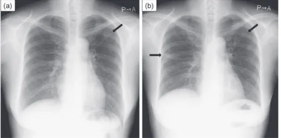
Fig. 2 Chest X-ray images 5 months before (a) and at the time of admission (b). (a) showed
trabecular shadow in the upper fi eld of the left lung. (b) showed patchy shadows in the middle fi eld of the right lung and small granular shadows in the upper fi eld of the left lung.
嗽,発熱が出現し,胸部 CT にて両肺上葉に小葉中心性 微細結節の集簇を認めたため,当科紹介となった。 身体所見:身長 161.0 cm,体重 56.1 kg,BMI 21.6 kg/ m2,体温37.6度,血圧113/70 mmHg,脈拍78回 ⁄分(整), 呼吸数 16 回 ⁄分,SpO2 96%(室内気)。意識清明,神経 学的所見に異常認めず,眼球,眼瞼結膜に貧血,黄疸認 めず。表在リンパ節触知せず。呼吸音清,心音整,腹部 平坦・軟,心窩部に圧痛あり,腹部手術創あり,下腿浮 腫なし。
検査所見(Table):白血球7600/μl(Neu 75.0%,Lym11.5 %),CRP 4.598 mg/dl と高 CRP 血症を認めた。また,空腹 時 血 糖 102 mg/dl,HbA1c 6.9%,T SPOT®.TB(T-SPOT) は陰性であった。
画像所見:当科受診時の胸部単純 X 線(Fig. 2b)では, 右中肺野に斑状影,左上肺野に小粒状影を認めた。胸腹 T2W イメージで高信号を呈する辺縁不整な腫瘤と,主
膵管と下部胆管の狭窄が認められた。超音波内視鏡下穿 刺 吸 引 細 胞 診(endoscopic ultrasound-guided fi ne needle aspiration : EUS-FNA)を 3 回施行したが,確定診断に至 らず,2013 年 7 月,膵癌の術前診断にて膵体尾部切除術 を行った。膵臓と傍大動脈リンパ節の病理組織にて,ラ 氏型巨細胞やリンパ球が浸潤した類上皮細胞肉芽腫を多 数認めた。大きな病変では中心部に壊死を伴っていた (Fig. 1b)。結核やサルコイドーシスが鑑別として考えら れたが,Ziehl- Neelsen 染色で抗酸菌は証明されず(結核 菌群 PCR や培養検査は行っていなかった),腫瘤は完全 に切除され,症状も改善していたため,経過観察となっ ていた。 約 4 カ月後,膵性糖尿病に対してインスリン導入のた め当院内分泌・代謝内科に入院。入院経過中に湿性咳
Table Laboratory data on admission
Fig. 3 Chest and abdominal computed tomography images on admission. (a) Chest computed tomography showed
multiple granular shadows in both upper lobes with a small volume of right pleural effusion. (b) Abdomial computed tomography showed multiple enlarged para-aortic lymph nodes with a small volume of ascitic fl uid.
[Hematology] RBC 4.52×10⁶ /μl Hb 10.0 g/dl Ht 32.3 % Plt 62.5×10⁴ /μl WBC 7600 /μl Neu 75.0 % Lym 11.5 % Mono 12.5 % Eos 1.0 % Baso 0.0 % [Serology] CRP 4.598 mg/dl IgG 2049 mg/dl IgA 506 mg/dl IgM 66 mg/dl ACE 17.2 IU/l CEA 0.7 pg/ml CA19-9 7.1 U/ml
HIV Ag-Ab negative T-SPOT®.TB negative [Biochemistry] TP 8.1 mg/dl alb 3.5 mg/dl AST 19 IU/l ALT 5 IU/l LDH 225 IU/l ALP 176 IU/l Amy 54 IU/l T.bil 0.5 mg/dl BUN 15 mg/dl Cr 0.65 mg/dl Na 136 mEq/l K 4.7 mEq/l Cl 99 mEq/l Glu 102 mg/dl HbA1c 6.9 %
Fig. 4 Chest and abdominal computed tomography images after anti-tuberculosis chemotherapy. (a) showed that
nodular opacities in the lung fi elds and pleural effusion had disappeared. (b) showed that para-aortic lymph nodes enlargement and ascites retention had improved.
部 CT(Fig. 3)では,両肺上葉に多発小粒状影,少量の 右胸水を認め,腹部において,少量の腹水と多発傍大動 脈リンパ節腫大を認めた。 入院後経過:気管支鏡検査を施行した。右 B3気管支 より洗浄を行い,抗酸菌塗抹は陰性であったが,培養陽 性,結核菌群抗原検査(キャピリア TB 法)陽性であっ た。ここで膵臓手術前の胸部単純 X 線(当科受診約 5 カ 月前)を参照すると(Fig. 2a),左上肺野に索状影を認 めており,この時点からの肺結核の存在が疑われた。従 って,膵臓の病理組織で壊死を伴う類上皮細胞肉芽腫を 認めていたことと合わせて考えると,膵臓の病変は,肺 結核に併存する膵結核であったと考えられた。 4 剤による標準治療(イソニアジド,リファンピシン, エタンブトール,ピラジナミド)を開始した。これによ り,微熱や咳嗽, 怠感などの自覚症状は改善し,画像 上,肺野陰影,胸腹水,腹部リンパ節の腫大も改善した (治療開始 6 カ月後:Fig. 4)。薬剤感受性は,全ての抗
結核薬に対して感受性であった。 6 カ月で治療終了し, その後も病状再燃は認めていない。 考 察 膵結核はきわめて低頻度である。臨床所見や画像上, 膵癌との鑑別が困難であることから,膵癌と術前診断さ れていた外科切除標本の病理組織診断で偶然に結核と判 明することがある3)。膵癌のほか,膵嚢胞性腫瘍,慢性 膵炎,自己免疫性膵炎などと鑑別が難しいこともある。 膵結核は男性に多いとされ,発症年齢は報告によって 様々であるが,比較的若年(40∼50 代)での罹患が多 い。臨床症状は多様であり,腹痛,食思不振,体重減少, 発熱と寝汗,胆汁鬱滞による黄疸,腹部腫瘤などが起こ りうる。膵臓以外の病変,特に胸部単純写真での異常の 発見は,膵病変が結核症の一部分であることを示す手が かりとなるが,併存する肺活動性病変の存在は,50% 程 度までと報告されている4)。 Xia らは,次のような所見があれば結核を疑うべきで あるとして,膵結核に特徴的な臨床所見を述べている。 ①若年者である,②結核の既往がある,または結核流行 地域出身である,③上腹部痛,発熱,体重減少の臨床所 見がある,④超音波や CT 検査で膵腫瘤と周囲リンパ節 の腫脹がある5)。また,膵結核の発症様式は以下のよう な経路が考えられている。①粟粒結核の一部として起こ る血行性散布,②未知の部位からの血行性・リンパ行性 進展,③近接臓器からの直接進展,の 3 つである6)。こ のうち,潜在する肺病変からの血行性,リンパ行性進展 が最も多いといわれている4)。本症例は,肺の活動性結 核の存在が後に判明したことから,結核菌が血行性に膵 臓に波及し,その後周囲リンパ節にも進展したと考えら れた。 膵癌と膵結核を明確に区別する画像所見はない。膵結 核の病変は,膵頭部・体部に最も頻度が高く,CT では, 低濃度の腫瘤として描出され,56% の症例で石灰化を伴 う。随伴所見として,造影 CT で周囲リング状に増強さ れたリンパ節を認めることがある7)。MRI では,T1W イ メージにおいて低∼等信号,T2W イメージにおいて不均 一な高信号を呈する8)。FDG-PET 検査は,結核を含む抗 酸菌症において偽陽性となることが知られており,結核 の存在診断の一助となる可能性は示唆されているが9), その所見は,膵癌と類似しているため,悪性との鑑別に は有用でないかもしれない。内視鏡的逆行性胆道膵管造 影(endoscopic retrograde cholangiopancreatography: ERCP) では,主膵管の変位・狭窄などが認められることがある 一方,病変が膵頭部に存在する場合でも,膵癌の所見と は異なって膵管は正常である例も多く報告されている8)。 また,ERCP による胆汁検体採取は抗酸菌の同定にも役 立つ。EUS では境界の明瞭な低エコー病変が認められ, 悪性との鑑別は困難であるとされている。悪性を示唆す る所見と考えられていた脈管侵襲は,膵結核でも起こり うることが示されている10)。 膵腫瘤を呈する患者について膵結核は鑑別診断の一つ として心にとめておくべきであるが,膵癌が膵結核の主 たる鑑別診断であることから,手術可能な膵癌の外科的 切除を遅らせるべきではなく,癌であった場合の播種の 危険を避けるため,EUS-FNA が望ましいアプローチで あると考えられている4)。肉芽腫は最もよく認められる 所見であり,症例の 50% 以上において証明される。抗酸 菌塗抹は外科検体においても限られた数でしか陽性にな らないが,EUS-FNA で得られた検体を用いると,47% の 症例で陽性所見が得られ,さらに TB-PCR 法を用いた場 合,43∼80% の患者で陽性が得られたという報告があ る4)。本例では EUS-FNA を 3 回行い,軽度異型を示す細 胞やリンパ球を中心とする炎症細胞の浸潤,壊死が認め られていたが,確定診断は得られなかった。抗酸菌の塗 抹は陰性であったが,培養検査,TB-PCR 検査は行って おらず,行うべきであったと考える。 インターフェロンγ遊離試験(IGRA)の一つである T-SPOT は,肺結核,肺外結核どちらに対しても優れた 感度をもつとされる。T-SPOT の感度を検討したメタア ナリシスによると,Diel らの報告では感度 87.5%11)であ り,その後の報告でも,肺結核で 88%,肺外結核で 90% であった12)。IGRA の偽陰性に影響を及ぼす因子に関し て は,QFT で 検 討 し た 報 告 が 多 く,高 齢,BMI 低 値 (BMI < 16 kg/m2),HIV 合併,免疫抑制治療,末梢血リ ンパ球減少,CD4 陽性リンパ球数減少などが偽陰性に関 連する因子として報告されている13)。T-SPOT の偽陰性 について検討した Pan らの報告では,肺結核における偽 陰性は 8.7%,肺外結核における偽陰性は 13.1% であり, 高齢が関連する因子であった14)。また,Liao ら15)は高齢 とステロイド使用が偽陰性に関連する因子であったと報 告している。本例においては,当科初診時の T-SPOT は 陰性であったが,結核治療開始約 6 カ月後に再度測定し たところ,今度は陽性であった。この患者は膵性糖尿病 であるが,糖尿病では IGRA の診断特性は影響を受けな いとされている16)。結核の感染後に,未治療の自然経過 で IGRA が陰転化することがあると報告されており17),本 例ではこれに当てはまる可能性はあるが,結核治療開始 後に陽転化していた理由については不明である。T-SPOT を活動性結核に対する補助診断として使用する際には, 常に偽陰性が存在しうることを理解したうえで検査結果 を評価する必要があると考えられた。 膵結核の治療に関しては,様々な研究において 6 ∼12 カ月の標準的な抗結核治療による良好な治療成功率が報
告されている。適切な追跡期間や間隔は明らかではない が,個々の患者の臨床データや画像所見の改善に基づい て決定されるべきである4)。本例では膵性糖尿病があっ たが,食事療法のみで血糖コントロールは良好となり, 糖尿病に対する投薬も最終的に不必要な状態となった。 よって,6 カ月間の抗結核治療を行い,治療後の経過も 良好であった。 結 語 膵結核は稀な疾患であり,臨床所見や画像上,膵癌と の鑑別が困難である。悪性腫瘍が疑われても,腫瘍マー カー陰性例,典型的な画像を呈さない症例,結核の既往 のある症例などでは,本疾患を念頭において診断をすす めるべきであると思われる。潜在する肺病変の検索も重 要である。まず結核の可能性を疑うことが大事で,診断 が遅れるのは結核を疑わないためであることが多い。早 期に診断されれば,膵結核は予後良好であり,不要な外 科的切除を避けることができる。
著者の COI(confl icts of interest)開示:本論文発表内 容に関して特になし。
文 献
1 ) 「結核の統計2015」. 公益財団法人結核予防会, 東京, 2015.
2 ) Zacharia GS, Antony R, Kolassery S, et al.: Isolated pan-creatic tuberculosis masquerading as panpan-creatic cancer. Gastroenterol Rep. 2014 ; 2 : 154 157.
3 ) Tan KK, Chen K, Liau KH, et al.: Pancreatic tuberculosis mimicking pancreatic carcinoma: series of three cases. Eur J Gastroenterol Hepatol. 2009 ; 21 : 1317 19.
4 ) Sharma V, Rana SS, Kumar A, et al.: Pancreatic tubercu-losis. J Gastroenterol Hepatol. 2016 ; 31 : 310 318. 5 ) Xia F, Poon RT, Wang SG, et al.: Tuberculosis of
pancre-as and peripancreatic lymph nodes in immunocompetent
patients: experience from China. World J Gastroenterol. 2003 ; 9 : 1361 1364.
6 ) Baraboutis I, Skoutelis A: Isolated tuberculosis of the pancreas. JOP. 2004 ; 5 : 155 158.
7 ) Lee WK, Van Tonder F, Tartaglia CJ, et al.: CT appearances of abdominal tuberculosis. Clin Radiol. 2012 ; 67 : 596 604. 8 ) Nagar AM, Raut AA, Morani AC, et al.: Pancreatic
tuberculosis: a clinical and imaging review of 32 cases. J Comput Assist Tomogr. 2009 ; 33 : 136 141.
9 ) 高倉裕樹, 砂田幸一, 清水邦彦:PETを契機に発見され た結核性収縮性心膜炎. 結核. 2016 ; 91 : 65 68 10) Rana SS, Sharma V, Sampath S, et al.: Vascular invasion
does not discriminate between pancreatic tuberculosis and pancreatic malignancy: a case series. Ann Gastroenterol. 2014 ; 27 : 395 398.
11) Diel R, Loddenkemper R, Nienhaus A: Evidence-based comparison of commercial interferon-γγ release assays for detecting active TB. Chest. 2010 ; 137 : 952 968.
12) Fan L, Chen Z, Hao XH, et al.: Interferon-γγ release assays for the diagnosis of extrapulmonary tuberculosis: a sys-tematic review and meta-analysis. FEMS Immunol Med Microbiol. 2012 ; 65 : 456 466.
13) 根本健司, 大石修司, 田口真人, 他:活動性結核に対す るT-Cell Xtend®使用下Tスポット®.TBの当院実地臨床に
おける有用性. 結核. 2016 ; 91 : 445 449.
14) Pan L, Jia H, Liu F, et al.: Risk factors for false-negative T-SPOT.TB assay results in patients with pulmonary and extra-pulmonary TB. J Infect. 2015 ; 70 : 367 380. 15) Liao CH, Lai CC, Tan CK, et al.: False-negative results
by enzyme-linked immunospot assay for interferon-gamma among patients with culture-confi rmed tuberculosis. J In-fect. 2009 ; 59 : 421 423.
16) Walsh MC, Camerlin AJ, Miles R, et al.: Sensitivity of interferon-γγ release assays is not compromised in tubercu-losis patients with diabetes. Int J Tuberc Lung Dis. 2011 ; 15 : 179 184.
17) Mori T, Harada N, Higuchi K, et al.: Warning of the specific interferon-gamma response after years of tuberculosis infec-tion. Int J Tuberc Lung Dis. 2007 ; 11 : 1021 1025.
Abstract A 52-year-old woman was referred to our hospital
presenting with epigastric pain and weight loss. A contrast-enhanced abdominal computed tomography (CT) scan showed a low-density mass in the body of the pancreas, indicative of a malignancy. Endoscopic ultrasound-guided fi ne needle aspiration of the pancreatic mass was performed three times and showed no specifi c fi ndings. A distal pancreatectomy was performed, and a pathological examination revealed epitheli-oid cell granulomas and necrosis. Ziehl-Neelsen staining did not reveal acid-fast bacilli in the pancreatic mass. A diagnosis of tuberculosis or sarcoidosis of the pancreas was con-sidered; however, the patient chose to undergo a follow-up examination without therapeutic intervention because the pancreatic mass had been removed completely and she had recovered well.
Four months after the operation, the patient was readmitted to our hospital for insulin therapy for pancreatic diabetes. She presented with a fever and a productive cough, and a chest CT scan showed multiple nodules in both upper lobes. A bronchoscopy was performed and bronchoalveolar lavage fl uid cultures for Mycobacterium tuberculosis were positive. The patient received antitubercular quadri-therapy and showed symptomatic and radiologic improvement.
At the initial examination, we had been unable to establish the correct diagnosis; however, the detection of pulmonary lesions led to the time-delayed diagnosis of pancreatic tuber-culosis. Owing to its rarity, it is diffi cult to diagnose pancreatic tuberculosis using clinical symptoms and radiological imaging modalities; thus, pathologic and bacteriologic confi rmation is essential. To avoid performing an unnecessary laparotomy in patients with pancreatic tuberculosis, increased vigilance and an accurate diagnostic approach are required.
Key words: Extrapulmonary tuberculosis, Pancreatic
tuber-culosis, Pancreatic cancer
1Department of Respiratory Medicine and Allergology, Kindai University Faculty of Medicine, 2Department of Medical Safety Administration, Division of Infection Control and Prevention, Kindai University Hospital
Correspondence to: Akiko Sano, Department of Respiratory Medicine and Allergology, Kindai University Faculty of Medicine, 377_2 , Ohnohigashi, Osakasayama-shi, Osaka 589_8511 Japan. (E-mail: sanoa@med.kindai.ac.jp)
−−−−−−−−Case Report−−−−−−−−
A CASE OF PULMONARY TUBERCULOSIS WITH PANCREATIC LESION
1Akiko SANO, 1Osamu NISHIYAMA, 1Hiroyuki SANO, 2Koichiro YOSHIDA, and 1Yuji TOHDA