Decrease in oxidized high-density lipoprotein is associated with slowed progression of coronary artery calcification: Subanalysis of a prospective multicenter study
Takashi Miki, MDa), Toru Miyoshi, MDa), Kazuhiko Kotani, MDb), Kunihisa Kohno, MDa), Hirohiko Asonuma, MDc), Satoru Sakuragi, MDd), Yasushi Koyama, MDe), Kazufumi Nakamura, MDa), Hiroshi Ito, MDa)
aDepartment of Cardiovascular Medicine, Okayama University Graduate School of Medicine, Density and Pharmaceutical Sciences, Okayama, Japan
bDivision of Community and Family Medicine, Jichi Medical University, Shimotsuke, Tochigi, Japan
cDepartment of Cardiology, Kasaoka Daichi Hospital, Kasaoka, Japan
dDepartment of Cardiovascular Medicine, Iwakuni Medical Center, Iwakuni, Japan
eDepartment of Cardiology, Sakurabashi Watanabe Hospital, Osaka, Japan
The number of figures:1 and the number of tables: 4 The number of supplemental figure: 1
Address for correspondence Toru Miyoshi, MD, PhD
Department of Cardiovascular Medicine, Okayama University 2-5-1 Shikata-cho, Kita-ku, Okayama 700-8558, Japan
Tel: +81-86-235-7351 Fax: +81-86-235-7353
E-mail: [email protected]
Key words:high-density lipoprotein; coronary artery calcification; atherosclerosis; oxidized lipoprotein; computed tomography
Abstract
Background and aims: Oxidized high-density lipoprotein (oxHDL) is characterized by reduced anti-inflammatory properties compared with HDL. However, the role of oxHDL in the
pathogenesis of coronary artery calcification (CAC), a marker of subclinical atherosclerosis, remains unclear. We prospectively investigated the association between the change in oxHDL and the progression of CAC in a substudy of a multicenter study.
Methods: In the principal study, patients with a CAC score of 1 to 999 were treated with pitavastatin with/without eicosapentaenoic acid. Measurement of CAC with multidetector-row computed tomography and a blood test were performed at baseline and at the 1-year follow-up. In the principal study, the increase in CAC did not differ among treatment groups. In this substudy, patients were divided into two groups: CAC progression (change in Agatston score of >0) and no CAC progression.
Results: In total, 140 patients were analyzed. OxHDL after treatments significantly decreased from 167 (132–246) to 122 (103–149) (median [25th–75th percentile], U/ml) (p<0.001). The annual change in CAC was significantly positively associated with changes in oxHDL (r=0.17, p=0.04), triglycerides (r=0.17, p=0.04), and high-sensitivity C-reactive protein (r=0.22, p=0.01) but was not associated with changes in low-density lipoprotein cholesterol or HDL-cholesterol.
Multiple logistic analysis demonstrated that the increase in oxHDL per 10 units/ml was independently associated with CAC progression (odds ratio, 1.06; 95% confidence interval, 1.02–1.11; p=0.008).
Conclusion: The decrease in oxHDL is associated with the attenuation of CAC progression, suggesting that oxHDL is a potential target for preventing atherosclerosis.
1. Introduction
Coronary artery calcification (CAC), as quantified by computed tomography (CT), is closely correlated with overall atherosclerotic plaque formation and predicts incident cardiovascular disease (CVD). 1, 2 In addition, serial assessment of CAC has been used to monitor the
progression of atherosclerosis and assess the effectiveness of medical therapies aimed at reducing cardiac risk. 3 We recently reported the results of a prospective multicenter study that examined the effects of intensive and standard pitavastatin treatment with or without eicosapentaenoic acid on the annual progression of CAC. 4 The principal study found that the progression of CAC in each patient group was not affected by the allocated treatments. Thus, determination of the factors involved in CAC progression is of great clinical interest.
As a residual cardiovascular risk factor, high-density lipoprotein cholesterol (HDL-C) is of great interest in lipid management. A lower HDL-C level is an independent risk factor for CVD.5 However, previous clinical trials have shown that cholesteryl ester transfer protein inhibitors or high-dose niacin significantly increased HDL-C levels but failed to reduce cardiovascular events.6-8 HDL can undergo a variety of modifications, including oxidation, making it dysfunctional and even proatherogenic.9 Emerging evidence shows that the HDL-C efflux capacity is inversely associated with the prevalence of obstructive coronary artery disease 10 and the incidence of cardiovascular events. 11 The main component of HDL, apolipoprotein A-I, is very easily oxidized, resulting in selective inhibition of ABCA1-dependent cholesterol efflux from macrophages. We have developed an enzyme-linked immunosorbent assay system for measuring oxidized HDL (oxHDL). 12 This assay showed that oxHDL is increased in diabetic patients 12 and is predictive of CVD outcomes in patients undergoing hemodialysis for chronic renal failure. 13, 14 However, the association between CAC progression and oxHDL remains unknown.
In this subanalysis, we investigated the association between changes in the oxHDL level and the annual progression of CAC in patients with hypercholesterolemia who were undergoing statin therapy.
2. Materials and Methods 2.1 Ethics
The principal study was a prospective, open-label, multicenter trial conducted from May 2010 to August 2011. That study was approved by the Ethics Committee of all involved hospitals. All participants provided written informed consent before enrolling. The study was conducted in accordance with the principles contained within the Declaration of Helsinki. The study is registered at the UMIN Clinical Trials Registry (UMIN000003171; Effect of pitavastatin and EPA on coronary artery calcification detected by computed tomography: PEACH study) (EPA, eicosapentaenoic acid).
2.2 Study protocol
The study design is shown in Supplemental Figure 1. The principal study protocol has been described previously.4 Patients were enrolled after evaluation of eligibility in each institution, including baseline multidetector row CT (MDCT) image acquisition. Eligible participants were over 20 year-old patients with Agatston score 1-999, hypercholesterolemia and no history of atherosclerotic cardiovascular disease. All patients were randomly allocated to the PIT2, PIT4, or PIT2+EPA group (PIT2, pitavastatin at 2 mg/day; PIT4, pitavastatin at 4 mg/day). Allocation was conducted by the Clinical Trials Unit based at Okayama University via a secure website and was stratified with the center using random permuted blocks. Baseline blood test data were obtained immediately before starting the allocated treatment. MDCT and blood tests were performed again at the 1-year follow-up. 4
Figure 1 is a flow diagram of the study design. In the principal study, we enrolled 217 patients at 27 centers in Japan. Among them, 157 patients were included in the primary analysis.
Seventeen patients were excluded because their stored blood samples were not available for measurement of the serum oxHDL level. Finally, 140 patients were included in this secondary analysis. The primary outcome of the sub-study was the association between the change in the oxHDL concentration and the annual CAC progression (Agatston score).
The patients were divided into two groups according to the annual change in the Agatston score: patients in the CAC progression group had an annual change in the Agatston score of >0, and patients in the CAC non-progression group had an annual change in the Agatston score of ≤0.
We then examined the association between the annual change in the oxHDL level and the progression of CAC.
2.3 MDCT imaging and CAC analysis
MDCT imaging was performed in a standardized fashion as previously described. 4 MDCT images were documented in a Digital Imaging and Communications in Medicine (DICOM) format, which was sent to the core laboratory at L&L Company (Osaka, Japan) for blinded analysis. As described by Agatston et al., the CAC score was determined by multiplying the area of each calcified lesion by a weighing factor corresponding to the peak pixel intensity for each lesion. 15 The sum of each lesion in all of the coronary arteries was used for analysis.
2.4 Risk factors and laboratory analyses
Data on demographics, smoking status, and medication were collected for each participant.
Current smoking was defined as a history of cigarette smoking during the past year. Diabetes was confirmed according to the criteria of the American Diabetes Association 16 or based on a history of diabetes mellitus treatment. Hypertension was defined as having a seated blood pressure of
≥140/90 mmHg or undergoing treatment with antihypertensive medication. All laboratory values
were determined at an independent central study laboratory (SRL Corp., Tokyo, Japan). Standard enzymatic methods were used to measure total cholesterol, HDL-C, LDL-C, and triglycerides.
The high-sensitivity C-reactive protein (hsCRP) level was determined with an assay by
Roche-Hitachi (Hitachi Ltd., Tokyo, Japan). Residual serum was separated and stored at −80°C, and the serum concentration of oxHDL was measured using an enzyme-linked immunosorbent assay (Ikagaku Corp., Kyoto, Japan) as previously described. 12 The intra- and inter-assay
coefficients of variation of the oxHDL assay were 8.2% and 10.0%, respectively. Human ApoA-1 was measured ~~.
2.5 Statistical analysis
Continuous variables are presented as mean ± standard deviation or median (interquartile range) as appropriate. Categorical variables are presented as frequency and proportion (%). Differences between any two groups were evaluated using the chi-square test for categorical variables and Student’s t-test for continuous variables. Multivariate logistic regression analyses of CAC progression were adjusted for age, sex, hypertension, diabetes mellitus, smoking, the change in hsCRP, the change in triglyceride, the change in LDL-C, and the change in HDL-C. A p value of
<0.05 was considered significant. All statistical analyses were performed using SPSS 27.0 for Windows (IBM, Armonk, NY, USA).
3. Results
3.1 Baseline patient characteristics
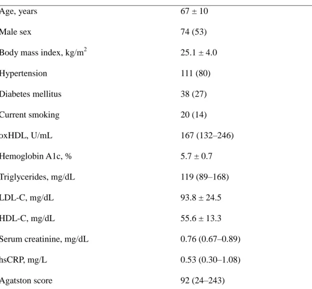
The baseline patient characteristics are summarized in Table 1. The mean age was 67 years, and 53% of patients were men. In terms of comorbidities, 80% of patients had hypertension and 27%
had diabetes mellitus. The median serum oxHDL concentration was 167 U/mL, and the median Agatston score was 92.
3.2 Association between baseline oxHDL level and metabolic parameters
The baseline log-transformed oxHDL level was correlated with total cholesterol (r=0.21, p=0.01), HDL-C (r=0.33, p<0.01), and log-transformed triglycerides (r=−0.21, p=0.01). There were no significant correlations between the baseline oxHDL level and age, body mass index, hemoglobin A1c, LDL-C, serum creatinine, or hsCRP.
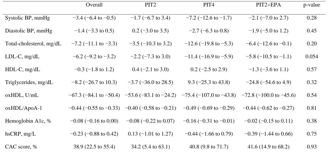
3.3 Change in blood pressure, biochemical parameters, and CAC score
Table 2 shows the mean annual change in oxHDL and other variables in each treatment group. In all three groups, the oxHDL level at follow-up was lower than that at baseline. Treatment with PIT4 and PIT2+EPA slightly reduced the oxHDL level compared with PIT2 alone, but not significantly. The annual change in the CAC score had weak, but significant correlation with the annual changes in oxHDL (r=0.17, p=0.04). The annual change in the CAC score was also significantly correlated with triglycerides (r=0.17, p=0.04), and hsCRP (r=0.22, p=0.01), but was not with the annual changes in systolic blood pressure (r=−0.06, p=0.51), LDL-C (r=0.05,
p=0.54), HDL-C (r=0.01, p=0.95), or hemoglobin A1c (r=−0.14, p=0.164).
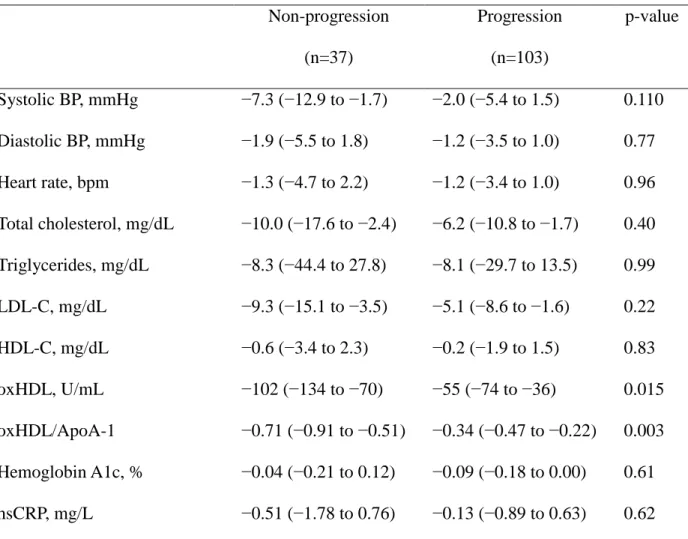
3.4 Comparison of variables according to CAC progression
At the 1-year follow-up, 103 patients (74%) showed CAC progression. When the annual changes in variables were compared between the non-CAC progression and CAC progression groups, the oxHDL concentration was significantly lower in the non-CAC progression group. No significant changes in the other variables were observed (Table 3).
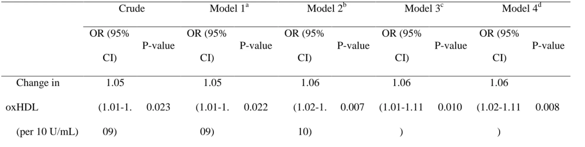
3.5 Association between annual change in oxHDL and CAC progression
The univariate logistic regression analysis showed that the odds ratio (OR) per 10-U/mL annual change in the oxHDL level was statistically significant (OR: 1.05, 95% CI: 1.01–1.09, p=0.02).
The multivariate logistic regression analysis revealed that the annual change in the oxHDL level was an independent determinant of CAC progression after adjusting for age, sex, hypertension,
diabetes mellitus, current smoking, change in LDL-C, change in HDL-C, change in triglycerides, change in hsCRP and allocated treatment groups (OR: 1.06, 95% CI: 1.02–1.11, p=0.008) (Table 4).
4. Discussion
The main finding of this study is that the decrease in the oxHDL level was significantly associated with the attenuation of CAC progression in patients with hypercholesterolemia and undergoing statin therapy. To our knowledge, this is the first study to evaluate the association between the change in the oxHDL level and CAC progression.
CAC has been strongly established as an independent predictor of adverse events, with a significant incremental prognostic value over traditional risk stratification algorithms. 1, 2 CAC progression has also been reported to be significantly associated with a higher rate of
cardiovascular events. 17, 1819, 20 A large observational study encompassing more than 20,000 participants demonstrated that an increase in CAC is associated with a 4- to 7-fold increase in cardiovascular events independent of the baseline CAC score, cardiovascular risk factors, and demographic variables. 17 Furthermore, studies have demonstrated that progression of CAC is significantly associated with an increase in both the calcified and noncalcified plaque volume, paralleling an increase in the atherosclerosis burden and cardiovascular risk. 21 Thus, an
increasing CAC score is undoubtedly associated with increasing overall atherosclerosis and more cardiovascular events. This study showed that a decrease in the oxHDL level was associated with attenuation of CAC progression. This finding suggests that oxHDL may be a potential target for slowing the progression of atherosclerosis.
Calcification is a common feature of atherosclerotic lesions. Several basic experiments have been performed to understand the underlying mechanism, and these experiments showed the role
of HDL in regulating calcification of vascular cells. 22-24 HDL protected against vascular calcification by regulating the osteoblastic differentiation and osteogenic activity of vascular smooth muscle cells that was induced by inflammatory cytokines such as interleukin-1β. 22 Furthermore, oxidation of HDL enhanced osteogenic activity in calcifying vascular cells. HDL is considered to be susceptible to conversion from an anti-atherogenic to pro-atherogenic state.
Sharma et al. reported that oxHDL induces proinflammatory effects on monocytes, including an increase in their binding to aortic endothelial cells. 25 Thus, HDL regulates vascular calcification through direct and indirect manners. In this context, HDL may be a promising target for vascular calcification associated with atherosclerosis.
In this study, oxHDL at follow-up decreased from baseline in all three groups without a reduction of the HDL-C level. This led to an improvement in the balance between oxHDL and HDL, which is beneficial for protecting atherosclerosis progression. A previous study involving patients with dyslipidemia showed that higher-dose statin therapy reduced oxidative stress markers more than lower-dose statin therapy. 26 Another study involving patients with coronary artery disease showed that treatment with EPA plus a statin reduced oxidative stress more than treatment with a statin alone. 27 Accordingly, statin therapy and a combination of statin + EPA therapy potentially reduce the oxHDL level independent of the HDL level, suggesting that these medications improve dysfunctional HDL. Future studies are required to identify a medical therapy that reduces oxHDL, leading to an improvement in the anti-atherosclerotic function of HDL.
This study has several limitations. First, because the study included only patients with hypercholesterolemia undergoing statin therapy, the results cannot be applied to the general population. Second, although the Agatston score is an excellent surrogate marker for the
prediction of CVD, we only analyzed CAC progression as the endpoint, not actual CVD events.
Therefore, we cannot conclude that there is an association between the increase in oxHDL and CVD events. Third, data on coronary CT angiography were not available in this study; thus, changes in plaque volumes and morphology could not be evaluated.
In concussion, this study demonstrated that a reduction in the oxHDL level is significantly associated with the attenuation of CAC progression in patients with hypercholesterolemia and undergoing statin therapy. Furtherer studies are required to identify the best method with which to improve HDL function for protecting against the development of atherosclerosis.
Conflicts of interest
HI received honoraria from Kowa Pharmaceutical Co. and Mochida Pharmaceutical Co. The other authors have no conflicts of interest in relation to the materials presented in this article.
Financial support
This study was funded by the Japan Heart Foundation (No. 12090021).
Acknowledgments
We thank Makoto Nakahama, MD, Yusuke Kawai, MD, Tadahisa Uesugi, MD, Takefumi Oka, MD, Mitsuru Munemasa, MD, Natsuki Takahashi, MD, Naoki Mukohara, MD, Seiji Habara, MD,Yusuke Katayama, MD, Ritsuko Terasaka, MD, Atsushi Mima, MD, Hitoshi Matsubara, MD, Shingo Hosogi, MD, Masayuki Doi, MD, Masayuki Ueeda, MD, Norio Urabe, MD, Kazufumi Takeuchi, MD, Yasuharu Namba, MD, Tetsuya Sato, MD, Nobuyuki Yamada, MD, Masahito Taniguchi, MD, Yutaka Kajikawa, MD, Kouki Watanabe, MD, Kenichi Hisamatsu, MD, Hiroo Kobayashi, MD, and Kiyoaki Maekawa, MD (the PEACH investigators). We also thank Kaoru Akazawa, Miyuki Fujiwara, and Masayo Ohmori for their technical assistance.
Author contributions
K Kohno, T Miyoshi, KN, and HI were involved in the conception, design, or planning of the study. T Miki, K Kotani, SS, YK, and HI were involved in the acquisition of data. T Miki and T Miyoshi were involved in the analysis of data. K Kotani, T Miyoshi, and HI were involved in the interpretation of results. T Miki, T Miyoshi, and HI substantially contributed to drafting of the manuscript.
References
[1] Osawa, K, Nakanishi, R and Budoff, M, Coronary Artery Calcification, Glob Heart, 2016;11:287-293.
[2] Greenland, P, LaBree, L, Azen, SP, Doherty, TM and Detrano, RC, Coronary artery calcium score combined with Framingham score for risk prediction in asymptomatic individuals, JAMA, 2004;291:210-215.
[3] Radford, NB, DeFina, LF, Barlow, CE, Lakoski, SG, Leonard, D, et al., Progression of CAC Score and Risk of Incident CVD, JACC. Cardiovascular imaging, 2016;9:1420-1429.
[4] Miyoshi, T, Kohno, K, Asonuma, H, Sakuragi, S, Nakahama, M, et al., Effect of Intensive and Standard Pitavastatin Treatment With or Without Eicosapentaenoic Acid on Progression of Coronary Artery Calcification Over 12 Months- Prospective Multicenter Study, Circulation journal : official journal of the Japanese Circulation Society, 2018;82:532-540.
[5] Stone, NJ, Robinson, JG, Lichtenstein, AH, Bairey Merz, CN, Blum, CB, et al., 2013 ACC/AHA Guideline on the Treatment of Blood Cholesterol to Reduce Atherosclerotic Cardiovascular Risk in Adults, Journal of the American College of Cardiology,
2014;63:2889-2934.
[6] Boden, WE, Probstfield, JL, Anderson, T, Chaitman, BR, Desvignes-Nickens, P, et al., Niacin in patients with low HDL cholesterol levels receiving intensive statin therapy, N Engl J Med, 2011;365:2255-2267.
[7] Schwartz, GG, Olsson, AG, Abt, M, Ballantyne, CM, Barter, PJ, et al., Effects of dalcetrapib in patients with a recent acute coronary syndrome, N Engl J Med,
2012;367:2089-2099.
[8] Barter, PJ, Caulfield, M, Eriksson, M, Grundy, SM, Kastelein, JJ, et al., Effects of torcetrapib in patients at high risk for coronary events, N Engl J Med, 2007;357:2109-2122.
[9] Navab, M, Reddy, ST, Van Lenten, BJ and Fogelman, AM, HDL and cardiovascular disease: atherogenic and atheroprotective mechanisms, Nat Rev Cardiol, 2011;8:222-232.
[10] Khera, AV, Cuchel, M, de la Llera-Moya, M, Rodrigues, A, Burke, MF, et al.,
Cholesterol efflux capacity, high-density lipoprotein function, and atherosclerosis, N Engl J Med, 2011;364:127-135.
[11] Rohatgi, A, Khera, A, Berry, JD, Givens, EG, Ayers, CR, et al., HDL cholesterol efflux capacity and incident cardiovascular events, N Engl J Med, 2014;371:2383-2393.
[12] Ueda, M, Hayase, Y and Mashiba, S, Establishment and evaluation of 2 monoclonal antibodies against oxidized apolipoprotein A-I (apoA-I) and its application to determine blood oxidized apoA-I levels, Clin Chim Acta, 2007;378:105-111.
[13] Honda, H, Ueda, M, Kojima, S, Mashiba, S, Suzuki, H, et al., Oxidized high-density lipoprotein is associated with protein-energy wasting in maintenance hemodialysis patients, Clin J Am Soc Nephrol, 2010;5:1021-1028.
[14] Honda, H, Ueda, M, Kojima, S, Mashiba, S, Michihata, T, et al., Oxidized high-density lipoprotein as a risk factor for cardiovascular events in prevalent hemodialysis patients,
Atherosclerosis, 2012;220:493-501.
[15] Agatston, AS, Janowitz, WR, Hildner, FJ, Zusmer, NR, Viamonte, M, Jr., et al., Quantification of coronary artery calcium using ultrafast computed tomography, J Am Coll Cardiol, 1990;15:827-832.
[16] Standards of medical care in diabetes--2008, Diabetes Care, 2008;31 Suppl 1:S12-54.
[17] Budoff, MJ, Hokanson, JE, Nasir, K, Shaw, LJ, Kinney, GL, et al., Progression of coronary artery calcium predicts all-cause mortality, JACC Cardiovasc Imaging,
2010;3:1229-1236.
[18] Raggi, P, Cooil, B, Ratti, C, Callister, TQ and Budoff, M, Progression of coronary artery
calcium and occurrence of myocardial infarction in patients with and without diabetes mellitus, Hypertension, 2005;46:238-243.
[19] Nakanishi, R, Ceponiene, I, Osawa, K, Luo, Y, Kanisawa, M, et al., Plaque progression assessed by a novel semi-automated quantitative plaque software on coronary computed
tomography angiography between diabetes and non-diabetes patients: A propensity-score matching study, Atherosclerosis, 2016;255:73-79.
[20] Budoff, MJ, Young, R, Lopez, VA, Kronmal, RA, Nasir, K, et al., Progression of coronary calcium and incident coronary heart disease events: MESA (Multi-Ethnic Study of Atherosclerosis), Journal of the American College of Cardiology, 2013;61:1231-1239.
[21] Ceponiene, I, Nakanishi, R, Osawa, K, Kanisawa, M, Nezarat, N, et al., Coronary Artery Calcium Progression Is Associated With Coronary Plaque Volume Progression: Results From a Quantitative Semiautomated Coronary Artery Plaque Analysis, JACC. Cardiovascular imaging, 2017.
[22] Ahotupa, M, Oxidized lipoprotein lipids and atherosclerosis, Free Radic Res, 2017;51:439-447.
[23] Nofer, JR, Walter, M, Kehrel, B, Wierwille, S, Tepel, M, et al., HDL3-mediated
inhibition of thrombin-induced platelet aggregation and fibrinogen binding occurs via decreased production of phosphoinositide-derived second messengers 1,2-diacylglycerol and inositol 1,4,5-tris-phosphate, Arterioscler Thromb Vasc Biol, 1998;18:861-869.
[24] Uittenbogaard, A, Shaul, PW, Yuhanna, IS, Blair, A and Smart, EJ, High density lipoprotein prevents oxidized low density lipoprotein-induced inhibition of endothelial nitric-oxide synthase localization and activation in caveolae, J Biol Chem,
2000;275:11278-11283.
[25] Sharma, N, Desigan, B, Ghosh, S, Sanyal, SN, Ganguly, NK, et al., The role of oxidized
HDL in monocyte/macrophage functions in the pathogenesis of atherosclerosis in Rhesus monkeys, Scand J Clin Lab Invest, 1999;59:215-225.
[26] Kei, A, Tellis, C, Liberopoulos, E, Tselepis, A and Elisaf, M, Effect of switch to the highest dose of rosuvastatin versus add-on-statin fenofibrate versus add-on-statin nicotinic
acid/laropiprant on oxidative stress markers in patients with mixed dyslipidemia, Cardiovasc Ther, 2014;32:139-146.
[27] Takaki, A, Umemoto, S, Ono, K, Seki, K, Ryoke, T, et al., Add-on therapy of EPA reduces oxidative stress and inhibits the progression of aortic stiffness in patients with coronary artery disease and statin therapy: a randomized controlled study, J Atheroscler Thromb,
2011;18:857-866.
Table 1. Baseline patient characteristics (n=140 patients)
Age, years 67 ± 10
Male sex 74 (53)
Body mass index, kg/m2 25.1 ± 4.0
Hypertension 111 (80)
Diabetes mellitus 38 (27)
Current smoking 20 (14)
oxHDL, U/mL 167 (132–246)
Hemoglobin A1c, % 5.7 ± 0.7
Triglycerides, mg/dL 119 (89–168)
LDL-C, mg/dL 93.8 ± 24.5
HDL-C, mg/dL 55.6 ± 13.3
Serum creatinine, mg/dL 0.76 (0.67–0.89)
hsCRP, mg/L 0.53 (0.30–1.08)
Agatston score 92 (24–243)
Data are presented as mean ± standard deviation, number (%), or median (25th–75th percentile), as appropriate. oxHDL, oxidized high-density lipoprotein; LDL-C, low-density lipoprotein cholesterol; HDL-C, high-density lipoprotein cholesterol; hsCRP, high-sensitivity C-reactive protein.
Table 2. Change in blood pressure, biochemical parameters, and CAC score from start of allocated treatment to 12-month follow-up
Overall PIT2 PIT4 PIT2+EPA p-value
Systolic BP, mmHg −3.4 (−6.4 to −0.5) −1.7 (−6.7 to 3.4) −7.2 (−12.6 to −1.7) −2.1 (−7.0 to 2.7) 0.28 Diastolic BP, mmHg −1.4 (−3.3 to 0.5) 0.2 (−3.0 to 3.5) −2.7 (−6.3 to 0.8) −1.9 (−5.0 to 1.2) 0.45 Total-cholesterol, mg/dL −7.2 (−11.1 to −3.3) −3.5 (−10.3 to 3.2) −12.6 (−19.8 to −5.3) −6.4 (−12.6 to −0.1) 0.20 LDL-C, mg/dL −6.2 (−9.2 to −3.2) −2.2 (−7.3 to 3.0) −11.4 (−16.9 to −5.9) −5.8 (−10.5 to −1.1) 0.054 HDL-C, mg/dL −0.3 (−1.8 to 1.2) 0.4 (−2.1 to 3.0) 0.2 (−2.5 to 2.9) −1.3 (−3.6 to 1.1) 0.57 Triglycerides, mg/dL −8.2 (−26.7 to 10.3) −3.7 (−36.0 to 28.5) 9.3 (−25.3 to 43.8) −24.8 (−54.6 to 4.9) 0.32 oxHDL, U/mL −67.3 (−84.1 to −50.4) −53.6 (−83.1 to −24.2) −75.4 (−107.0 to −43.8) −72.8 (−100.0 to −45.6) 0.54 oxHDL/ApoA-1 −0.44 (−0.55 to −0.33) −0.40 (−0.58 to −0.21) −0.49 (−0.69 to −0.29) −0.44 (−0.62 to −0.27) 0.81 Hemoglobin A1c, % −0.08 (−0.16 to 0.00) −0.08 (−0.22 to 0.07) −0.16 (−0.31 to −0.01) −0.02 (−0.15 to 0.11) 0.38 hsCRP, mg/L −0.23 (−0.88 to 0.42) 0.13 (−1.01 to 1.27) −0.44 (−1.66 to 0.79) −0.39 (−1.44 to 0.66) 0.75 CAC score, % 38.9 (22.5 to 55.4) 34.2 (5.4 to 63.1) 40.8 (9.8 to 71.7) 41.6 (14.9 to 68.2) 0.93 Data are presented as estimate (95% confidence interval). PIT2, pitavastatin at 2 mg/day; PIT4, pitavastatin at 4 mg/day; EPA,
eicosapentaenoic acid; BP, blood pressure; LDL-C, low-density lipoprotein cholesterol; HDL-C, high-density lipoprotein cholesterol;
oxHDL, oxidized high-density lipoprotein; ApoA-1, apolipoprotein A-1; hsCRP, high-sensitivity C-reactive protein; CAC, coronary artery calcification
Table 3. Comparison of annual changes in variables among patients with non-CAC progression and CAC progression over 1 year
Non-progression (n=37)
Progression (n=103)
p-value
Systolic BP, mmHg −7.3 (−12.9 to −1.7) −2.0 (−5.4 to 1.5) 0.110 Diastolic BP, mmHg −1.9 (−5.5 to 1.8) −1.2 (−3.5 to 1.0) 0.77 Heart rate, bpm −1.3 (−4.7 to 2.2) −1.2 (−3.4 to 1.0) 0.96 Total cholesterol, mg/dL −10.0 (−17.6 to −2.4) −6.2 (−10.8 to −1.7) 0.40 Triglycerides, mg/dL −8.3 (−44.4 to 27.8) −8.1 (−29.7 to 13.5) 0.99 LDL-C, mg/dL −9.3 (−15.1 to −3.5) −5.1 (−8.6 to −1.6) 0.22 HDL-C, mg/dL −0.6 (−3.4 to 2.3) −0.2 (−1.9 to 1.5) 0.83
oxHDL, U/mL −102 (−134 to −70) −55 (−74 to −36) 0.015
oxHDL/ApoA-1 −0.71 (−0.91 to −0.51) −0.34 (−0.47 to −0.22) 0.003 Hemoglobin A1c, % −0.04 (−0.21 to 0.12) −0.09 (−0.18 to 0.00) 0.61 hsCRP, mg/L −0.51 (−1.78 to 0.76) −0.13 (−0.89 to 0.63) 0.62 Data are presented as estimate (95% confidence interval). CAC, coronary artery calcification; BP, blood pressure; LDL-C, low-density lipoprotein cholesterol; HDL-C, high-density lipoprotein cholesterol; oxHDL, oxidized high-density lipoprotein;ApoA-1, apolipoprotein A-1; hsCRP, high-sensitivity C-reactive protein.
Table 4. Determinants of coronary artery calcification progression
Crude Model 1a Model 2b Model 3c Model 4d
OR (95%
CI)
P-value
OR (95%
CI)
P-value
OR (95%
CI)
P-value
OR (95%
CI)
P-value
OR (95%
CI)
P-value
Change in oxHDL
(per 10 U/mL)
1.05 (1.01-1.
09)
0.023
1.05 (1.01-1.
09)
0.022
1.06 (1.02-1.
10)
0.007
1.06 (1.01-1.11
)
0.010
1.06 (1.02-1.11
)
0.008
a) Adjusted for age and male sex; b) adjusted for model 1 plus presence of hypertension, diabetes mellitus and current smoking; c) adjusted for model 2 plus annual changes in lipid parameters and high-sensitivity C-reactive protein; d) adjusted for model 3 plus allocated treatment groups.
OR, odds ratio; CAC, coronary artery calcification; oxHDL, oxidized high-density lipoprotein; CI, confidential interval.
Figure legends
Figure 1. Flow diagram of patients in this study
EPA, eicosapentaenoic acid at 1,800 mg/day; MDCT, multidetector row computed tomography;
PIT2, pitavastatin at 2 mg/day; PIT4, pitavastatin at 4 mg/day.
Supplemental Figure 1. Protocol of the principal study
Patients were enrolled after determination of the first coronary artery calcification score in each facility. One year after the end of the allocated treatment phase, the coronary artery calcification score was redetermined. EPA, eicosapentaenoic acid; MDCT, multidetector row computed tomography.