A Bombyx homolog of ovo is a segmentation gene that acts downstream of Bm‑wnt1 ( Bombyx wnt1 homolog)
journal or
publication title
Gene Expression Patterns
volume 27
page range 1‑7
year 2018‑01
URL http://id.nii.ac.jp/1578/00002416/
doi: 10.1016/j.gep.2017.10.002
Creative Commons : 表示 ‑ 非営利 ‑ 改変禁止 http://creativecommons.org/licenses/by‑nc‑nd/3.0/deed.ja
A Bombyx homolog of ovo is a segmentation gene that acts downstream of Bm-wnt1(Bombyx wnt1 homolog)
Hajime Nakao
Insect Genome Research and Engineering Unit, Division of Applied Genetics, Institute of Agrobiological Sciences, National Agriculture and Food Research Organization (NARO), 1-2 Oowashi, Tsukuba, Ibaraki 305-8634, Japan
Correspondence to Hajime Nakao
Insect Genome Research and Engineering Unit, Division of Applied Genetics, Institute of Agrobiological Sciences, National Agriculture and Food Research Organization (NARO), 1-2 Oowashi, Tsukuba, Ibaraki 305-8634, Japan
Tel.: +81-29-838-6127, Fax: +81-29-838-6028, E-mail: nakaoh@ affrc.go.jp
Keywords
Bombyx, ovo, wnt1, RNAi, segmentation, pair-rule genes
Abstract
Insect embryogenesis is divided into long and short/intermediate germ types. The long germ type may exhibit Drosophila-like hierarchical segmentation mechanisms, whereas the short/intermediate type assumes some repeating mechanisms that are considered to be ancestral. Embryogenesis in Bombyx mori possesses both characteristics. Here, Bombyx ovo homolog (Bm-ovo) was identified as a gene involved in segmentation. ovo is a Drosophila gene that encodes a zinc finger transcription factor and studies on its homolog functions in other systems have suggested that it acts as a switch to enable the initiation of differentiation from a progenitor cell state. This is the first description for ovo homologs being involved in insect segmentation. Bm-ovo is expressed dynamically during embryogenesis in a pattern that resembles that of gap and pair-rule genes. In Bm-ovo RNAi knockdown embryos, posterior segmentation does not proceed. In addition, defects in anterior segments are observed. In Bm-wnt1 knockdown embryos, the Bm-ovo expression pattern was changed, suggesting that Bm-wnt1 is an upstream regulator of Bm-ovo. The involvement of Bm-ovo may represent a novel ancestral step under the control of wnt genes in insect segmentation: this step may resemble those operating in cell differentiation processes.
Introduction
In insect embryos, maternal, gap, and pair-rule genes are successively expressed to produce segments along the anterior-posterior (AP) axis. Maternal genes dictate the AP axis. Gap genes subdivide embryos into broad domains according to their expression profiles. The expression of seven to eight stripes of pair-rule genes serves as a template for future segments. Insect embryogenesis has been largely categorized into long and intermediate/short germ types (Davis and Patel, 2002; Liu and Kaufman, 2005; Peel et al., 2005). In the long germ type, as represented by Drosophila, the combinatorial input of maternal and gap genes directs the expression of the individual stripes of pair-rule genes, which leads to the nearly simultaneous expression of pair-rule stripes, i.e., segment formation (Pankratz and Jäckle, 1993). In contrast, in the short/intermediate type, posterior segments are sequentially added, in which some repeating segmentation mechanisms are likely to operate. In the short germ-type insect Tribolium, the wave-like expression of pair-rule genes even-skipped (eve) and odd-skipped (odd) has been observed in the pre-segmental area (Sarrazin et al., 2012; El-Sherif et al., 2012). The pair-rule genes, eve, odd, and runt (run) interact positively and negatively to form a circuit that appears to be responsible for the striped expression of these genes. Thus, the wave-like expression of a subset of pair-rule genes may be passed onto the pair-rule gene circuit, leading to the production of segments (pair-rule gene stripes) in a clock-like manner (Choe et al., 2006; Lynch et al., 2012).
It is commonly acknowledged that the latter Tribolium-like segmentation mode is ancestral, while the former Drosophila-like hierarchical segmentation mode is derived.
Recent evidence suggests that both modes may operate in some species (Rosenberg et al., 2014), such as Bombyx mori.
B. mori is a lepidopteran insect, the segmentation mechanisms of which have remained elusive. Its embryogenesis has long and short/intermediate-like features. The egg has a large embryonic primordium, within which individual segments are fate mapped without a growth zone early in embryogenesis similar to long germ insects (Myohara, 1994). However, when pair-rule gene interactions were examined, B. mori exhibited Drosophila- and Tribolium-like characteristics (Nakao, 2015). Furthermore, the roles of Bombyx wnt1/wingless and the Krüppel homolog resemble those in short/intermediate insects: embryos show truncated phenotypes in the posterior region when these gene
(family) activities are perturbed (Beermann et al., 2011; Bolognesi et al., 2008, 2009;
Miyawaki et al., 2004; Nakao, 2010, 2015; Yamaguchi et al., 2011; Cerny et al., 2005;
Liu and Kaufman, 2004; Mito et al., 2006). Embryonic Bm-wnt1 is considered to have two functions: its organizing function in posterior development, as reflected in the knockdown phenotype as described above, and its function as a segment polarity gene, similar to that in Drosophila. During normal embryogenesis, Bm-wnt1 is expressed in the large posterior domain in the early stages, the expression domain then recedes posteriorly, and segmental stripes begin to appear in an anterior to posterior sequence (Nakao, 2010). Posterior expression appears to be responsible for posterior organization functions, while segmental expression appears to exhibit a segment polarity function that has not yet been experimentally validated in Bombyx. Furthermore, the addition of supernumerary posterior segments is induced at the posterior terminus in a manner that resembles that of short/intermediate insects by the knockdown of the hunchback homolog (Bm-hb) (Nakao, 2016). These findings strongly suggest that the short/intermediate type mode of segmentation is operating in normal Bombyx embryogenesis.
An ovo homolog from B. mori (Bm-ovo) was described herein. ovo is a Drosophila gene that encodes an evolutionarily conserved zinc finger transcription factor with various biological functions. It is required in the female germ line for proper oogenesis: the mutants display egg chambers filled with excess undifferentiated germ cells, an ovarian tumor phenotype (Oliver, 1987). ovo is also involved in early germ line development:
the transcript is deposited in the germ plasm as a maternal factor, regulating the expression of vasa (Yatsu et al., 2008). In the zygote, often referred to as shavenbaby (svb) at this stage, it is necessary and sufficient to cell-autonomously direct the formation of denticles (trichome-cytoplasmic extrusion of epidermal cells) in the ventral epidermis of the embryo abdomen, in which there is a segmentally repeating denticle pattern separated by naked cuticle, thereby contributing to the binary choice to produce either a naked cuticle or denticles. The upstream regulation of svb has been investigated.
The high mobility group (HMG)-domain protein SoxNeuro (SoxN) is necessary and sufficient to cell-autonomously direct the expression of svb. SoxN, in turn, receives positive and negative inputs from the epidermal growth factor receptor (Egfr) ligand Spitz (Spi) and Wingless (Wg), respectively. These mechanisms result in the restriction of trichome-producing cells. The closely related protein of Spi, Dichaete is co-regulated
with Spi and has a partially redundant function in the activation of svb, albeit to a lesser extent. However, these regulatory relationships are not strictly hierarchical, but complex feedback mechanisms are involved (Overton et al., 2007).
Apart from Drosophila, ovo homologs are isolated from diverse animals. The mouse homolog also controls germline and epidermis differentiation. In C. elegans, the ovo homolog lin-48 is required for the development of hindgut. These findings point to the role of ovo homologs in the differentiation and maintenance of specific cell types (Wieschaus, 1984; Oliver, 1987; Dai, 1998; Johnson, 2001). More recently, human homolog of ovo (OVOL1) was shown to regulate the transition of progenitor to differentiated trophoblast cells (Renaud et al., 2015).
In the present study, the embryonic functions of Bm-ovo were examined. Bm-ovo expression during the embryonic stage resembled that of gap and pair-rule genes.
Embryonic Bm-ovo RNAi embryos showed segmentation defects in the gnathal/thoracic region and posterior abdomen. In the posterior body part, the abnormal expression of some pair-rule genes that are crucial for establishing pair-rule gene expression patterns was observed: the posterior part did not produce pair-rule stripes, but a broad band of expression was observed in the affected region for the pair-rule genes examined. In Bm-wnt1 knockdown embryos, alternation in Bm-ovo expression pattern was observed, suggesting that Bm-wnt1 is an upstream regulator of Bm-ovo. This interaction may be related to those operating in Drosophila embryonic epidermal patterning as described above.
The involvement of Bm-ovo may represent a novel ancestral step under the control of wnt genes in insect segmentation, and this step may resemble those operating in cell differentiation processes.
Materials and Methods
Silkworm strains, rearing, and development
The B. mori strain pnd-2 was used in this study. Silkworms were reared on an artificial diet (Nippon Nosanko). Refer to Nagy et al. (1994) for a general description of early Bombyx development.
Identification and isolation of Bm-ovo cDNA
The Drosophila ovo sequence was used to search the Bombyx cDNA database (Mita et al., 2003) for Bombyx ovo homologs and one wing-disk-derived cDNA was identified.
cDNA was obtained and initial analyses were performed using this cDNA. The annotated genome database (KAIKObase; Mita et al., 2004) subsequently became available, which revealed that the open reading frame of Bm-ovo comprises four exons (E1, E2, E3 and E4; gene ID: BMgn000987). The cDNA clone described above was found to lack the second exon. To obtain cDNA species expressed during early embryogenesis, a PCR amplification procedure was employed. Total RNAs in embryos 14 and 20 hours AEL were prepared using TRIZOL reagent (Invitrogen). Single-strand cDNAs were synthesized using total RNAs as templates by PrimeScriptTMII 1st strand cDNA Synthesis Kit (TAKARA) according to the manufacturer’s instructions. With these cDNAs, PCR amplification was performed using PrimeSTAR GXL DNA Polymerase (TAKARA). A primer pair was set at the sequence near the start and stop codons, respectively, so as to amplify the full open reading frame. PCR conditions were 40 cycles at 98°C for 10 s; at 55°C for 15 s; at 68°C for 3 min. The primer pair was the Bm-ovoCp, 5’-primer and Bm-ovoE3-4p, 3’-primer (see below). The amplified product was analyzed using 1% agarose gel electrophoresis. The results obtained revealed the predominant amplification of an approximately 2.6-kbp product from both templates (Supplementary Fig. 1). The amplified product was cloned and sequenced. The sequence analysis revealed that it had all four exons. Subsequent analyses were performed using this cDNA.
In situ hybridization
Fixation and in situ hybridization were performed as previously described (Nakao, 1999, 2010). Probes for Bm-eve and Bm-wnt1 were previously described (Nakao, 2010).
Probes for Bm-ovo, designated as Bm-ovoCp, Bm-ovoE1p, Bm-ovoE2p and Bm-ovoE3-4p, were synthesized using cloned cDNAs. cDNAs were PCR fragments amplified using the primers described below. The primer pairs used for the amplification of these fragments were as follows: Bm-ovoCp, 5’-primer: 5’-
GGGGGATCCAGTCCTAACGAAGCGGCCAA-3’, 3’-primer: 5’-
CCCAAGCTTTTTATACGGTCTGACTCCGG-3’; Bm-ovoE1p, 5’-primer: 5’-
GGGGGATCCAGTCCTAACGAAGCGGCCAA -3’, 3’-primer 5’-
CCCAAGCTTCAGTGCATTCCTTTTCTTTATCC -3’; Bm-ovoE2p, 5’-primer: 5’-
GGGGGATCCCGCACAAAAGAAACTAGACG -3’, 3’-primer 5’-
CCCAAGCTTAGCACTAAAACAGGTCGTGC -3’; Bm-ovoE3-4p, 5’-primer: 5’-
GGGGGATCCTAGGACTACCAGCAGAGCTT -3’, 3’-primer 5’-
CCCAAGCTTAATTGTGTACTGGCATGGGC -3’. The Bm-ovoCp primer pair was used for amplification from the cloned cDNA template (fufe-P20_F_P18), and the amplified product comprised approximately 0.3-kb exon 1 and 0.3-kb exon 3 (see the Results section). Bm-ovoE1p, E2p and E3-4p primer pairs were used for amplification from the cDNA prepared using PrimeScriptTM II 1st strand cDNA Synthesis Kit (TAKARA) and total RNA at 20 hours AEL. The amplified products were approximately 0.3, 0.5, and 0.6 kbp, which corresponded to parts of exons 1, 2, and 3-4, respectively. After amplification, cDNAs were cloned into pBluescript vectors. In order to obtain probes, plasmid DNA was cut with an appropriate enzyme and RNA probes were synthesized using either T3 or T7 polymerase, depending on the direction of the insert using DIG RNA Labeling Kit (SP6/T7) (Roche).
RNAi
The RNAi procedure was described previously (Nakao, 2012). The templates used for in vitro transcription were PCR fragments of the corresponding genes, flanked by T7 promoter sequences. Regarding Bm-ovo RNAi, three different dsRNAs, designated as Bm-ovoCRNAi, Bm-ovoE1RNAi and Bm-ovoE3-4RNAi, were used in analyses. The primer pairs used for amplification were as follows: Bm-ovoCRNAi, 5’-TAATACGACTCACTATAGGGAGAAGTCCTAACGAAGCGGCCAA-3’,
5’-TAATACGACTCACTATAGGGAGATTTATACGGTCTGACTCCGG-3’;
Bm-ovoE1RNAi, 5’-
TAATACGACTCACTATAGGGAGAAGTCCTAACGAAGCGGCCAA-3’, 5’-TAATACGACTCACTATAGGGAGACAGTGCATTCCTTTTCTTTATCC-3’;
Bm-ovoE3-4RNAi,
5’-TAATACGACTCACTATAGGGAGATAGGACTACCAGCAGAGCTT-3’,
5’-TAATACGACTCACTATAGGGAGAAATTGTGTACTGGCATGGGC. Bm-ovoC,
ovoE1, and E3-4RNAi targets corresponded to the regions amplified by the Bm-ovoCp, Bm-ovoE1p, and Bm-E3-4p primer pairs described above, respectively. For Bm-wnt1 RNAi, two independent dsRNA corresponding to different part of the gene was used for the analysis. The primer pairs were as follows: Bmwnt1RNAi1, 5’-TAATACGACTCACTATAGGGAGAGCAGAATGAAGTGTCTGTGG-3’,
5’-TAATACGACTCACTATAGGGAGAAAGCCGATGTTGTCGCTGCA-3’, Bmwnt1RNAi2,
5’-TAATACGACTCACTATAGGGAGAAGGGAATTCGTTGATACCGG-3’, 5’-TAATACGACTCACTATAGGGAGAACCTCGCAACACCAATGGAA-3’.
Results
Identification of a Bombyx ovo homolog
A recent study described the ovo homolog from Bombyx (Bm-ovo); multiple forms of spliced variants were differentially expressed among the tissues examined (Xue et al., 2014). The identification of Bm-ovo in the present study was performed independently in our laboratory. After cDNA database search (Mita et al., 2003), a cDNA derived from wing-disk-expressed mRNA was obtained (fufe-P20_F_P18). Initial analyses were performed using a probe (Bm-ovoCp) or double-stranded RNA (Bm-ovoCRNAi) made from this cDNA as a template. However, a subsequent analysis revealed that this mRNA species lacked exon E2 and was poorly represented in early embryonic stages (see Materials and Methods). After obtaining embryonically expressed cDNA, which comprised exons E1, E2, E3 and E4, analyses were also performed using a few mutually independent probes or dsRNA in addition to the analyses described above. The results of these analyses were essentially the same.
Bm-ovo embryonic expression
A gene expression analysis of Bm-ovo was performed by in situ hybridization using the probes, Bm-ovoCp, E1p, E2p and E3-4p (see Materials and Methods). Irrespective of probes used, the results were essentially the same (Supplementary Fig. 2). Just after egg laying, weak expression was observed in the germ anlage, suggesting its maternal load
(Fig. 1A, B). Regional heterogeneities in expression were not detected within the germ anlage, suggesting that the transcript did not accumulate in the putative germ plasm, although the existence of germ plasm was suggested by Bm-nanosO (Bombyx nanosO) expression pattern (Nakao et al., 2008). Specific accumulation in the germ line was not observed during the time period examined in the present study, i.e., up to 5 days AEL (After Egg Laying), which was in contrast with previous findings obtained using Drosophila (Yatsu et al., 2008). Expression was dynamic more than 22 hours AEL and resembled that of the gap and pair-rule genes. At 22 hours, expression was detected in one anterior domain (head) and one large posterior domain (Fig. 1C). Later, the posterior domain resolved into three strong stripes (bands) and two weak stripes (bracketed) (Fig. 1D, E). The three strongly expressed stripes remained thereafter, and overlapped with Bm-eve stripes #2, #6, and #8 (Fig. 1F, G). Although it could not be determined due to technical difficulties which pair-rule stripes the weak Bm-ovo stripes corresponded to or which pair-rule stripes the broad strong Bm-ovo stripes exactly cover, a (morphological) comparison of single- and double-stained embryos suggested that stripes #1, #3, and #7 escaped Bm-ovo coverage at the later stages. However, the expression pattern of Bm-ovo suggested that the Bm-ovo posterior expression domain covered all the Bm-ovo stripes at least once, except for stripe #1. As the development proceeded, the background signal intensity transiently increased, region-specific staining became obscured (Fig. 1H), and background staining then decreased again (Fig.
1I).
Analysis of Bm-ovo functions using embryonic RNAi
Embryonic RNAi was used to analyze Bm-ovo functions. dsRNAs were injected into embryos at 0~2 hours AEL. As described above, three dsRNAs were used to examine phenotypes. Target gene expression was markedly reduced for all the dsRNAs used, suggesting that the RNAi procedure was effective (Supplementary Fig. 3).
The phenotypes of knockdown embryos were examined for their morphologies at 72 hour AEL. In RNAi-treated embryos, anomalies were observed in the anterior and posterior of embryos (Fig. 2). Anterior phenotypes represented defects in the morphology of the region encompassing gnathal/thoracic segments, with the mesothoracic segment being the most frequently affected. In these regions, the observed
phenotypes included irregular morphologies or the deletion of segments and/or appendages, or fused or disorganized gnathal/thoracic segments (Fig. 2B, C). In the posterior, the fusion of posterior abdominal segments was observed for all dsRNA species: in the most severe cases, a total of six abdominal segments were observed (Fig.
2B, C, Table 1). The identities of the remaining abdominal segments were not entirely clear. However, based on the observation that three proleg-bearing segments remained in RNAi treated embryos (Fig. 2B), the identities of A1 to A5 appeared to be preserved in the correct order (in the wild-type, prolegs were observed in A3-A6, also see below).
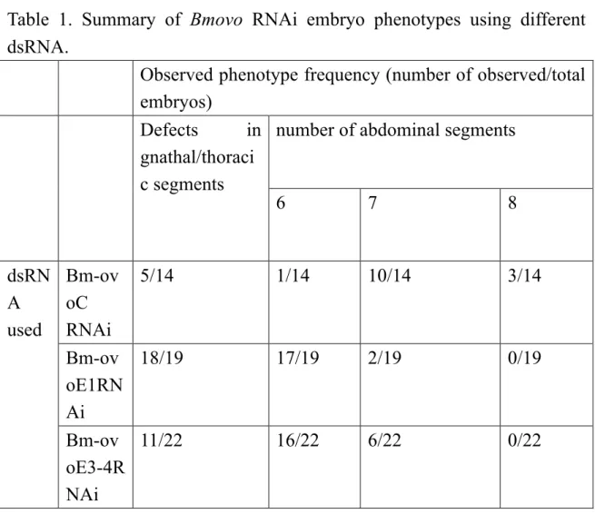
In both phenotype classes, the severity of the phenotype appeared to differ between the dsRNAs used (Table 1); for example, the number of abdominal segments was significantly lower in Bm-ovoE1RNAi or Bm-ovoE3RNAi embryos than in Bm-ovoCRNAi embryos (P<0.05), reflecting the incomplete nature of the RNAi knockdown. Therefore, the knockout phenotype may differ from those observed here by the RNAi knockdown.
In order to elucidate the cause of these morphological consequences, embryos were analyzed molecularly. Since pair-rule genes were responsible for segment formation, the expression of three key pair-rule genes, Bombyx even-skipped (Bm-eve), runt (Bm-run), and odd-skipped (Bm-odd) was examined at 23~28 hours AEL when the full complement of each pair-rule gene stripe was expressed in wild-type embryos (Fig. 3A, C, E; Nakao, 2015). The most notable change was the broad expression of these pair-rule genes in the posterior region without the resolution of stripes after stripe #5, which is consistent with posterior abdominal segment fusion morphologies (Bm-eve, n=29/31; Bm-run, n=27/28; Bm-odd, n=50/54). In addition, the weak expression of the Bm-eve stripe #3 (and less frequently, stripe #2) was sometimes observed (n=15/21), together with the weak expression of Bm-run and Bm-odd stripe #2 (Bm-run, n=5/25;
Bm-odd, n=16/51; Fig. 3B, D, F, Supplementary Fig. 4). However, it is important to note that although the intensity of expression was changed, patterning in the anterior region was essentially not affected by Bm-ovo RNAi, which is in contrast to posterior patterning in the affected embryos.
Bm-ovo is regulated by Bm-wnt1
The present findings show that Bm-ovo is involved in Bombyx body segmentation.
Bm-wnt1 also functions in the establishment of segments. These homologs are known to interact in Drosophila embryonic epidermal patterning as described in the Introduction section. Therefore, embryonic RNAi was used to test whether Bm-ovo and Bm-wnt1 are interact and, if so, how. Bm-ovo RNAi was performed as described above and Bm-wnt1 expression in the resulting embryos was compared with that in wild type embryos. As examined at ~23h AEL, Bm-wnt1 expression in RNAi-treated embryos was similar to that in wild type embryos (n=14/14; Fig. 4A, B). However, at approximately 26h AEL, when more anterior segmental stripes of Bm-wnt1 became visible, these stripe boundaries in treated embryos were more irregular, except for a few anterior-most stripes (n=20/20; black arrows in Fig. 4D), than those in wild type embryos, whereas differences were not observed in the posterior expression domain (n=34/34; white arrows in Fig. 4A-D). Next, Bm-wnt1 RNAi was performed to examine changes in Bm-ovo expression. The in situ hybridization of RNAi treated embryos using the Bm-wnt1 probe indicated that the target gene expression was markedly reduced, suggesting that the RNAi was effective (Supplementary Fig. 3). A previously study reported that Bm-wnt1 knockdown resulted in severely truncated embryos (Yamaguchi et al., 2011). Similar truncated embryos were observed in the RNAi experiment in the present study (n=26/26). However, the phenotypes appeared to be more severe than those previously reported. Bm-wnt1RNAi embryos in this study comprised head with some epidermal structures attached. In these embryos, appendages other than the labrum and antenna were not clearly identifiable: thoracic legs were not observed, which contrasted with to the existence of immature three thoracic legs in the previous study (Fig. 5, Supplementary Fig. 5, Yamaguchi et al., 2011). Bm-ovo RNAi expression was then compared between wild type and RNAi-treated embryos fixed at about 26h AEL.
In RNAi treated embryos, changes in Bm-ovo expression pattern were observed. While stripes that were clearly visible in the middle part of wild type embryos were absent in RNAi treated embryos, weak expression was noted from middle to the posterior.
However, expression in anterior and posterior body region remained in treated embryos (n=19/19; Fig. 4E, F).
Discussion
Implications of the ovo homolog in Bombyx segmentation
This study revealed embryonic Bm-ovo expression patterns and some functions. Bm-ovo was dynamically expressed along the AP axis in a manner that resembled gap and pair-rule genes, and RNAi-treated embryos showed anomalies in anterior and posterior body parts, together with alterations in pair-rule gene expression patterns. The expression pattern and results of a molecular analysis of RNAi appeared to be roughly consistent with the morphological changes observed in Bm-ovo RNAi embryos.
Assuming that, as a rough estimate, Bm-eve stripe #3 coincides with the posterior mesothoracic (T2) segment and the Bm-eve stripe # 2 posterior labium and Bm-run and Bm-odd stripe #2 exist in between, the weak expression of Bm-eve stripe #2 and #3 and Bm-run and Bm-odd stripe #2 may lead to aberrant gnathal/thoracic morphologies. For the observed posterior phenotypes, only the strongly expressed Bm-ovo domain appeared to be responsible. If this is the case, the abdominal phenotypes may be interpreted as the correct formation of the A1 to A5 segments with fused subsequent segments, and the region corresponding to the expression of Bm-eve stripe #7 contributes to the formation of the seventh abdominal segment because Bm-eve stripes#6 and #8, which strongly expressed the Bm-ovo domain cover, are expected to be mainly expressed in the posterior parts of A5 and A7, respectively.
Therefore, it is reasonable to assume that the changes observed in the expression of pair-rule genes are responsible for the observed morphological consequences. However, the mechanisms that cause these phenotypes appear to differ between the anterior and posterior regions because while the posterior phenotype was caused by deficiencies in pair-rule patterning, the pair-rule gene expression pattern in the anterior suggested that the pair-rule patterning itself is normal. The weak expression of the specific stripes of pair-rule genes is likely to be a secondary effect, i.e., caused by some mechanisms after normal patterning is achieved, such as an effect on mRNA stability.
On the other hand, given the broad expression along the AP axis of Bm-ovo, the observed phenotypes appeared to be modest; anomalies in the head or anterior abdominal segments, corresponding to strong head and weak expression around Bm-eve stripe #4 or #5, may be expected. The failure to detect corresponding phenotypes may reflect inefficiency in RNAi or the presence of redundant systems masking their manifestations.
Regulation of Bm-ovo expression
Although some irregularities were observed in segmental Bm-wnt1 stripes in Bm-ovo RNAi embryos, no difference was noted from wild type embryos in the posterior expression domain. Since Bm-ovo influences pair-rule gene expression as described above, the irregular stripe phenotype may be mediated through its effects on pair-rule gene functions. However, Bm-ovo does not appear to influence posterior organizing function of Bm-wnt1. On the other hand, Bm-wnt1 RNAi resulted in a change in Bm-ovo patterning. These results suggest that the regulation of Bm-ovo is controlled by the posterior organizing function of Bm-wnt1, appears to be complex, and may also be under the control of signaling pathways other than that of Bm-wnt1 because Bm-wnt1 RNAi did not result in a simple reduction in the expression of Bm-ovo. In Drosophila epidermal patterning system, ovo/svb receive inputs from Egfr pathway as well as Wingless signaling, albeit indirectly. Moreover, with its more immediate regulators, SoxN and Dichaete, ovo/svb has feedback relationships. It currently remains unclear whether similar regulatory events are operating in Bombyx segmentation process.
However, changes in the patterning observed in Bm-ovo expression suggests a similarity in the system operating in Bombyx with those of Drosophila epidermal patterning, in which Wg represses ovo/svb expression and contributes to spatially restricting the ovo/svb expression domain. Drosophila wg is a segment polarity gene that is not involved in early segmentation events observed in short germ insects and in Bombyx.
The involvement of Wg pathway in early segmentation appears to represent an ancestral condition. On the other hand, Dichaete is involved in Drosophila segmentation, with a dynamic expression pattern reminiscent of gap and pair-rule genes as in the case of Bm-ovo (Nambu and Nambu, 1996; Russell et al., 1996). The role of Dichaete in Drosophila segmentation may be an evolutionary remnant of the ancestral Wg pathway, possibly involving an ovo homolog. In this perspective, it is interesting that preliminary results suggested that Bombyx Dichaete (Bm-Dct) is also involved in segmentation (Supplementary Fig. 6).
Information obtained in Drosophila or other systems suggests the involvement of ovo homologs in cell differentiation and maintenance. More recently, a study of a human homolog of ovo indicated that it may act as a switch to enable the initiation of differentiation from the progenitor cell state by repressing stem-like properties (Renaud
et al., 2015). The involvement of Bm-ovo in Bombyx segmentation suggests the existence of a critical step common to transition from an undifferentiated to differentiated state or a cell fate decision process in ancient insect segmentation mechanisms, which is under control of wnt homologs.
References
Beermann, A., Prühs, R., Lutz, R. and Schröder, R. (2011) A context-dependent combination of Wnt receptors controls axis elongation and leg development in a short germ insect. Development 138: 2793-2805.
Bolognesi, R., Farzana, L., Fischer, T. D. and Brown, S. J. (2008). Multiple Wnt genes are required for segmentation in the short-germ embryo of Tribolium castaneum.
Curr. Biol. 18: 1624-1629.
Bolognesi, R., Fischer, T. D. and Brown, S. J. (2009). Loss of Tc-arrow and canonical Wnt signaling alters posterior morphology and pair-rule gene expression in the short-germ insect, Tribolium castaneum. Dev. Genes Evol. 219: 369-375.
Cerny, A., Bucher, G., Schröder, R. and Klingler, M. (2005). Breakdown of abdominal patterning in the Tribolium Krüppel mutant jaws. Development 132, 5353-5363.
Choe, C. P., Miller, S. C. and Brown, S. J. (2006). A pair-rule gene circuit defines segments sequentially in the short-germ insect Tribolium castaneum. Proc. Natl. Acad.
Sci. USA 103, 6560-6564.
Dai, X., Sconbaum, C., Degenstein, L., Bai, W., Mahowald, A. and Fuchs, E. (1998) The ovo gene required for cuticle formation and oogenesis in flies is involved in hair formation and spermatogenesis in mice. Genes Dev. 12 (21): 3452-3463.
Davis, G.K. and Patel, N.H. (2002). Short, long, and beyond: molecular and embryological approaches to insect segmentation. Annu. Rev. Entomol. 47, 669-699.
El-Sherif, E., Averof, M. and Brown, S. J. (2012). A segmentation clock operating in blastoderm and germband stages of Tribolium development. Development 139, 4341-4346.
Johnson, A. D., Fitzsimmons, D., Hagman, J. and Chamberlin. (2001) EGL-38 Pax regulates the ovo-related gene lin-48 during Caenorhabditis elegans organ development.
Development 128, 2857-2865.
Liu, P.Z., Kaufman, T.C. (2004) Krüppel is a gap gene in the intermediate germband insect Oncopeltus fasciatus and is required for development of both blastoderm and germband-derived segments. Development 131, 4567–4579.
Liu, P. Z. and Kaufman, T.C. (2005) Short and long germ segmentation: unanswered questions in the evolution of a developmental mode. Evol. Dev. 7: 6, 629-646.
Lynch, J.A., El-Sherif, E., Brown, S.J. (2012) Comparisons of the embryonic development of Drosophila, Nasonia, and Tribolium. WIREs Dev. Biol. 1, 16-39.
Mito, T., Okamoto, H., Shinahara, W., Shinmyo, Y., Miyawaki, K., Ohuchi, H. and Noji, S. (2006) Krüppel acts as a gap gene regulating expression of hunchback and even-skipped in the intermediate germ cricket Gryllus bimaculatus. Dev. Biol. 294, 471-481.
Mita, K., Kasahara, M., Sasaki, S., Nagayasu, Y., Yamada, T., Kanamori, H., Namiki, N., Kitagawa, M., Yamashita, H., Yasukochi, Y., Kadono-Okuda, K., Yamamoto, K., Ajimura, M., Ravikumar, G., Shimomura, M., Nagamura, Y., Shin-I, T., Abe, H., Shimada, T., Morishita, S. and Sasaki, T. (2004). The genome sequence of silkworm, Bombyx mori. DNA Res. 11(1), 27-35.
Mita, K., Morimyo, M., Okano, K., Koike, Y., Nohata, J., Kawasaki, H., Kadono-Okuda, K., Yamamoto, K., Suzuki, M. G., Shimada, T., Goldsmith, M. R.
and Maeda, S. (2003) The construction of an EST database for Bombyx mori and its application. Proc. Natl. Acad. Sci. USA 100 (24): 14121-12146.
Miyawaki, K., Mito, T., Sarashina, I., Zhang, H., Shinmyo, Y., Ohuchi, H. and Noji, S. (2004). Involvement of Wingless/Armadillo signaling in the posterior sequential segmentation in the cricket, Gryllus bimaculatus (Orthoptera), as revealed by RNAi analysis. Mech. Dev. 121: 119-130.
Myohara, M. (1994). Fate mapping of the silkworm, Bombyx mori, using localized irradiation of the egg at fertilization. Development 120, 2869-2877.
Nagy, L., Riddiford, L. and Kiguchi, K. (1994). Morphogenesis in the early embryo of the Lepidopteran Bombyx mori. Dev. Biol. 165, 137-151.
Nakao, H. (1999). Isolation and characterization of a Bombyx vasa-like gene. Dev.
Genes Evol. 209, 312-316.
Nakao, H., Matsumoto, T., Oba, Y., Niimi, T. and Yaginuma, T. (2008). Germ cell specification and early embryonic patterning in Bombyx mori as revealed by nanos orthologues. Evol. Dev. 10:5, 546-554.
Nakao, H. (2010). Characterization of Bombyx embryo segmentation process:
Expression profiles of engrailed, even skipped, caudal and wnt1/wingless homologues.
J. Exp. Zool. PartB Mol. Dev. Evol. 314B, 224-231.
Nakao, H. (2012). Anterior and posterior centers jointly regulate Bombyx embryo body segmentation. Dev. Biol. 371, 293–301.
Nakao, H. (2015). Analyses of interactions among pair-rule genes and the gap gene Krüppel in Bombyx segmentation. Dev. Biol. 371, 293–301.
Nakao, H. (2016). Hunchback knockdown induces supernumerary segment formation in Bombyx. Dev. Biol. 413, 207-216
Nambu, P. A. and Nambu, J. R. (1996) The Drosophila fishhook gene encodes a HMG domain protein essential for segmentation and CNS development. Development 122, 3467-3475.
Oliver, B., Perrimon, N. and Mahowald, A. P. (1987) The ovo locus is required for sex-specific germ line maintenance in Drosophila. Genes Dev. 1: 913-923.
Overton, P. M., Chia, W. and Buescher, M. (2007) The Drosophila HMG-domain proteins SoxNeuro and Dichaete direct trichome formation via the activation of shavenbaby and the restriction of Wingless pathway activity. Development 134 (15):
2807-2813.
Pankratz, M. J. and Jäckle, H. (1993). Blastoderm segmentation. In The development of Drosophila melanogaster (ed. M. Bate and A. M. Arias), pp. 467-516. New York:
Cold Spring Harbor Laboratory Press.
Peel, A.D., Chipman, A.D. and Akam, M. (2005). Arthropod segmentation: Beyond the Drosophila paradigm. Nature Rev. Genet. 6, 905-916.
Renaud, S. J., Chakraborty, D., Mason, C. W., Karim Rumi, M. A., Vivian, J. L.
and Soares, M. J. (2015) OVO-like 1 regulates progenitor cell fate in human trophoblast development. Proc. Natl. Acad. Sci. USA 112 (45): E61275-E6184.
Russell, S. R. H., Sanchez-Soriano, N., Wright, C. R. and Ashburner, M. (1996) The Dichaete gene of Drosophila melanogaster encodes a SOX-domain protein required for embryonic segmentation. Development 122, 3669-3676.
Rosenberg, M.I., Brent, A. E., Payre, F. and Desplan, C. (2014). Dual mode of embryonic development is highlighted by expression and function of Nasonia pair-rule genes. eLife 3: e01440.
Sarrazin, A. F., Peel, A. D. and Averof, M. (2012). A segmentation clock with two-segment periodicity in insects. Science 336, 338-341.
Wieschaus, E., Nüsslein-Volhard, C. and Jürgens, G. (1984) Mutations affecting the pattern of the larval cuticle in Drosophila melanogaster. Part III. Aygotic loci on the X-chromosome and fourth chromosome. Roux Arch. Dev. Biol. 193: 296-307.
Xue, R., Hu, X., Cao, G., Huang, M., Xue, G., Qian, Y., Song, Z. and Gong, C.
(2014) Bmovo-1regulates ovary size in the silkworm, Bombyx mori. PLoS One 9(8):
e10492B.
Yamaguchi, J., Mizoguchi, T. and Fujiwara, H. (2011) siRNAs induce efficient RNAi response in Bombyx mori embryos. PLoS One 6, e25469.
Yatsu, J., Hayashi, M., Mukai, M., Arita, K., Shigenobu, S. and Kobayashi, S.
(2008) Maternal RNAs encoding transcription factors for germline-specific gene expression in Drosophila embryos. Int. J. Dev. Biol. 52, 913-923.
Figure Legends
Figure 1. Bm-ovo expression at early embryonic stages. Embryos were fixed at the indicated AEL times and stained with the Bm-ovo probe (A, C, D, F, H and I; blue) or double stained together with the Bm-eve probe (E, G; Bm-ovo: red, Bm-eve: blue). (B) Propidium Iodide (PI) image of A. Nuclei were not observed at this stage as shown. The anterior is to the left. (A-C) Ventral view. (D-I) Lateral view. (D-G) The ventral side is up. (H-I) The dorsal side is up. Digits indicate Bm-eve stripe numbers. Two weakly expressed bands are in brackets. The Bm-ovoCp probe was used in this experiment.
Figure 2. Morphologies of Bm-ovo RNAi embryos at 72 hours AEL. Wild-type (A) or Bm-ovo RNAi-treated embryos (B, C) were fixed at 72 hours AEL and stained with PI.
Lateral view. The anterior is to the left. The dorsal side is up. Asterisks indicate prolegs.
Wild-type embryos possess four proleg-bearing segments (A), whereas Bm-ovo RNAi-treated embryos have three (B). Note that in RNAi-treated embryos, the posterior abdominal segments and dorsal side of the meso- (T2) and meta-thoracic (T3) segments were fused (B). The dorsally fused T2 and T3 segments are in brackets. In C, irregularly shaped segments were observed (arrows). at: antenna. md: mandible. mx: maxilla. li:
labium. T1: prothorax. T2: mesothorax. T3: metathorax. A6~A9: sixth, seventh, eighth, and ninth abdominal segments, respectively.
Figure 3. Pair-rule gene expression was altered in Bm-ovo RNAi-treated embryos.
Wild-type (A, C, E) or Bm-ovo RNA-treated embryos (B, D, F) were fixed at approximately 25-27 hours AEL and subjected to in situ hybridization in order to examine pair-rule gene expression patterns: (A, B) Bm-eve, (C, D) Bm-run and (E, F) Bm-odd. Lateral view. The anterior is to the left. The ventral side is up. Digits indicate the stripe number of the indicated pair-rule genes. See the text for details. The Bm-ovoCp probe was used in this experiment.
Figure 4. Interactions between Bm-ovo and Bm-wnt1. (A-D) Wild type and Bm-ovo RNAi treated embryos were fixed (A, B: at 23h AEL, C, D: at 26h AEL) and stained for Bm-wnt1. (E, F) Wild type and Bm-wnt1 treated embryos were fixed at 26h AEL and stained for Bm-ovo. Lateral view. The anterior is to the left. The ventral side is up. The arrow in (F) indicates the anterior boundary of the middle expression domain. See the text for details. The Bm-ovoCp probe was used in this experiment.
Figure 5. Morphologies of wild type (A) and Bm-wnt1RNAi treated (B) embryos.
Embryos were fixed after blastokinesis (~5d AEL) and stained with PI. Lateral view. at:
antenna. lr: labrum. md: mandible
Acknowledgment
I thank the anonymous reviewers and Editor for their valuable comments and suggestions. I also thank Dr. Kakeru Yokoi at NARO for his assistance.
Table 1. Summary of Bmovo RNAi embryo phenotypes using different dsRNA.
Observed phenotype frequency (number of observed/total embryos)
Defects in gnathal/thoraci c segments
number of abdominal segments
6 7 8
dsRN A used
Bm-ov oC RNAi
5/14 1/14 10/14 3/14
Bm-ov oE1RN Ai
18/19 17/19 2/19 0/19
Bm-ov oE3-4R NAi
11/22 16/22 6/22 0/22
Figure'1 A
D E
F G
ovo ovo
ovo
ovo
eve/ovo
eve/ovo
1
2 3 4 5 6 7
8 1 2 3 4 5
22h
24h 24h
25h 25h
0~2h
A B C
H ovo I ovo
ovo
72h 4d
Figure'2
⭐ ⭐ ⭐ ⭐
⭐ ⭐ ⭐
at
at md
md mx
mx li T1
T1 T2
T2 T3
T3
A
B
wild
ovo RNAi
A7 A8
A9
A6 A7?
ovo RNAi
C
A6
T2
T3 at
md
Figure'3
1 1
1 1
1 1
2 2
2 2
2 2
3
3
3 3
3 3
4
4
4 4
4 4
5
5
5 5
5 5
6
6
6 7
7
7 8
8
A B
C D
E F
eve eve
run run
odd odd
wild
wild
wild
ovo RNAi
ovo RNAi
ovo RNAi
wild
wild
wild
ovo RNAi
ovo RNAi
wnt1 RNAi ovo ovo
wnt1 wnt1
wnt1 wnt1
A
C
B
D
E F
Figure'4
A
B
wild
wnt1 RNAi
lr at
md
lr
at md?
Figure5
Supplementary Figure 1. An analysis of embryonically expressed Bm-ovo open reading frame (ORF) species. The PCR primer pair was prepared around DNA regions corresponding to the start and stop codons in order to amplify the full ORF, and amplification was performed using cDNAs prepared at the indicated AEL times. In both cases, a strong band of approximately 2.6 kbp was detected (arrow). Other weak bands were also faintly observed; however, their identities currently remain unknown.
Supplementary Figure 2. Bm-ovo expression at 26h AEL stained with Bm-ovo probes corresponding to different regions of the Bm-ovo gene. Embryos were fixed and stained using the probes indicated. Lateral view. The anterior is to the left. The ventral side is up.
Essentially the same staining pattern was observed for all the probes used. Scale bar:
0.2mm.
Supplementary Figure 3. Bm-wnt1 RNAi and Bm-ovo RNAi were effective. Wild type, Bm-wnt1RNAi-, and Bm-ovoRNAi-treated embryos were fixed and stained with probes targeted by RNAi. (A-C) Wild type embryos (A) and embryos subjected to Bmwnt1RNAi1(B) and Bmwnt1RNAi2 (C) were fixed at 26h AEL and stained with the Bm-wnt1 probe. Wild type embryos (D) and embryos subjected to BmovoCRNAi1(E) were fixed at 26h AEL and stained with Bm-ovoCp. Wild type embryos (F) and embryos treated with BmovoE1RNAi1(G) and BmovoE3-4RNAi2 (H) were fixed at 23h AEL and stained with Bm-ovoE2p. The anterior is to the left. (A-E) Lateral view. The ventral side is up (F-H) Ventral view. Scale bar: 0.2mm. Dashed lines approximate the boundaries of germ bands.
Supplementary Figure 4. An example of Bm-ovo RNAi treated embryos fixed at 23h AEL and stained for Bm-run. The anterior is to the left. Lateral view. The ventral side is up.
Note that the intensity of #2 stripe is weaker than those of the other stripes (stripes #1, 3 and 4).
Supplementary Figure 5. Examples of mature Bm-wnt1 RNAi treated embryos. (A) A comparison of the morphologies of a wild type (left) and Bm-wnt1 RNAi embryo (right).
In Bm-wnt1 RNAi-treated embryos, incomplete head capsules with some epidermal structure attached posteriorly was observed. (B, B’) An example of an RNAi treated embryo. Dorsal or ventral views are shown, respectively. (C, C’) show another example.
The anterior is to the left. As shown, gnathal appendage structures were not observed in Bm-wnt1 treated embryos and thoracic legs were absent. Arrows indicate head capsules.
Scale bar in A indicates 0.5mm.
Information on Bombyx Dichaete (Bm-Dct) has been deposited in KAIKObase (gene ID:
BMgn003079). Based on sequence information, dsRNA targeting at Bm-Dct was prepared as described in the Materials and Methods section and subjected to an RNAi analysis. The sequences of the primers used for the amplification of the dsRNA template were: 5’-TAATACGACTCACTATAGGGAGATGTCGACTTTGAGCCATCAC-3’, 5’- TAATACGACTCACTATAGGGAGAGGTGCCATTAGCTTGGAGAA-3’.
Supplementary Figure 6. Morphology of a Bm-Dct RNAi-treated embryo. Wild-type (A) or Bm-Dct RNAi-treated (B) embryos were fixed at 72 hours AEL and stained with PI.
Although some normal morphogenesis appears to occur in the head region in the Bm-Dct RNAi-treated embryo, the trunk region is aberrant (n=20/20). lr: labrum. at: antenna. md:
mandible. mx: maxilla. li: labium. T1: prothorax. T2: mesothorax. T3: metathorax.
Arrows indicate appendages of unknown identities.