ORIGINAL ARTICLE
PRODUCTION OF GROWTH-RELATED ONCOGENE PROTEIN-α IN A HUMAN ORAL SQUAMOUS CELL CARCINOMA CELL LINE STIMULATED
WITH TUMOR NECROSIS FACTOR-α: ROLE IN TUMOR ANGIOGENESIS AND TUMOR PROLIFERATION
Norihiko Narita
1),Tomoh Matsumiya
2),Takao Kon
1),Ryo Hayakari
2), Ryohei Itoh
1),Kosei Kubota
1),Hirotaka Sakaki
1),Ken Furudate
1),
Hidemi Yoshida
2),Tadaatsu Imaizumi
2),Wataru Kobayashi
1)and Hiroto Kimura
1)Abstract The CXC chemokine growth-related oncogene protein-α (GRO-α) has a wide variety of biological
activities including as neutrophil trafficking or migration of vascular endothelial cells. In addition, studies have shown a crosstalk between tumor cells and vascular endothelial cells; GRO-α released by endothelial cells induces invasion of tumor cells toward endothelial cells, indicating an importance of GRO-α in a tumor environment. Oral squamous cells are reported to produce GRO-α in response to cytokines such as tumor necrosis factor-α (TNF-α). However, little is known about how GRO-α is involved in oral cancer. Here, we investigated the biological role of GRO-α for both tumor growth and angiogenesis in oral squamous cell carcinoma cells. We first evaluated the effect of TNF-α on GRO-
αexpression in three oral cancer cells from different origins. Among the cell lines we used, KOSC-2 cells expressed the highest amount of GRO-
αmRNA in response to TNF-
α. TNF-
α-treated condition medium from KOSC- 2 cells enhanced endothelial cell chemotaxis and the chemotactic activity was partially inhibited by the addition of neutralizing anti-GRO-α antibody. In addition, GRO-α exerted tumor cell migration of KOSC-2. From these results, we conclude that GRO-α may contribute to both angiogenesis and proliferation in oral cancer.
Hirosaki Med.J. 65:147―155,2014 Key words: growth-related oncogene protein-α (GRO-α) ; tumor necrosis factor-α (TNF-α) ;
oral squamous cell carcinoma cells.
1) Department of Dentistry and Oral Surgery, Hirosaki University Graduate School of Medicine, Hirosaki, Japan.
2) Department of Vascular Biology, Institute of Brain Science, Hirosaki University Graduate School of Medicine, Hirosaki, Japan.
Correspondence: T. Matsumiya
Received for publication, December 18, 2013 Accepted for publication, December 26, 2013
1) 弘前大学大学院医学研究科医科学歯科口腔外科学講座
2) 弘前大学大学院医学研究科医科脳血管病態学講座
別刷請求先:松宮朋穂 平成25年12月18日受付 平成25年12月26日受理
原 著
培養ヒト口腔扁平上皮癌細胞における TNF-α 依存的な GRO-α の誘導:
GRO-α による血管新生作用と腫瘍増殖作用について
成 田 紀 彦
1)松 宮 朋 穂
2)今 敬 生
1)早 狩 亮
2)伊 藤 良 平
1)久保田 耕 世
1)榊 宏 剛
1)古 舘 健
1)吉 田 秀 見
2)今 泉 忠 淳
2)小 林 恒
1)木 村 博 人
1)抄録 CXC ケモカインファミリーである GRO-α
は,好中球走化性因子として知られている他,腫瘍増殖能や血管新生
能を有することが明らかとなっている.これまでに口腔粘膜上皮の GRO-α 産生は報告されていたが,GRO-α の口腔癌
における役割は不明である.そこで本研究では口腔癌における GRO-
αを介した血管新生作用や腫瘍増殖効果について実
験的に検討した. 3 種類の口腔扁平上皮癌由来細胞に TNF-α 処理をしたところ,GRO-α の発現量は細胞間で大きく異
なっており,TNF-α 依存的な GRO-α 産生は個々の腫瘍細胞の性質に依存することが示唆された.GRO-α を最も多く産
生した KOSC-2(舌癌由来細胞株)の TNF-α 処理後の培養上清は,血管内皮細胞の走化性を亢進し,GRO-α 特異的な中和
抗体の添加はその亢進を部分的に抑制した.さらに,ヒト組み換え型 GRO-α は KOSC-2 の増殖を促進した.これらの結
果から,口腔癌において GRO-α は腫瘍の増悪因子である可能性が示唆された.
弘前医学 65:147―155,2014
キーワード:GRO-α;腫瘍壊死因子(TNF-α);口腔扁平上皮癌.
is shown to be correlated with both tumor angiogenesis and lymph node metastasis in oral cancer
14). Furthermore, microarray analysis revealed that GRO-α is more markedly expressed in oral cancer cells than in normal oral epithelial cells
15). These suggest the essential role of GRO-α in oral cancer cells.
Endothelial cells express NF-κB-dependent GRO-α, mostly in response to TNF-α
16). CXC chemokines including GRO-α and IL-8 secreted by endothelial cells have been shown to induce tumor cell invasion
17). On the other hand, the role of GRO-α, which is produced from oral squamous carcinoma, is incompletely understood.
We have been studying the effect of TNF-α on human oral squamous cell carcinoma, and here we report the expression of GRO-α is cell line-specific, even in response to TNF-α.
We also studied the effect of GRO-α on tumor growth and endothelial cell chemotaxis.
Materials and Methods
Reagents
Cell culture medium Humedia EB-2 and its supplements were purchased from Kurabo
(Osaka, Japan). Primer oligo(dT)
12-18and M-Mulv reverse transcriptase were from GIBCO- BRL (Gaithersburg, MD, USA). Digoxigenen
(DIG)-labeling and detection systems were obtained from Boehringer Mannheim (Mannheim, Germany) and a GRO- α enzyme-linked immunosorbent assay (ELISA) kit from R&D Systems (Minneapolis, MN, USA). An RNeasy total RNA isolation kit and Taq DNA polymerase were from Qiagen (Hilden, Germany). A Northern Max kit and a Lig’nScribe kit were from Ambion (Austin, TX, USA).
Cell culture
A cell line of human oral squamous cell carcinoma, KOSC-2, was a generous gift from
Introduction
Tumor necrosis factor-α (TNF-α) regulates a variety of biological functions related to inflammatory reactions, cell growth and apoptosis; and the most important source of TNF-α is macrophages
1). TNF-α affects carcinoma cells to induce the expressions of many cytokines
2,3). Constitutive activation of nuclear factor-κB (NF-κB) is observed in many types of cancer cells, strongly suggesting a critical role in cancer development and progression
4). Among several carcinogens, TNF-α is thought to be the most potent activator of NF-κB
5). In the tumor mass, tumor- associated macrophage (TAM) should be major source of TNF-α
6).
Growth-related oncogene protein-α (GRO-α)
/ CXCL1 was first identified as a growth factor of melanoma
7,8). GRO-α belongs to the C-X-C chemokine family and has chemotactic activity for neutrophils
9). Some types of the C-X-C chemokine family, which contain the sequence Glu-Leu-Arg (the ELR motif) in front of the C-X-C motif, have been shown to possess a potent angiogenic property
10). Interleukin-8 (IL- 8) / CXCL8, epithelial and neutrophil activating protein-78 (ENA-78) /CXCL5, and GRO-α are the members of this group
10). A variety of chemokines including GRO-α are rapidly and markedly induced by TNF-α
11). This indicates that GRO-α acts as the secondary mediator in response to TNF-α. TNF-α has also be reported to induce GRO-α normal oral keratinocytes;
however, the role of GRO-α in oral squamous cells has not been proven by experimental analysis
12).
In healthy oral mucosa, IL-8 and monocyte
chemotactic protein-1 (MCP-1) /CCL2 mRNA
are constitutively expressed whereas mRNA
expression of GRO-γ /CXCL3, a member of
GRO family chemokine, is significant lower
13).
In contrast, high level of GRO-α expression
the National Institute of Health Science (Tokyo, Japan)
18). The other human oral squamous cell carcinoma cell lines, HSC-3 and Ca9-22 were purchased from JCRB Cell Bank (Osaka, Japan). The cells were cultured using RPMI- 1640 (KOSC-2) or DMEM (HSC-3 and Ca9- 22) supplemented with 10% fetal bovine serum
(FBS) and penicillin/streptomycin. The cells were subjected to the stimulation with TNF-α when they reached about 80% confluence.
Human umbilical vein endothelial cells
(HUVECs) were purchased from KURABO Tokyo, Japan). The cells were cultured in Humedia EB-2 supplemented with 2% FBS, 10 ng/mL recombinant human (r(h)) epidermal growth factor, 5 ng/mL r(h) basic fibroblast growth factor, 1μg/mL hydrocortisone and 10 μg/mL heparin. CD45+ cells were found in the cultures.
RNA extraction and quantitative reverse transcription- polymerase chain reaction (qRT-PCR)
Total RNA was extracted from the cells using an RNeasy total RNA isolation kit. Single- strand cDNA was synthesized from 1 μg of total RNA using primer oligo(dT)
12-18and M-Mulv reverse transcriptase. A CFX96 Real-Time PCR System (Bio-Rad) was used for quantitative analyses of GRO-α and 18S rRNA expression.
The sequences of the primers were:
GRO-α-F (5’-ATGGCCCCGCGTGCTCTCTCC-3’), GRO-α-R (5’-GTTGGATTTGTCACTGTTCAG-3’), 18S rRNA-F: 5’-ACTCAACACGGGAAACCTCA-3’, and rRNA-R: 5’-AACCAGACAAATCGCTCCAC-3’.
Amplifications were performed using iQ SYBR Green Supermix (Bio-Rad), according to the manufacturer’s specifications. Cycling conditions were as follows: 50°C, 2 min; 95°C, 3 min; 40 cycles of 95°C (15s) + 58°C (30 s) + 72°C (30 s). A melting curve was generated by acquiring fluorescence measurements while slowly heating to 95°C at a rate of 0.1°C per second. Melting curves and quantitative
analysis of the data were performed using a CFX manager, as previously reported
19).
ELISA for GRO-α
After the treatment with TNF-α, the KOSC- 2 cells were washed twice with RPMI- 1640 and incubated for 2 h in RPMI-1640 containing 0.5% human serum albumin (RPMI-HSA). The medium was collected and subjected to ELISA for GRO-α.
Endothelial cell chemotaxis
Endothelial cell chemotaxis was examined using a 24-well chemotaxis chamber as described previously
20). Briefly KOSC-2 cells were grown to confluence and stimulated for 4 h with 10 ng/mL TNF-α. Then the medium was replaced with Medium 199 containing 0.5% HSA
(M199-HSA), and the cells were conditioned for 2 h. Aliquots (100 μL) of the conditioned medium, M199-HSA containing 1 ng/mL r(h)
GRO-α, 10 pg/mL vascular endothelial growth factor (VEGF), or control medium were placed in lower chambers and upper chambers filled with 100 μL of HUVEC suspension (1x10
5cells/
mL M199- HSA). When indicated, an anti- GRO-α neutralizing antibody was added to the medium. After incubating for 4 h at 37°C, the membrane from each chamber was fixed with methanol and stained with Giemsa solution.
Transmigrated cells in random four low-power fields were counted under a microscope.
Wound assay
Confluent monolayers of KOSC-2 cells
were wounded using a scalpel and a rubber
policeman as described
21). Then the cultures
were washed with 20 mM phosphate-buffered
saline, pH 7.4 (PBS), and further incubated in
the conditioned medium of the cells stimulated
for 4 h with 10 ng/mL TNF-α. The cells were
washed with PBS, fixed with 10% formaldehyde,
and photographed under a microscope. Control
medium and the medium containing 1 ng/mL r(h)GRO-α were also tested in parallel.
Statistics
For chemotaxis assay (Fig.3.), data were analyzed using one-way analysis of variance
(ANOVA) to compare the treatment effects.
Tukey's post-hoc analyses were applied for multiple comparisons, with the statistical significance set at P<0.05.
Results
Expression of GRO-α in oral squamous cell lines stimulated with TNF-α
We first asked whether most of the oral squamous cancer cells can induce GRO-α in response to TNF-α. In this study, we used three oral cancer cells from different donors to observe GRO-α expression in response to TNF-α.
TNF-α (10 ng/mL) transiently expressed
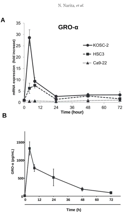
Fig. 1 Time course of the expression of GRO-α in KOSC-2 cells stimulated with TNF-α. (A) KOSC-2
(●), HSC-3 (■), and Ca9-22 (▲) cells were incubated with 1 ng/mL of TNF-α for 4-72 h. mRNA expression of GRO-α or 18s rRNA were analyzed by real-time RT-PCR. (B) KOSC-2 cells were incubated with 1 ng/ml of TNF-α for 4-72 h. The conditioned medium was collected and ELISA was performed. Means (±SD) of three experiments shown.
22 32)
0 500 1000 1500
0 12 24 36 48 60 72
GRO-α(pg/mL)
Time (h)
A
B
Fig. 1
0 5 10 15 20 25 30 35
0 12 24 36 48 60 72
mRNA expression (fold increase)
Time (hour)
GRO- α
KOSC-2 HSC3 Ca9-22
GRO-α in KOSC-2 and HSC-3 cells. In both cells, GRO-α mRNA reached the maximal level 4 h after the stimulation with TNF-α (Fig 1A).
The induced levels of mRNA levels of GRO-α in KOSC-2 were markedly higher than that in HSC-3 cells. In contrast, no such increase of GRO-α was observed in Ca9-22 (Fig 1A). These observations suggested that the induction of GRO-α in response to TNF-α varies depending on the cell type. The time course of GRO-α protein secretion corresponded with that of the mRNA expression (Fig. 1B).
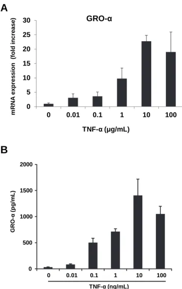
TNF-α enhanced GRO-α mRNA expression of KOSC-2 cells in a concentration- dependent manner (Fig. 2A). The expression of GRO-α was observed from the treatment with 0.1 ng/
mL TNF-α. TNF-α also stimulated the secretion of GRO-α protein and the maximal effect was observed at 10 ng/mL (Fig. 2B).
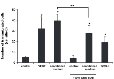
GRO-α has chemotactic activity for endothelial cells
The results of endothelial cell chemotaxis are summarized in Fig. 3. VEGF is known as
Fig. 2 Concentration-dependent induction of GRO-α by TNF-α. KOSC-2 cells were incubated with 0.01- 100ng/
mL TNF-α for 4 h. (A) The expression of mRNA for GRO-α or GAPDH was analyzed by RT-PCR. (B)
The conditioned medium of KOSC-2 cells was collected and subjected to ELISA for GRO-α. Means (±
SD) of three experiments are shown.
23
0 500 1000 1500 2000
GRO-α(pg/mL)
0 0.01 0.1 1 10 100 TNF-α(ng/mL)
A
B
Fig. 2
0 5 10 15 20 25 30
0 0.01 0.1 1 10 100
mRNA expression (fold increase)
TNF-α (μg/mL) GRO-α
a potent angiogenic factor, and thus we used VEGF as a positive control for this migration assay for endothelial cells. As we expected, only small amount of VEGF could induce chemotaxis in HUVECs. The conditioned medium from TNF-α-treated KOSC-2 cells significantly
enhanced the transmigration of endothelial cells, and r(h) GRO-α was also found to be active in this assay. To evaluate the possible role for GRO-α in the TNF-α-treated conditioned medium, we added anti-neutralizing antibody against GRO-α in the conditioned medium, and
Fig. 3 Endothelial cell transmigration in response to GRO-α. Control medium (M199-HSA), VEGF (100 pg/mL, in M199-HSA). GRO-α (1 ng/mL, in M199-HSA) or conditioned medium (from KOSC-2 stimulated with TNF-α for 4 h) was placed in lower chambers, and upper chambers were filled with HUVEC suspension. When indicated, anti-neutralizing GRO-α antibody was added to the conditioned medium. After incubating for 4 h at 37°C, the membrane was fixed and stained with Giemsa solution.
Transmigrated cells in random four fields were counted under a microscope. *P<0.05 statistically significant difference compared with the control, **P<0.05 vs TNF-α-treated conditioned medium.
24
Number of transmigrated cells (cells/field)Fig. 3
0 10 20 30 40 50
control VEGF conditioned control conditioned GRO-α medium medium
+ anti-GRO-αAb
* *
*
*
**
Fig. 4 KOSC-2 cells migration in the presence of GRO-α. Confluent monolayers of KOSC-2 cells were wounded as described in “Materials and Methods”. The cells were incubated in the presence or absence of 1 ng/
mL GRO-α, or conditioned medium (from KOSC-2 cells stimulated with TNF-α for 4 h) for 20 h, then fixed and photographed.
The arrows point to the original edge of the wound. Data shown represent from two independent experiments.