ICU
-acquired weakness in a pediatric patient with bronchial asthma after mechanical ventilation and corticosteroids therapy
Masaru SHIMURA, Soupei GO, Yasuyo KASHIWAGI, Hisashi KAWASHIMA
Department of Pediatrics, Tokyo Medical University
Abstract
We report a 15
-month
-old boy with bronchial asthma who developed severe muscle weakness after mechani- cal ventilation suspected to be intensive care unit acquired weakness (ICU
-AW). Before muscle weakness appeared, he underwent corticosteroid therapy for more than 4 weeks and mechanical ventilation under sedation for 18 days. He was also in a state of malnutrition. Muscle biopsy revealed mild atrophy of type 2 fiber. Mus- cular symptoms improved in 4 weeks without any sequelae. Little has been reported on pediatric patients with ICU
-AW. Indeed, much of the mechanisms and pathophysiology involved remains to be clarified, making a mis- diagnosis common. Therefore, ICU
-AW should be suspected in critically ill pediatric patients showing muscle weakness.
Introduction
Patients with bronchial asthma developing myopathy following mechanical ventilation and administration of corticosteroids and neuromuscular blocking agents have been documented for more than forty years
1-3). Poly- neuropathy in intensive care unit (ICU) patients affected by sepsis and multiple organ failure has been also recog- nized over the past three decades
4-6). Such weakness syndromes associated with critically ill patients have been referred to as ICU acquired weakness (ICU
-AW), and include critical illness myopathy (CIM) and critical illness polyneuropathy (CIP)
7). While most studies of ICU
-AW have been conducted in adult patients, very few studies in pediatric patients have been reported. There- fore, the epidemiology, characteristics, and risk factors of ICU
-AW in childhood remain to be clarified, and the diagnostic criteria in children remain to be established.
In this report, we describe a 15
-month
-old Japanese patient with suspected ICU
-AW who developed severe muscle weakness after mechanical ventilation and long
-term administration of corticosteroids and sedative agents.
Case
A 15
-month
-old boy presented with a terrible cough and wheezing for 3 days, symptoms which appeared the day following a house cleaning. Although he exhibited wheezing and a pallid complexion, the symptoms improved after nebulization with β2 stimulants, and he was able to return home temporarily. However, his respiratory status deteriorated, and he was brought to us again at night on the same day. On physical examina- tion, his state of consciousness was clear, but he looked pale and was febrile, with a temperature of 37.5°C. He was tachycardic (respiratory rate : 42/min) and had an J. Tokyo Med. Univ., 76 (1) : 64
-69, 2018
Case Report
This work was not supported by any grant, and there is no conflict with any employment. The family concerned in this case provided informed consent for its publication.
Received March 6, 2017, Accepted October 13, 2017
Key words : ICU
-acquired weakness, critical illness myopathy, muscle weakness, bronchial asthma, mechanical ventilation
Corresponding author : Hisashi Kawashima M.D., PhD, Department of Pediatrics, Tokyo Medical University, 6
-7
-1 Nishishinjuku, Shinjuku
-ku, Tokyo160
-0023, Japan
TEL : 03
-3342
-6111 (Ex 62440) FAX : 03
-3344
-0643
O2 saturation of 93% in room air. Severe wheezing and prolonged expiration were observed. He had no medi- cal history of note, except for urticaria after eating eggs.
His growth and development were normal, and he was able to walk well unassisted before admission. His father had atopic dermatitis and mother had bronchial asthma in childhood. Blood examination showed an increase in the number of white blood cells (WBC 16,300/µL, neutrophils 80.7%, lymphocytes 15.6%, basophils 0.2%, eosinophils 0.2%, monocytes 3.3%) and a slight increase in C
-reactive protein (CRP 0.6 mg/dl), but the rest of the results, including those for blood gas and liver transaminase, were normal. He had an ele-
vated total IgE (434.0 IU/L) and positive RAST reaction against mites (class 5), house dust (class 4), egg (class 4), and ovomucoid (class 4). The results for various viral and bacterial tests, including for respiratory syncytial virus, Bordetella pertussis, chlamydia pneumonia, and mycoplasma pneumonia were all negative. Chest X
-ray findings revealed a mediastinal shift to the right side with the over
-inflation of the left lung, with mediastinal and subcutaneous emphysema (Figure 1A). He was admit- ted due to a diagnosis of status asthmaticus and mediasti- nal emphysema.
His clinical course after admission is shown in Figure 2. Humidified oxygen, inhalation of short
-acting β2
Figure 1 A, B : A. Chest X
-ray on admission, B. Computed tomography of chest on day3
Figure 2 Clinical course after admission
ChE : serum cholinesterase, TP : total serum protein, Alb : serum albumin, mPSL : methylprednisolone, PSL : predniso-
lone, DTR : deep tendon reflex, MMT : manual muscle testing
agonist, and venous injection of corticosteroids were started. However, his respiratory status, including retraction and nasal alar breathing, and subcutaneous emphysema worsened. His blood oxygen levels were not maintained by face mask administration of oxygen, and, moreover, chest CT showed a deterioration of medi- astinal emphysema (Figure 1B). Mechanical ventilation was initiated under sedation with midazolam on day 3.
Theophylline was administered via injection. His respi- ratory condition, mediastinal emphysema, and subcuta- neous emphysema improved gradually. On day 9, arti- ficial ventilation was stopped and he was extubated. As he showed improvement in his respiration and oxygen- ation, corticosteroids and oxygen were tapered. An attempt was made to feed him per os, but he was hardly able to take any food. Weight reduction (-21%, com- pared with on admission) and decreased serum nutri- tional markers, including those for cholinesterase, pro- tein, and album were marked. His respiration status deteriorated again and his fever returned on day 15. He became restless and was not able to maintain eye contact with his mother on day 17, so he was re
-intubated for reintroduction of mechanical ventilation. He was sedated with a combination of midazolam and fentanyl during the second period of mechanical ventilation.
Additionally, he started to undergo central venous hyper- alimentation on day 18. His respiration and nutritional status showed a tendency toward improvement, and he
was extubated on day 27. Corticosteroids were admin- istered from admission to day 32. During the clinical course, serum glucose and theophylline levels were maintained within normal limits, and no neuromuscular blocking agents were used.
Severe muscle weakness appeared after withdrawal of mechanical ventilation for the second time. On day 27, he could move his head, but was not able to move his extremities and had a weak deep tendon reflex (DTR) bilaterally. Manual muscle testing of the upper and lower extremities yielded values of 0 and 1, respectively.
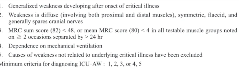
Sensory disturbance could not be evaluated because of his age. There were no elevations of creatine kinase or aldolase. The cell count, protein, and pressure of cere- brospinal fluid were all normal. Electromyography and motor sensory nerve conduction tests showed no obvious abnormalities on day 58. A muscle biopsy on day 59 showed no necrotic or regenerating fibers. There was no inflammation or perifascicular atrophy, except for mild atrophy of type II fibers. His symptoms and exam- ination findings fulfilled the criteria for a diagnosis of ICU
-AW, and probably also CIM (Table 1, 2)
7). The muscle weakness gradually improved without any spe- cific treatment. He could move his extremities and had a normal DTR on day 30. On day 31 he was able to take food well per os. He could sit up without support on day 36 and walk by himself on day 54. Manual muscle testing of the extremities yielded values of 4 or 5
Table 1 Diagnostic criteria for ICU
-AW 1. Generalized weakness developing after onset of critical illness
2. Weakness is diffuse (involving both proximal and distal muscles), symmetric, flaccid, and generally spares cranial nerves
3. MRC sum score (82) < 48, or mean MRC score (80) < 4 in all testable muscle groups noted on ≧ 2 occasions separated by > 24 hr
4. Dependence on mechanical ventilation
5. Causes of weakness not related to underlying critical illness have been excluded Minimum criteria for diagnosing ICU
-AW : 1, 2, 3, or 4, 5
ICU
-AW : intensive care unit
-acquired weakness, MRC : Medical Research Council Adapted from Stevens RD et al
7Table 2 Diagnostic criteria for CIM 1. Patient meets criteria for ICU
-AW
2. Sensory nerve action potential amplitudes are > 80% of lower limit of normal in ≧ 2 nerves 3. Needle electromyogram in ≧ 2 muscle groups demonstrates short
-duration, low
-amplitude
motor unit potentials with early or normal full recruitment with or without fibrillation poten- tials
4. Direct muscle stimulation demonstrates reduced excitability (muscle/nerve ratio > 0.5) in ≧ 2 muscle groups
5. Muscle histology consistent with myopathy Probable CIM : criteria 1, 2, 3 or 4 ; or 1 and 5 Definite CIM : criteria 1, 2, 3 or 4, 5
CIM : critical illness myopathy, ICU
-AW : intensive care unit
-acquired weakness
Adapted from Stevens RD et al
7on day 63 of discharge. At a 1
-year follow
-up exami- nation, his growth, motor, and intellectual development were within the normal range.
Discussion
We encountered a pediatric patient who developed muscle weakness possibly due to ICU
-AW. The muscle weakness appeared following a total of 18 days of mechanical ventilation under sedation with corticoste- roids. He was also in a state of malnutrition, leading to a catabolic state before weakness developed. The mus- cular symptoms fully recovered within 4 weeks.
The diagnostic criteria of ICU
-AW, CIM, and CIP, which were established based on evidence from adult patients, are shown in Table 1
-3
7). Determining whether a patient meets these criteria includes invasive tests, and they are therefore difficult to apply in children. In pre- vious studies, 50% of adult patients in the ICU developed ICU
-AW, commencing within the first few days of ill- ness
8-10). On the other hand, it was reported that the incidence of ICU
-AW in pediatric patients was 1.7% in a prospective study
11)and 0.02% in a retrospective study
12). Apparent differences in the incidence of this condition between children and adults may be due to under
-recog- nition of ICU
-AW, CIM, and CIP in critically ill pediatric patients, as well as related to the problem of diagnostic criteria.
The pathophysiological mechanisms underlying ICU
-AW are complex, although a number have been sug- gested, including dysfunctional microcirculation leading to neuronal injury and axonal degeneration, the inactiva- tion of sodium channels, mitochondrial dysfunction, and oxidative stress
13). Furthermore, sepsis, a catabolic state, multi
-organ system failure, systemic inflammatory response syndrome, long duration of mechanical ventilation, immobility, hyperglycemia, corticosteroids, neuromuscular blocking agents, and female have been considered as risk factors
13). However, several risk factors, including corticosteroids and neuromuscular blocking agents, were reported to be unrelated to ICU
-AW in some trials
14)15). Based on these findings, it is conceivable that the pathogenic mechanism is multifacto- rial and diverse in each case. In the present case, no
central venous hyper
-alimentation was given until the 18
thhospital day. Therefore, the patient showed severe malnutrition, which contributed to the catabolic state.
The muscle weakness in this case might also have been due to malnutrition in addition to the long duration of mechanical ventilation, corticosteroids, and immobility.
In terms of muscle histopathology, type II fiber atrophy, which was observed here, and thick filament loss have been reported in CIM
16)17). Reduced compound muscle action potential amplitudes and decreased muscle membrane excitability in electrodiagnostic testing are usually seen in CIM. No such abnormal findings were observed here, however
18). This may have been due to the late timing of the muscle biopsy, which was conducted after recovery of muscular symptoms, and possibly differences between children and adults.
Although there is no specific treatment for ICU
-AW, we should be aware that it can develop in critically ill pediatric patients. We should prevent and remove risk factors, avoiding excess sedation and unnecessary medication. Little evidence is available with regard to ICU
-AW in children. Therefore, most patients might be overlooked. Further study is required to elucidate the mechanism of ICU
-AW during childhood.
References
1) MacFarlane IA, Rosenthal FD : Severe myopathy after status asthmaticus. Lancet (London, England) 2 : 615, 1977
2) Leatherman JW, Fluegel WL, David WS, Davies SF, Iber C : Muscle weakness in mechanically ventilated patients with severe asthma. American journal of respiratory and critical care medicine 153 : 1686
-1690, 1996
3) Behbehani NA, Al
-Mane F, D’Yachkova Y, Pare P, FitzGerald JM : Myopathy following mechanical ventilation for acute severe asthma : the role of mus- cle relaxants and corticosteroids. Chest 115 : 1627
-1631, 1999
4) Bolton CF, Gilbert JJ, Hahn AF, Sibbald WJ : Poly- neuropathy in critically ill patients. Journal of neu- rology, neurosurgery, and psychiatry 47 : 1223
-1231, 5) Bolton CF, Laverty DA, Brown JD, Witt NJ, Hahn 1984 Table 3 Diagnostic criteria for CIP
1. Patient meets criteria for ICU
-AW
2. Compound muscle action potential amplitudes are decreased to < 80% of lower limit of nor- mal in ≧ 2 nerves
3. Sensory nerve action potential amplitudes are decreased to < 80% of lower limit of normal in
≧ 2 nerves
4. Normal or near
-normal nerve conduction velocities without conduction block 5. Absence of a decremental response on repetitive nerve stimulation
CIP : critical illness polyneuropathy, ICU
-AW : intensive care unit
-acquired weakness
Adapted from Stevens RD et al
7AF, Sibbald WJ : Critically ill polyneuropathy : elec trophysiological studies and differentiation from Guillain
-Barre syndrome. Journal of neurology, neurosurgery, and psychiatry 49 : 563
-573, 1986 6) Zochodne DW, Bolton CF, Wells GA, et al : Critical
illness polyneuropathy. A complication of sepsis and multiple organ failure. Brain : a journal of neu- rology 110 : 819
-841, 1987
7) Stevens RD, Marshall SA, Cornblath DR, et al : A framework for diagnosing and classifying intensive care unit
-acquired weakness. Critical care medicine 37 : S299
-308, 2009
8) Hermans G, De Jonghe B, Bruyninckx F, Van den Berghe G : Clinical review : Critical illness polyneu- ropathy and myopathy. Critical care (London, Eng- land) 12 : 238, 2008
9) de Letter MA, Schmitz PI, Visser LH, et al : Risk factors for the development of polyneuropathy and myopathy in critically ill patients. Critical care medicine 29 : 2281
-2286, 2001
10) Puthucheary ZA, Rawal J, McPhail M, et al : Acute skeletal muscle wasting in critical illness. Jama 310 : 1591
-1600, 2013
11) Banwell BL, Mildner RJ, Hassall AC, Becker LE, Vajsar J, Shemie SD : Muscle weakness in critically ill children. Neurology 61 : 1779
-1782, 2003 12) Field
-Ridley A, Dharmar M, Steinhorn D, McDonald
C, Marcin JP : ICU
-Acquired Weakness Is Associated With Differences in Clinical Outcomes in
Critically Ill Children. Pediatric critical care medicine : a journal of the Society of Critical Care Medicine and the World Federation of Pediatric Inten- sive and Critical Care Societies 17 : 53
-57, 2016 13) Kress JP, Hall JB : ICU
-acquired weakness and
recovery from critical illness. The New England journal of medicine 370 : 1626
-1635, 2014
14) Hermans G, Wilmer A, Meersseman W, et al : Impact of intensive insulin therapy on neuromuscular com- plications and ventilator dependency in the medical intensive care unit. American journal of respiratory and critical care medicine 175 : 480
-489, 2007 15) Papazian L, Forel JM, Gacouin A, et al : Neuromus-
cular blockers in early acute respiratory distress syn- drome. The New England journal of medicine 363 : 1107
-1116, 2010
16) Lacomis D, Giuliani MJ, Van Cott A, Kramer DJ : Acute myopathy of intensive care : clinical, electro- myographic, and pathological aspects. Annals of neurology 40 : 645
-654, 1996
17) Bierbrauer J, Koch S, Olbricht C, et al : Early type II fiber atrophy in intensive care unit patients with non- excitable muscle membrane. Critical care medicine 40 : 647
-650, 2012
18) Shepherd S, Batra A, Lerner DP : Review of Critical
Illness Myopathy and Neuropathy. The Neurohospi-
talist 7 : 41
-48, 2017
気管支喘息重積発作に対する人工呼吸管理、ステロイド療法後に ICU
-acquired weakness と考えられる筋力低下を呈した男児例
志 村 優 呉 宗 憲 柏 木 保 代 河 島 尚 志
東京医科大学小児科
【要旨】 気管支喘息重積発作に対する人工呼吸管理およびステロイド療法後に、
ICU
-acquired weakness
(ICU
-AW
) と考えられる筋力低下を呈した、15ヶ月男児例を経験したので報告する。筋力低下を認める前には、4週間以上の ステロイド投与、18日間の鎮静下での人工呼吸管理を受け、低栄養状態にあった。筋生検では2
型筋線維の軽度の 萎縮を認めた。筋症状は後遺症なく、約4
週間で改善した。ICU-AW
の小児例の報告は稀であるため、小児期ICU
-AW
の発症機序や病態生理の多くはまだ解明されていない。治療経過中に筋力低下を呈す小児の重症例では、ICU-AW
を鑑別する必要がある。〈キーワード〉 ICU-