Nanostructure Control and Photochemistry in Inorganic Nano-sheet Materials
March, 2015
FUJIMURA, Takuya
Preface
The studies presented in this thesis were carried out under the guidance of Professor Shinsuke Takagi at Department of Applied Chemistry, Graduate Course of Urban Environmental Sciences, Tokyo Metropolitan University.
Takuya FUJIMURA Department of Applied Chemistry
Graduate Course of Urban Environmental Sciences Tokyo Metropolitan University
1-1 Minami-ohsawa, Hachiohji
Tokyo 192-0364, JAPAN
Contents
Chapter 1. General Introduction 1.1. Photosynthesis
1.2. Functionality and structure of the light harvesting system and reaction center 1.3. Dependency of the inter distance and relative orientation of the molecules on
photochemical reactions
1.4. Assemblies of the organic molecules and their photo-functionality 1.5. Gold nano particles
1.6. Host materials as a reaction field
1.7. Assembly of the dyes on the clay surfaces 1.8. Purpose of this thesis
1.9. References
Chapter 2. Generation of the Monodispersed Gold Nano Particles on the Clay Surfaces with Deposition Reduction Method
2.1. Introduction
2.2. Experimental section 2.3. Result and discussion
Effect of the mixing method to deposit the gold nano particles on Montmorillonite surfaces
Effect of the concentration of gold precursor to deposit the gold nano particles on the Montmorillonite surfaces
Stability of the gold nano particles deposited on the Montmorillonite surfaces 2.4. Conclusion
2.5. References
Chapter 3. Generation of Small and Non-Aggregated Gold Nano-Particles on the Clay Surface Modified with Porphyrin Molecules
3.1. Introduction
3.2. Experimental section
3.3. Result and discussion
Generation of the gold nano particles on the MNT surface modified with co-adsorbed TMPyP
Effect of the loading level of TMPyP on MNT surfaces
Thermal Stability of gold nano particles deposited on the MNT surfaces modified with TMPyP
Effect of the loading level of gold precursor and estimation of the appropriate gold precursor amount
3.4. Conclusion 3.5. References
Chapter 4. Formation of Assembled Structure of Gold Nano Clusters on the Clay Surface Utilizing Porphyrin/Clay Complex as a Template
4.1. Introduction
4.2. Experimental section 4.3. Result and discussion
Deposition of the Gold NPs on MNT Surfaces via PTR Method TEM observation of Gold NPs deposited via PTR Method 4.4. Conclusion
4.5. References
Chapter 5. Construction of Layered Structure of Porphyrin − Saponite Clay Complex 5.1. Introduction
5.2. Experimental section 5.3. Result and discussion
Absorption Spectra and Stability of TMPyP/SSA Thin Hybrid Film
Absorption Spectra and X-ray Diff raction of TMPyP/SSA Hybrid Thin Film at Various Loading Levels
Fluorescence Behavior of TMPyP/SSA Hybrid Film 5.4. Conclusion
5.5. References
Chapter 6. Environmentally Responsive Chromism of Hybrid Thin Film Composed of Porphyrin and Clay Mineral
6.1. Introduction
6.2. Experimental section 6.3. Result and discussion
Intercalation reaction of MgTMPyP into interlayer space of SSA
UV-Vis. absorption and Fluorescence spectra of MgTMPyP/SSA hybrid film Chromism of the MgTMPyP/SSA Hybrid Film Depending on Relative Humidity Vapochromism of the MgTMPyP /SSA Hybrid Film
6.4. Conclusion 6.5. References
Chapter 7. Energy Transfer and Subsequent Electron Transfer between Assembled Dyes on the Clay Surfaces
7.1. Introduction
7.2. Experimental section 7.3. Result and discussion
Absorption spectra of AA@OAm
216+∩clay, ZnTMPyP
4+∩clay, DNPV
2+∩clay Energy transfer reactionin in (AA@OAm
216+(EnD)−ZnTMPyP
4+(EnA))∩clay system
Energy Transfer Reaction and Subsequent Electron Transfer Reaction in AA@OAm
216+(EnD)−ZnTMPyP
4+(EnA as well as eD)−DNPV
2+(eA) ∩Clay System
7.4. Conclusion 7.5. References Summary
Acknowledgement
Chapter 1 General Introduction
1.1. Photosynthesis
1-4Energy issue is one of the big problems which human beings have to solve. Lots of countries depend on the fossil fuels as main energy source, however fossil fuels are limited resources, thus the development of the alternative energy resources is crucial important question. To solve this problem, solar energy, geothermal energy, wind power energy, tidal energy and other alternative energy are receiving a lot of attention as new energy sources. Especially, solar energy has attracted great attention as a new clean energy resource, and several types of the system, such as solar cells, have been
developed and have attracted great research interest in several decades. However, one of the weak points of the solar energy cannot be stored, and energy conversion systems in which sunlight energy is transferred to storable energies are eagerly anticipated.
The photosynthesis in the plant is one of the great processes to utilize the sun light energy as an energy resource. The reaction of photosynthesis is as follows.
The carbohydrates synthesized by photosynthesis reaction are important energy sources to creatures, which are not only the plants but also animals. It should be noted that the
6 H
2O + 6 CO
2→ 6 O
2+ ( CH
2O )
6solar energy, which will not be able to stored, is converted to the chemical energy, which has chemical stability and can be stored. This energy conversion system would be expected as a model for effective utilization of the solar energy to solve the energy resources problem. A general outline of photosynthesis is (i) A light-harvesting complexes in a thylakoid membrane absorb the sunlight. (ii) The harvested excitation energy is transferred to a reaction center by an energy transfer reaction. (iii) The
excitation energy is converted to electrochemical energy by a charge separation reaction in the reaction center. (iv) NADPH and ATP are synthesized by the result of proton transfer coupled with electron transportations. (v) NADPH and ATP induce the synthesis of carbohydrate from CO
2and H
2O. However, the detail mechanism of photosynthesis abundantly complicated, many researchers have investigated the mechanism of each step of photosynthesis by each original approaches to reveal the detail mechanism of photosynthesis.
1.2. Functionality and structure of the light harvesting system and reaction center
5-12,115As described in 1.1 in chapter 1, lots of researchers have investigated to reveal the mechanism of the photosynthesis. Especially the initial process of the photosynthesis has attracted great research interests because of the uniqe photo-functionality. Sunlight energy is absorbed in the light harvesting system at initial process of photosynthesis, and the excitation energy is transported to reaction center via energy transfer reaction.
The light harvesting complexes consist of the core-antenna and peripheral antenna. The
core antenna was constructed around the reaction center, and peripheral antenna inhibits around this reaction center – core antenna complex. The peripheral antenna has crucial roles to adapt successfully to the environment changes, such as the strength of the light.
These light-harvesting systems consist of chlorophylls, bacteriochlorophylls, and carotenoids which have a large molar extinction coefficient. Recently, the structure of the light harvesting complexes has been investigated by X-ray and microscopy analysis, and it is revealed that assembled dyes compose the light harvesting system. The image of the light harvesting system in Rps. acidophyla bacteria was shown in Figure 1.
Figure 1. Light harvesting system in Rps. acidophyla bacteria
7To realize the functionality as the light harvesting system, not only the appropriate functional dyes, but also this beautiful assembled structure composed of functional dyes are necessary.
On the other hand, electron transfer reaction, which is thought to be the essential for the light reaction of photosynthesis, proceeds in the reaction center. This process is extremely important from the point of view of the energy conversion, and there are lots
0.92%nm%0.94%nm%
1.8%nm%
1.8%nm%
2.1%nm%
Energy''
''Transfer'
of researches regarding to the reaction center of the photosynthesis. The structure of the reaction center has been also investigated with X-ray or other techniques, and the dynamics of electron transfer reaction in the reaction center has been investigated with the laser flash photolysis and time resolved electron spin resonance. The structural image of the reaction center is shown in Figure 2.
Figure 2. Image of the reaction center
115The photoinduced electron transfer reaction has researched as one of the fundamental
photoreaction for a long time. Photoinduced electron transfer is an excited state electron
transfer process by which excited electron is transferred from donor to acceptor. Due to
this reaction, the radical ion pair (donor cation radical and acceptor anion radical) is
generated, and it is called charge separation state. Generally, charge separation state is
promptly decreased via back electron transfer or charge recombination, which is the
electron transfer reaction from acceptor anion radical to donor cation radical, thus life
time of the charge separation state is not long. However, long-lived charge separation state is achieved in the reaction center by suppression of the charge recombination of radical ion pair. The electron transfer reaction in the reaction center is the successive reaction, that is (i) excitation of the special pair which would be dimer of
chlorophyll-a,(ii) photoinduced electron transfer reaction to bacteriopheophytin, (iii) electron transfer reaction from bacteriopheophytin to quinone-a, (iv) electron transfer reaction from quinone-a to quinone-b. The time scale of the each electron transfer reaction in the reaction center is summarized in Figure 3.
Figure 3. Energydiagram of the electron transfer reaction in the reaction center
As summarized in Figure 3, the stepwise electron transfer reactions proceed and the charge recombination reactions are suppressed in every process. One of the important factors to suppress the charge recombination is the apposite reorganization energy,
(i) (ii)
(iii)
(iv)
which is required energy for the reorganization of the solvent around the ion and reorganization of the molecule structures. In addition, the stepwised charge
transportation is also crucial important point to achieve the long-lived charge separation state, and assembly of the molecules would be also important to achieve this stepwise electron transfer reaction as described in later.
1.3. Dependency of the inter distance and relative orientation of the molecules on photochemical reactions
13-26Energy transfer and electron transfer reaction are two of the most important interaction between excited molecules and other grand state molecule. The paradigm of the energy transfer and electron transfer reaction is shown in Scheme 1.
A * +B → A + B * (a) A * +B → A
+•+ B
−•(b)
Scheme 1. Paradigm of the energy transfer reaction (a) and electron transfer reaction (b)
where * indicates electrically excited state, A and B indicates donor and acceptor in each reaction, respectively. The energy transfer reaction can be categorized into three types by their mechanism. In this section, the energy transfer reaction by the
dipole-dipole interaction is mainly described. The energy transfer reaction by the
dipole-dipole interaction is known as fluorescence resonance energy transfer (generally
called FRET) or Förster-type energy transfer. This dipole-dipole interaction occurs
through oscillating electrical field, and it should be noted that FRET does not involve the overlap of the orbital. The theoretical energy transfer rate constant for Förster-type is expressed in following equation 1.
(eq. 1)
where φ
Dis the fluorescence quantum yield of the donor molecules, n is reflective index of the medium, N is Avogadro’s number, is the orientation factor, τ
Dis the excited life-time of donor in the absence of acceptor, R is the inter molecular distance between energy donor and acceptor (center to center), and the integral part represents the spectral overlap between absorption spectrum of energy acceptor and fluorescence spectrum of energy donor, respectively. On the other hand, electron transfer reaction whose
paradigm is showed in scheme 1-(b) requires the orbital overlap. The inter molecular distance dependency of the electron transfer reaction has been investigated for a long time, the theory of the electron transfer has been developed by Murcus, Libby, Rehm, Weller and other huge number of researchers. It has revealed the dependency of the electron transfer reaction on inter distance and relative orientation of the molecules between electron donor and acceptor as shown in following equation 2.
kel =kel0exp
[
−β
(d−d0)] (eq. 2)
∫
=
5 4 6 42
) ( ) 128 (
10 ln 9000
ν ν ν ε τ ν
π
φ
κ f d
R N
k
ETn
D D Awhere k
elindicates electron transfer rate constant, β is the orbital parameter which is in inverse proportion to overlap integration of the orbital of electron donor and acceptor, d is the inter molecular distance between electron donor and acceptor, d
0is the van der Waals distance. Considering these dependencies of the energy transfer and electron transfer reaction on the inter distance and relative orientation between donor and acceptor molecules, control of these parameter is thought to be the crucial point to control these reaction and to realize the excellent photo-functionalities such as light harvesting system and reaction center.
1.4. Assemblies of the organic molecules and their photo-functionality
27-52As described in above section, assemble structures of photo-functional dyes show the unique photochemical or photophysical proterties such as light harvesting system and reaction center in natural photosynthesis. Inspired these researches, the assembly of the organic molecules has been investigated in several decades, and photo-functionalities of the assembled functional dyes have been demonstrated in the several system. One of the central systems to assemble the functional dyes is the covalent linked system where functional dyes are linked via covalent bond. The functional dyes are linked via covalent bond in this system, thus inter-molecular distances and relative orientation between energy donor and acceptor were strongly immobilized. The energy transfer or/and electron transfer reaction have been investigated between covalent linked a donor molecule and an acceptor molecule in several decades. These crucial works have
developed the theories of these reactions and achieved the efficiently energy transfer
reaction and long-lived charge separation state. In the context of these great works, the energy / electron transfer reaction has been investigated between several donors and acceptors in recent years. Gust et al. investigated an antenna property of molecule shown in Figure 4, and they revealed the pH dependency of the energy transfer behavior this dye.
Figure 4. Structure of the antenna molecule synthesized and investigated by Gust et al.
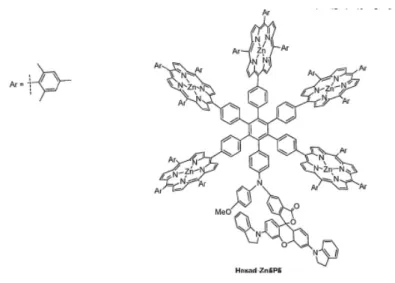
32The molecule featured five porphyrin antenna moieties and a pH-sensitive dye. The
energy transfer behavior was significantly depends on the pH values. Okada and
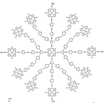
co-workers synthesized a huge antenna molecule, as it is called dendrimer, as shown in
Figure 5.
Figure 5. Dendrimer type antenna molecule reported by Okada et al.
35The energy transfer reaction took place from peripheral porphyrin molecules to the central porphyrin molecule judging from time resolved fluorescence spectroscopy.
These researches constitute a new and important step to achieve the artificial light harvesting system. However, the synthesis and purification of the molecules are
generally complicated, and these complications are bottlenecks for this covalent bonded
system. On the other hand, supra molecules system would be another central system to
construct the assembly of the functional dyes. It is supposed that dye aggregation,
whose theory was established by Kasha et al., in solution is the pioneer works for
construction of the dye assembly in solution. This supra molecules assembly is thought
to be the flagship to construct the dyes assembly which has superior photo-functionality
such as artificial light harvesting system. For example, Kobuke and co-workers have
reported the ring shaped supra molecules system similar to the light harvesting system
composed of the Zinc porphyrin derivatives.
Figure 6. Supramolecular porphyrin macro-rings reported by Kobuke et al.
44Tamiaki et al. reported the supramolecular chlorin assembly as artificial lightharvesting device (Figure 7). An efficient energy transfer between porphyrins took place in the dye assembly.
Fig. 14 Complementary coordination polymers 26P and 27P, and the unit molecules.
dependent on the concentration. The average number has been estimated to produce more than a 700-mer for
26Pin CHCl
3from gel permeation chromatography (GPC) data, and a 45-mer for
27Pin 5.8
×10
−5M CHCl
3from the self-association constant.
The absorption spectrum of polymer
26Pshows split Soret bands at 408.5 and 489.5 nm, which are blue- and red-shifted by
−528 and 1357 cm
−1, respectively, compared to the Soret band of the corresponding monomer
26M-Py(Fig. 15). Polymeric
27Palso shows split Soret bands at 432 and 490.5 nm, whereas the dissociated monomer
27M-Pyshows split Soret bands at 437 and 473.5 nm. The absorption spectrum of both polymers covers a wide wavelength range of visible light. The fluorescence spectrum of polymeric
26Pdoes not show any obvious quenching,
55indicating a potential use in light-harvesting antennae. The fluorescence was quenched efficiently when an acceptor molecule was introduced at the terminal position.
47This means that excitation takes place at any location in the assembly, and that the energy is transferred to the terminal acceptor.
Fig. 15 UV–vis absorption spectra of26M-Py,26P,27M-Py, and27P.
(Key: polymer and dimer=black, monomer=grey.)
When complementary dimer motifs are connected by non- linear spacers, macrocyclic oligomers are produced spontaneously under diluted conditions without the formation of polymeric side products. The size of the macroring is easily controlled by the angle between the two connecting bonds. In the case of
m-phenylene-linked bis(imidazolyl-zinc-porphyrins), pentamericand hexameric supramolecular macrorings,
C-P5and
C-P6, areformed
56(Fig. 16). To determine the exact assembly number using mass spectrometry, the macrorings were covalently linked using a ring-closing metathesis reaction.
56b,57The covalently linked macrorings did not dissociate under the conditions used in the mass spectrometer, not even when using coordinating solvents, such as pyridine. Two different sized macrorings were separated using GPC.
Fig. 16 Supramolecular porphyrin macroringsC-P5,C-P6,C-EP5, and C-EP6with reference porphyrinsA,B, andC.
The photophysical dynamics of
C-P5and
C-P6were determined using reference porphyrins
A, B, and Cthrough nanosecond time-resolved fluorescence and fluorescence decay anisotropy, and femtosecond transient absorption (TA) and transient absorption anisotropy (TAA).
58The fast excitation-energy hopping (EEH) rates in the rings were also determined using the latter two methods. The TAA decay reflects the depolarization of the exciton that is initially localized in weakly coupled multi-chromophores.
The EEH parameters between the two zinc porphyrin monomers in the cofacial dimer and the cofacial porphyrin dimers in the macrorings were obtained from the data. Exciton–exciton annihilation processes occur in light-harvesting antenna systems under multi-photon absorption conditions. These processes are also associated with the EEH value.
59Pump power-dependent
1686 | Org. Biomol. Chem., 2007,5, 1679–1691 This journal is©
The Royal Society of Chemistry 2007Published on 27 April 2007. Downloaded on 26/11/2014 07:23:29.
View Article Online
Figure 7. The assembly strucuture of chlorins investigated by Tamiaki and co-workers
31The constructions of the supramolecular assembly in the solid state and their
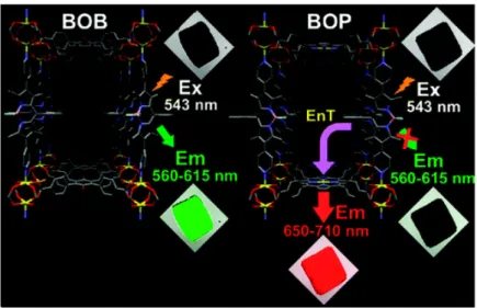
photo-functionalities have been demonstrated. Hupp and co-workers have reported the energy transfer and energy migration proceed in the ordered metal organic frameworks system. The structural image and photograph of obtained crystal is shown in Figure 8.
Figure 8. Structural image and photograph of the metal organic frameworks reported by
Hupp and co-workers
48The excitation energies were migrated between energy donor (bodipy), which show the green fluorescence, and energy transfer took place between bodipy and energy acceptor molecule (porphyrin), which shows the red fluorescence. Kuhn, Moebius and
co-workers focus attention on self-assembled monolayer dyes obtained by Langmuir- -Blodgett (LB) technique. They have investigated the electron and energy transfer reaction between J-aggregate (head to tail type aggregate) of amphiphilic cyanines in LB film. These crucial works have verified the dependency of the energy transfer reaction on distance. Whitten and co-workers reported the “super quenching”, that is the quenching of the large number of donor molecules by the small number of quencher molecules, in the J-aggregate type assembly system. These crucial works, in which the properties of Frenkel exciton was deftly utilized, have much greater hope for
construction of the artificial light harvesting system, because the essence of this investigation, that is the concentration of light energy absorbed by donor molecules to the small number of acceptor molecules, is resemble to the light harvesting system.
1.5. Gold nano particles
53-82The above section explains the assembly structure of organic molecules and
functionality of them, but the assembly structure of inorganic nano-particles is another interesting topic investigated in recently years. It has been revealed that the nanometer order inorganic nano particles show the interesting optical, electrical and magnetical properties which are different from the properties of bulk materials in several decades.
Especially, gold nano particle has been gathering the attention since Haruta and
co-workers reported the catalysis activity of the gold nano particles deposited on the supporting materials surfaces. In addition to this pioneer works, the synthesis of the gold nano cluster stabilized by protective reagent reported by Schumid and Brust et al.
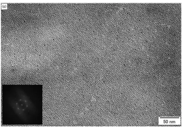
would be the breakthrough for the investigation of gold nano clusters. Recently, there are huge number of the reports of gold nano cluster and their properties. Schmid defined the cluster as the nano particles which was exactly defined in chemical composition and structure. On the other hand, assembly of the gold nano clusters also has investigated with the development of the synthesis technique of the gold nano clusters. Teranishi and co-workers demonstrate the excellent assembly of gold nano cluster at the air-liquid interface by π-π interaction of the ligand as shown in Figure 9.
Figure 9. The gold nano cluster assembly reported by Teranishi et al.
(Inset is the fast Fourier transform spots)
70assembled into a number of isolated hcp domains (∼100 nm×
∼100 nm, Figure 7). The mean interparticle spacing between the neighboring particle edges is estimated from the TEM image to be 1.9(0.3 nm, which is compatible with that for the self- assembled superlattices directly formed on an amorphous- carbon-coated copper grid, indicating that the interligandπ-π
interaction also induces the formation of hcp superlattices at the air-water interface. The interligand π-πinteraction was also implied by the small-angle X-ray diffraction of 1.5 nm TC8- BIP-Au nanoparticle powder (Figure S1 in the Supporting Information), which was obtained by the precipitation from a TC8BIP-Au DMF solution with methanol. The mean interpar- ticle spacing was calculated as 2.2 nm from a diffraction peak at 2.06°assigned to the (111) plane for the 3D superlattice of 1.5 nm TC8BIP-Au nanoparticles. This value is roughly consistent with that for a 2D superlattice within error, indicating that there is an interdigitation or overlap of BIP groups (0.8 nm).
The UV-vis spectra of different amounts of TC8BIP-Au nanoparticles on quartz substrates were measured to spectro- scopically confirm the existence of aπ-πinteraction (Figure 8). TC8BIP-Au nanoparticle chloroform solutions with two concentrations (3 and 30 mM) were deposited onto quartz substrates to form self-assembled domains of nanoparticles. The absorption peak of the BIP groups for 3 mM TC8BIP-Au nanoparticles was centered at 324 nm, whereas that of the 30 mM nanoparticles occurs at 321 nm. Since highly concentrated nanoparticles form multilayer topology, the extent of π-π stacking between the BIP rings is much higher in the vertical
Figure 8. UV-vis spectra of TC8BIP-Au nanoparticles on quartz substrates. TC8BIP-Au nanoparticle chloroform solutions of (a) 3 mM (dotted line) and (b) 30 mM (solid line) concentration were deposited. The spectra were normalized with BIPπ-π* absorbance.
Figure 9. (a) Low-magnification TEM image of long-range-ordered hcp 2D superlattices of 1.5 nm TC8BIP-Au nanoparticles after annealing at 50°C for 12 h. The dark domains indicated by arrows are bilayers. (b) Enlarged TEM image of (a). (c) TEM image of a nearly perfectly ordered hcp 2D superlattice of 1.5 nm TC8BIP-Au nanoparticles. The insets show FFT spots of each superlattice.
A R T I C L E S Kanehara et al.
13090 J. AM. CHEM. SOC.9VOL. 128, NO. 40, 2006
The well ordered gold nano clusters attracted much attention for their potential in single-electron-tunneling devices. However, the protective ligand which requires to stabilize and to assemble the gold nano cluster affect the properties of gold nano clusters. Teranishi and co-workers demonstrated that, for ∼ 2 nm alkanethiol-protected Au nanoparticles, the tunneling resistance of the alkanethiol layer changes from 8.5 GΩ (1-octanethiol) to 500 MΩ (1-hexanethiol), implying that the shorter ligand is desirable for a drastic reduction of the tunneling resistance. Considering this demonstration, the utilization of strong protecting reagents, such as thiol derivatives, is not favored for the some further applications.
1.6. Host materials as a reaction field
83-100Various host materials, such as micelle, clathrate, mesoporous materials, zeolites and nano-sheet materials have been investigated in several decades, and the guest organic molecules in the nano-spaces in these host materials shows interesting photo-physical and photochemical behaviors which are different from photochemical or photochemical behaviors in solution. Especially, layered material is one of the interesting host
materials because of the unique nano spaces supplied by the nano-sheets. We focused
on the clay minerals, which is one of the well-known inorganic layered materials, as the
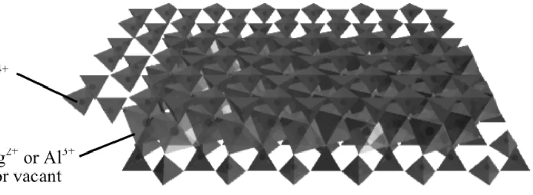
pho-chemical reaction fields. Typical clay layer consists of two sheets of tetrahedral
layer and an octahedral layer as shown in Figure 10.
Figure 10. The structure of saponite clay mineral
The nano-sheet of the clay minerals is two dimensionally expanded with 10-1000 nm (diameter), and the thickness of the neno-sheets is 0.97 nm. The surfaces of the clay minerals have negatively charge by the isomorphous-replacement of element in the layer. Owing to the charges, the interlayer of the clay minerals can swelled, and each nano-sheets can be exfoliated each other in some conditions. This swelling and
exfoliation ability depends on the exchangeable cation and the type of the clay minerals.
Some researchers revealed dependency of the interlayer distance on the relative humidity and counter cation. Clay minerals are classified by the their structure,
chemical formulation and the charge exchangeable capacity. A general classification of
clay minerals is shown in Table 1.
Table 1. Classification of the clay minerals
85Lots of the clay minerals has been reported several production area. Also clay minerals can be synthesized via hydrothermal reaction, and some researchers reported the novel mild condition synthesis method of the clay minerals. The chemical structure and composition of synthetic clay minerals is changeable by the synthetic procedure and the precursors. Bujdak et al. has been reported a unique technique to control the charge
T. Shichi, K. Takagi / Journal of Photochemistry and Photobiology C: Photochemistry Reviews 1 (2000) 113–130 115 Table 1
Classification of clay minerals
Clay Ideal composition Mint(Moct)(Mtetra)O1(OH)m·nH2Oa Layer type Schematic structureb 1:1
Kaolinite group (charge density∼0 per unit)
Kaolinite (Al2)(Si2)O5(OH4) Dioctahedral
Halloysite (Al2)(Si2)O5(OH4)·2H2O Dioctahedral
Serpentine group (charge density∼0 per unit)
Serpentine (Mg6)(Si4)O10(OH)8 Trioctahedral
2:1
Pylophyllite group (charge density∼0 per unit)
Pyrophyllite (Al2)(Si4)O10(OH)2 Dioctahedral
Talc (Mg3)(Si4)O10(OH)2 Trioctahedral
Smectite group (charge density:x∼0.2–0.6 per unit)
Montmorillonite Mx(Al2−xMgx)(Si4)O10(OH)2·nH2O Dioctahedral Beidellite Mx(Al2)(Si4−xAlx)O10(OH)2·nH2O Dioctahedral Nontronite Mx(Fe23+)(Si4−xAlx)O10(OH)2·nH2O Dioctahedral Saponite Mx(Mg3)(Si4−xAlx)O10(OH)2·nH2O Trioctahedral Hectorite Mx(Mg3−xLix)(Si4)O10(OH)2·nH2O Trioctahedral Vermiculite group (charge density:x∼0.6–0.9 per unit)
Dioctahedral vermiculite Mx(Al2−yFe43+)(Si4−xAlx)O10(OH)2·nH2O Dioctahedral Trioctahedral vermiculite Mx(Mg3)(Si4−xAlx)O10(OH)2·nH2O Trioctahedral
Mica group (charge density:x∼0.6–1 per unit)
Mica (muscovite) K(Al2)(Si3Al)O10(OH)2 Dioctahedral
Illite Kx(Al2)(Si4−xAlx)O10(OH)2 Dioctahedral
Biotite K[(Mg, Fe2+)(Fe3+, Al, Ti)](Si, Al)4O10(OH)2 Trioctahedral
Chlorite group
Cookeite [LiAl2(OH)6][(Al2)(Si3Al)O10(OH)2] Dioctahedral Clinochlore [Mg2Al (OH)6][(Mg3)(Si3Al)O10(OH)2] Trioctahedral Chamosite [Fe2Al (OH)6][(Mg3)(Si3Al)O10(OH)2] Trioctahedral Channel type
Sepiolite M2(x+y+2z)/2+ (Mg8−y−zMy3+!z) (Si12−xMx3+)O30(OH)4(OH2)4·8H2O Palygorskite M2+(x−y+2z)/2(Mg5−y−zMy3+!z)
(Si8−xMx3+)O20(OH)2(OH2)4·4H2O
aMint: intercalated metal cations; Moct: cations occupying octahedral position; Mtetra: cations occupying tetrahedral position.
b : tetrahedral sheet; : octahedral sheet; : OH surface;#: H2O;⊕: interlayer cation.
1.2. Intercalation of the organic guest molecules in clays
Montmorillonite can efficiently adsorb various organic compounds, ionic or neutral, within its interlayers. The in- tercalation is quite easily accomplished by magnetic stirring of the clay dispersed aqueous solution with an appropriate amount of guest ions dissolved in water or sometimes added directly as a powder. Such intercalation can be utilized as a probe for the identification of 2:1 type clay minerals [7,11].
Due to the ionic exchange with the exchangeable alkali ions,
generally, more than 95% of the guest ions are incorporated on the basis of the CEC of the clay [12,13]. Also, non-ionic guests with large dipole moments such as ketones and nitriles can be adsorbed to the exchangeable metallic ions within the layers according to their coordination with the ionic sites in the interlayers [6].
The anisotropic nature of intercalated guests molecules has been observed since the beginning of clay chemistry.
The adsorption property in clays permits the mutual interac- tion of the incorporated guests which form self-assembling
density of Montmorillonite, which is one of the typical clay minerals. The details of the clay minerals (structure, composition and so on) is characterized by NMR, X-ray diffraction, elemental analysis, electron microscopy, scanning probe microscopy, and other techniques. The clay minerals have been attracted attention as a host materials and reaction fields same as other layered materials. One of the interesting properties of the layered materials is the interlayer space between the stacked nano-sheets. As described above, this interlayer space is changeable by their medium and guest materials. For example, it has been revealed that the intercalation of the cationic surfactant induces the expanding of the interlayer spaces. It has been researched not only these intercalation reactions of the surfactant but also the intercalation reaction of the guest dyes and the photochemical / photo-physical properties of these hybrid materials. In addition, the clay nano-sheets can be exfoliated and well dispersed in some of the solvents. Synthetic saponite is exfoliated and dispersed in water, and this exfoliated clay nano-sheets supply the two-dimensional flat interface. These exfoliated or stacked clay nano-sheets
performs as the host materials for cationic dyes which adsorbs on clay surfaces via
electrostatic interaction, and some of the these guest dyes on clay minerals shows the
unique photo-physical or photochemical properties which is different from the
properties in solution.
1.7. Assembly of the dyes on the clay surfaces
101-114The adsorption behavior and photo-physical properties of cationic dyes on the surfaces / interlayer spaces of exfoliated clay have been investigated in several decades. The cationic dyes can adsorb on the clay surfaces via electrostatic interaction and
hydrophobic interaction in aqueous media. The researches demonstrated by Takagi and co-workers have revealed the adsorption behaviors of the multivalent cationic dyes on saponite clay surfaces. In their works, they demonstrate the formation of the unique hybrid materials composed of multivalent cationic dyes and saponite clay. The chemical composition of the saponite clay is expressed as
[(Si
7.20Al
0.80)(Mg
5.97Al
0.03)O
20(OH)
4]
−0.77(Na
0.77)
+0.77. The saponite clay is one of the trioctahedral smectite, and their diameter and thickness is ca. 20 nm and 0.97 nm, respectively. The replacement of Al
3+with Si
4+in tetrahedoral layer produces the anionic charges on the surfaces of clay nano-sheets, and counter cation of these anionic sites (generally Na
+) are exchangeable other cation. The average inter anionic distance is calculated as 1.2 nm from the cation exchange capacity of the synthetic saponite (1.00
× 10
-3eq g
-1) and their theoretical surface area (7.5 × 10
2m
2) on the basis of hexagonal array. This saponite clay is characterized as (i) transparency in the visible region
because of not including the transition metal and weak Rayleigh / Mie scatter, (ii) cation
exchangeable ability, (iii) flat and wide surfaces area, (iv) exfoliation or stack ability
(depending on their medium). Takagi and co-workers have demonstrated the formation
of the unique complex composed from multivalent cationic dyes and saponite clay, and
they have investigated photochemical and photo-physical properties of the adsorbed
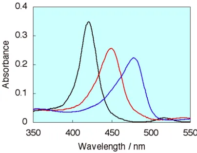
dyes on the saponite clay surfaces. Kerry, Takagi and Kawamata and other researchers have reported that the absorption spectra for adsorbed cationic porphyrin molecules (tetrakis(1-methylpyridinium-4-yl) porphyrin, abbreviated as TMPyP) on clay / intercalated in clay nano-sheets shows the red shift as shown in Figure 11.
Figure 11. Absorption spectra of TMPyP (black line) in water, (red line) adsorbed on the clay surfaces, (blue line) intercalated in clay nano-sheets
116These spectral shifts of porphyrin molecules are ascribed to the coplanarization of the porphyrin ring and meso substituted pyridinium ring. The adsorbed/non-adsorbed porphyrin can be realized from this absorption spectral change. The absorption spectra of the TMPyP at various loading level and concentration−absorbance plot
(Lambert-Beer like plot) was shown in Figure 12.
Figure 12. Left: Absorption spectra for TMPyP on the clay surface at various CEC conditions. Right: Absorbance concentration plot for p-TMPyP at 450 nm and 420 nm.
The shapes of absorption spectra was not changed up to 100% cation exchange capacity (CEC) of the saponite, and the plot linearity also kept up to 100% CEC of the saponite.
The non-adsorbed TMPyP was observed over 100% versus CEC as shown in both of absorption spectra and Lambert-Beer like plot. These results indicate that TMPyP molecules adsorbed on saponite surfaces without aggregation till 100% versus CEC, and it also indicated that the TMPyP adsorb on the saponite surfaces with high density even at high dye loadings although the dyes adsorbed on solid surfaces tend to
aggregation. The schematic representation for the TMPyP/clay complex is shown in Figure 13.
27
that there is no aggregation (aggregation is defined as the interaction between transition dipole moments of molecules). While organic molecules tend to be aggregated on the solid surfaces mainly due to the hydrophobic interaction in general, cationic porphyrin molecules can exist as a single molecule unit. The schematic representation for the p- TMPyP/clay complex is shown in Figure 13.
Figure 12. Left: Absorption spectra for p-TMPyP on the clay surface at various CEC conditions. Right: Absorbance concentration plot for p-TMPyP at 450 nm and 420 nm.
0!
0.6!
1.2!
300! 400! 500! 600!
Absorbance!
Wavelength / nm!
20% vs. CEC!
140% vs. CEC!
0 0.2 0.4 0.6 0.8 1 1.2
0 50 100 150
[H2TMPyP] / % vs. CEC of the
Absorbance
420 nm 450 nm
Loading level of porphyrin / % vs. CEC!
Absorbance!
Figure 13. The schematic representation for TMPyP/clay complex. The average intermolecular distance under 100% CEC condition is 2.4 nm on the basis of a
hexagonal array.
Takagi and co-workers have investigated to reveal the mechanism of this unique adsorption behavior and the formation of the dyes assembly on clay surfaces by the systematic experiments using several types of porphyrin derivatives and several aniconic charge density saponite clay. It was revealed that the 100% CEC adsorption without aggregation is observed only for limited porphyrins having limited molecular structures via those investigations. They indicates the crucial point for this unique adsorption behavior of the multivalent cationic dyes on saponite clay surfaces is the matching of the inter charge distances of the clay minerals and of the multivalent cationic dyes, and this effect was named as “Size-Matching Effect”. The saturated absorption amount dependency on the range difference between inter cationic distance of porphyrin molecules and inter anionic distance on the saponite clay was summarized in Figure 14.
28
Figure 13. The schematic representation for p-TMPyP/clay complex. The average inter- molecular distance under 100% CEC condition is 2.4 nm on the basis of a hexagonal array.
Takagi et al. revealed the mechanism of this nonaggregated high-density adsorption of p-TMPyP on the clay surface by the systematic experiments using several types of
porphyrin derivatives. As a result, it was revealed that the 100% CEC adsorption without aggregation is observed only for limited porphyrins having limited molecular structures.
When the inter-cationic charge distance in the porphyrin molecules matches well to the inter-anionic charge distance on the clay surface, the 100% CEC adsorption without aggregation was realized. Takagi et al. named this effect a “Size-Matching Effect”
(Figure 14).