Acta Med. Nagasaki 43: 34-39
Studies of the Cell Proliferating Ability of Flat Type Colorectal Cancer by using CD44 Immunostaining Method and Double Staining Method of
Ki67-AgNORs
Shiro NAKAMURA
First Department of Surgery, Nagasaki University School of Medicine
Author investigated the cell infiltrative activity, cell growth and cell proliferative activity of flat type colorectal
cancer. Tissue specimens were obtained by surgery. These specimens were examined in expression of CD44 for the evaluation of cell infiltrative activity and Ki67 for growing cell, and then double-staining for AgNORs and Ki67 was performed to evaluate proliferative activity in growing cells.
Analysis was performed by determining the labeling index (LI) of CD44 and Ki67, and the number of AgNORs in total 19 cases of type Is, type IIa, type IIa+IIc and type IIc.
CD44 LI was 43% for type Is, 47% for type IIa, 55% for type IIa+IIc plus IIc, and a tendency of higher expression was presented in flat-elevated-with-depressed and flat-with- depressed tumors of type IIa+IIc plus IIc. Ki67 LI showed almost parallel data in three types. By double-staining method, the results of AgNORs counts in Ki67 negative cells were similar to those of Ki67 LI indicating that the proliferative activity was not different between tumors. In contrast, AgNORs counts in Ki67 positive cells were 8.48 in type Is, 7.67 in type IIa, and 10.75 in type IIa+IIc plus IIc, and the highest proliferative activity was presented in flat- elevated-with-depressed and flat-with-depressed tumors of type IIa +IIc plus IIc. (p<0.05)
Author's results suggest that the flat-elevated-with-depressed and flat-with-depressed tumor have a different characteris- tics compared with two types and is higher malignant and potentially invasive colorectal cancers than the other types.
Key words : flat type colorectal cancer, CD44, Ki67, AgNORs
Address Correspondence : Shiro Nakamura, M.D.
First Department of Surgery,
Nagasaki University School of Medicine, 1-7-1 Sakamoto, Nagasaki 852-8501, Japan
Introduction
Recent studies have demonstrated the presence of a separate entity of colorectal carcinoma; the flat type
colorectal cancers. Furthermore, the diagnosis of flat type has become easier in recent years due to ad- vances in large bowel endoscopy.
The clinicopathological characteristics of flat type colorectal cancers are different from those of polypoid cancers." Compared with the polypoid cancers, the flat types are small, confined to the mucosa and submucosa and are characterized as slightly elevated, often flat, or sometimes with a central shallow depression. The sev- eral cancers of flat type have shown frequent deep in- vasion and infiltration of the lymphatics and blood vessels." Other studies have suggested that the origin of these tumors might be different from that of polypoid tumors." Thus, whereas the most polypoid tumors origi- nate from a pathway that includes adenoma-carcinoma sequence, the flat type colorectal cancers are thought to arise de novo. The morphological differences between both types of cancers might reflect differences in the biological characteristics of cancer cells. Furthermore, the morphogenesis and progression of different types of colorectal cancers might reflect the level of cell ki- netics.
CD44 was a glycoprotein expressed in a variety of cell types. Recent studies have demonstrated that the expression of alternatively spliced variants of CD44 transcripts (CD44v) correlates with tumor progression and metastasis.' In this regard, a number of recent studies have shown that the expression of CD44v, par- ticularly CD44v6, may reflect a tendency for metastasis and might be used as a marker for invasion of tumor in colorectal cancer.") , 6>
Ki67 was one marker of cell growth, and only ap- pears during early GI to G2/M phases of the cell cycle, so the appearance of this might reflect the cell
growth fraction rate of cycling cell.
AgNORs (argyrophilic nuclear organizer lesions) was ribosomal RNA homologous protein, and have been usually used to determine the cell proliferative activity of carcinomas in our laboratory." Using double-staining for Ki67 and AgNORs, author could analyze the cell proliferative activity of cycling and non-cycling cells.
In the present study, author used CD44 and Ki67 immuno-staining methods, and double-staining meth- ods for Ki67 and AgNORs to compare the proliferative activities of flat colorectal cancers, including early ele- vated tumors for reference of flat type, early flat- elevated, early flat-elevated-with-depressed and early flat-with-depressed tumors.
Materials and Methods
Samples.
Author studied 19 early colorectal cancer specimens collected surgically at the First Department of Surgery, Nagasaki University School of Medicine between 1992 and 1996. The tissue sections were fixed immediately in 10% neutral buffer formalin. Histological diagnosis was established following examination of tissue sec- tions stained with hematoxylin and eosin and was based on the General Rules for Clinical and Pathological Studies on Cancer of the Colon, Rectum and Anus."
Author defined the tumor as flat when the thickness of mucosa of the lesion was less than double the nor- mal mucosa in the same section. Using the above clas- sification, type IIa represented flat-elevated tumor that did not contain a depression component. On the other hand, type IIb represented true flat tumor which did not contain elevated or depression component (none of the tumors in the present study was of this type).
Type IIc represented the flat-with-depressed tumor which did not contain an elevated component. Finally, type IIa+IIc represented a combination of flat-elevated-with- depressed tumor. Additionally, author examined a ele- vated early cancer type Is (sessile) for comparison with flat types. Specimens with early cancer consisted of 6 elevated sessile cancers (type Is), 5 flat-elevated cancers (type IIa), 6 flat-elevated-with-depressed can- cers (type IIa+IIc), and 2 flat-with-depressed cancers (type IIc). All cancers consisted of tumors less than 20 mm in diameter with no lymph node metastasis.
In the present study, we considered the flat-elevated- with-depressed tumor and the flat-with-depressed tumor to be similar. The study protocol was approved by the Human Ethics Review Committee and a signed consent was obtained from each patient.
The resected specimens were fixed with 10% of neu- ral buffer formalin and embedded in paraffin. For as- sessment of each study, 5,u m sections were prepared, deparaffinized by xylene twice and rehydrated in a se- ries of ethanol solutions (100%, 90%, 80%), followed by rinsing in distilled water.
CD44 immunostaining
Tissue sections were placed in 0.01M of trisodium dehydrate buffer (PH = 6.0) and treated in microwave for 15 minutes using an electronic oven, and cooled to room temperature. Sections were then immersed in 1 % H202 with phosphate buffered saline (PBS) for 10 minutes to inactivate endogenous peroxidase. In the next step, they were exposed for one hour at 371C to 1:100 diluted, anti-CD44 variant 6 monoclonal antibody (a product of BMS) as the primary antibody. Sections were reacted with biotinylated anti-immunoglobulin and reagent using labeled streptoavidin-biotin peroxidase
(LSAB) kit (a product of DAKO). Sections were treated with 0.01% H202 and 3,3' diaminobenzidine, and examined under a light microscope (X400). The percentage of positive cells showing the staining of membrane was calculated relative to the total number of cells in four microscopic fields at random, and author estimated by CD44 labeling index (LI).
Ki67 immunostaining
Tissue sections were treated with 0.01M of trisodium dehydrate buffer (PH = 6.0) and placed in a microwave oven for 15 min, then cooled to room temperature.
Immunostaining was performed by exposure for one hour at 37°C to 1:100 diluted, mouse anti-monoclonal Ki67 antibody (MIB-1: a product of DAKO) as the pri- mary antibody. Sections were then reacted with biotinylated anti-immunoglobulin and reagent using Alkalifosphotase LSAB kit (a product of DAKO) and examined under a light microscope (X400). Finally, author determined the Ki67 LI by counting the num- ber of cells with positively stained nuclei among 1000 nuclei.
Ki67-AgNORs double staining
Following the Ki67 immunostaining method, author performed the AgNORs staining using modified one step silver staining technique described by Ploton et.al.9) The reagent was prepared in the dark by combining
100ml of 2 % gelatin solution in 1 % formic acid with 200m1 of 50% silver nitrate. The mixture was then spread over the sections at room temperature for 30
min. Thereafter, the sections were washed and destained with 5% sodium thiosulfate to detect AgNORs dots.
AgNORs dots per nucleus was counted in 100 nuclei of Ki67 positive or negative cells under a light micro- scope (x1000).
Statistical analysis
Data were expressed as mean ± SD. Differenced be- tween groups were examined for statistical signifi- cance using the Student t-test(stadt view). A p value less than 0.05 denoted the presence of a statistically significant difference.
Results
Characteristics and clinicopathological parameters of 19 cases were showed in Table 1. There were no dif-
Table 1. Characteristics and clinicopathological parameters of 19 cases
No Type Sex Age Location Invasion Diff Ly V
1 Is M 65 R m well IYO A
2 Is F 56 A m well IYO A
3 Is M 68 R sm well Iy2 A
4 Is M 67 S sm well IYO A
5 Is F 76 S sm well IYO A
6 Ila M 59 D m well IYO A
7 Ila M 70 R m well IYO A
8 Ila M 69 S sm mod ly2 v2
9 Ila M 58 R sm well IYO v0
10 Ila F 65 C m well IYO v0
11 Ila F 66 A m well IYO v0
12 IIa+IIc M 63 R m well IYO v0
13 IIa+IIc M 73 C sm well IyO v1
14 IIa+IIc F 63 S sm mod IYO v0
15 IIa+IIc M 77 R m well IYO v0
16 lla+llc F 65 R m well IYO v0
17 IIa+IIc M 38 C sm well lyl v0
18 IIc M 51 R sm well IYO vO
19 IIc M 72 S m well IYO A
(M: male, F: Female
C: cecum, A: ascending colon, D: descending colon S: sigmoid colon R: rectum m: mucosal invasion, sm: submucosal invasion
well: well differentiated adenocarcinoma, mod: moderatery differentiated adenocarcinoma ly: lymph duct invasion, v: vein invasion )
ferences of sex, age, location, wall invasion, differenti- ated type of histology and ly,v factors of vascular in- vasion in each type. The result of CD44 LI was showed in Figure 1. In 19 cases, CD44 LI was 43.0%
for type Is, 47.1 % for type IIa and 55.5 % for type IIa+IIc pluse IIc. A tendency of high LI of CD44 ex- pression was indicated in flat-elavated-with-depressed plus flat-with-depressed tumor (IIa+IIc pluse IIc), al- though there was not statistically significant difference between three types.
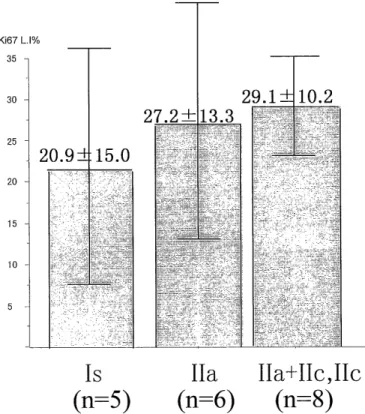
Ki67 was showed in Figure 2. Ki67 LI was 20.9%
for type Is, 27.2% for type IIa and 29.1% for type IIa+IIc plus IIc. Ki67 LI indicated the fraction rate of growing cells was not significant difference between
CD44index
55.5-1-12.2
70
47.1 ± 12.4
60
43.0±9.5
50
40
30
20
10
Is IIa IIa+IIc, IIc
(n=5) (n=6) (n=8)
Figure 1. CD44 LI in three types of early cancers. A tendency of high CD44 expression was presented in flat- elevated-with-depressed and flat-with-depressed tumors of type IIa+IIc plus IIc. (p=0.08)
Ki67 L 1%
35
30 2 29.1+-10. -2
7.2±13.3 == =~=_ -=
25
20.9 15.0
20 = - _
15
10
Is IIa IIa+IIc, IIc
(n=5) (n=6) (n=8)
Figure 2. Ki67 LI in three types of early cancers. High
activity of cell growth was not presented among 3
tumor types.
three types. For detailed analysis, double staining of Ki67 and AgNORs were perfomed for estimatiom of proliferative activity in non-growing and growing cells. A microphotography by double staining method was showed in Figure 3. Many dot spots of AgNORs consisted in Ki67 positive cells. AgNORs counts in Ki67 positive cells were showed in Figure 4. AgNORs counts were 8.5 dots for type Is, 7.7 dots for type IIa
and 10.7 dots for type IIa+IIc plus 11c. AgNORs in Ki67 positive cells showed more dot spots in flat- elevated-with-depressed plus flat-with-depressed can- cers (IIa +IIc pluse IIc) than that in elevated or flat- elevated tumors. (p<0.05)
AgNORs counts in Ki67 negative cells were showed in Figure 5. AgNORs counts was 3.2 dots for type Is, 2.8 dots for type IIa and 3.2 dots for type IIa+IIc plus IIc.The result of a staining for AgNORs number in Ki67-negative cells showed a similar the result of Ki67 LI by single staining, there was not significant differ- ence.
Figure 3. A microphotography of a flat type carcinoma (IIa+IIc) by Ki67-AgNORs double staining method. Several nuclei was stained Ki67 with fast-red. Ki67 positive cells contains many dot spots of AgNORs.
AgNOR dot
p<0.05
count/nuclei 14
12 10
.7 2.6
10 8.5 2.3
7.7 1.4
8 4
2
Is IIa IIa+IIc, IIc
(n=5) (n=6) (n=8)
AgNOR dot count/nuclei
14
12
10
8
6
3.2-!-0.8 2.8±0.4 3.2±0.7
4 2
Is IIa IIa+IIc, IIc
(n=5) (n=6) (n=8)
Figure 4. AgNORs counts in Ki67 positive cells. The high- est value was presented in flat-elevated-with-depressed and flat-with-depressed tumors of type IIa+IIc plus IIc.
(p=0.02)
Figure 5. AgNORs number in Ki67 negative cells. The proliferative activity was not presented among 3 tumor types.
Discussion
The adenoma-carcinoma sequence theory that ex- plains the natural history of colon cancers was first proposed in 1986 by Vogelstein et al.10' In this theory, intra-mucosal adenomas undergo cellular changes and transforms to elevated adenomas, which then grow as a polyp as well, invaded the submucosa, to become ad- vanced cancer."' However, this theory did not explain the transformation of elevated carcinoma to depressed advanced cancer. Furthermore, the recent recognition of flat type colorectal cancer had also led a number of in- vestigators to suggest that the adenoma-adenocarcinoma sequence theory did not explain this type of tumors,
and proposed the "de novo" theory."' In this theory, small lesions might become advanced cancers directly without transformation to adenomotous polyp.
Recent advances in large bowel endoscopic techniques have allowed easy diagnosis of flat and depressed colorectal
tumors that are smaller than lcm in diameter. These le- sions were locally invasive through the submucosa as well as distant metastasis, without increasing in size, in sharp contrast to the adenocarcinomatous polyps."'
Several types of classifications have been used to de- scribe these flat-elevated and flat-depressed lesions.
These were based on either the horizontal development of tumor glands or the height of the mucosa, prolifera- tion of tumor glands, macroscopic configuration or mi- croscopic findings."', 15)
Author used CD44 variant 6 to examine the inva- siveness of the flat type tumors. CD44 received modifi- cation of glucose chain from exon more than 20 with selective splicing mechanism of ten exon of the inter- nal with cap formation transfix type web glucose pro-
tein at least, and various isoforms exists. As biological activity, it was the ligand of hyaluronic acid, type I, type IV collagen, fibronectin and had action of adhe- sion factor between cell matrix between contiguity cell, and was suggested the lymphocyte homing recep- tor.") Expression of CD44 had been detected in several malignant tumors and its level in cancer cells corre- lates with infiltration and metastasis.") Furthermore, CD44V6 had been detected in breast cancer and corre- late with the invasiveness in gastric cancers."),"' In co-
lorectal tumors, previous studies had shown the ex- pression of CD44 variants, including CD44 v2-10 in carcinomas of the colon over a wide area, including
cancer cells, adenomas, normal mucosal goblet cells.20' However, high levels of expression of CD44v6 had been detected in advanced cancers, and correlated with Dukes histological stage. Expression of CD44v6 have also been described in early flat type cancer. In the present study, author determined the expression of CD44v6 to examine the invasiveness of early carci- noma of colon and rectum. Authors' results showed a
tendency of high labeling index of CD44 in flat- elevated-with-depressed plus flat-with-depressed tumors compared with the elevated or flat-elevated tumors.
These results might indicate that the flat-elevated- with-depressed and flat-with-depressed cancers were potentially more invasive than two other types.
Ki67 is nucleoprotein that appears during the G2/M phase from G1 phase in the cell cycle. Ki67 is
used a marker of growing cells in cell population.
Additionally, the proportion of Ki67-positive cells in cancer tissue correlated with malignant potential and was considered as a manifestation of invasiveness and
metastasis .21' However, in this study of Ki67 single staining, there was not statistically significant differ- ence among three types.
As for AgNORs, argentation does the nuclear body build-up area which considered of ribosomal DNA loop that existed in the short arm of acrocentric chromo- some. Ribosomal DNA is copied in ribosomal RNA by RNA polymerase 1, and it is thought when participate in growth of cell and nuclear body related to closely, build-up of ribosome and protein synthesis deeply.
And AgNORs becomes one index of cell proliferative activity. In our laboratory, double staining method for Ki67 or PCNA and AgNORs is usually used for the es- timation of malignant behavior in colon and gastric cancers, 22), 23), 24) and this method allow identification of high malignant potential cells for both Ki67 and AgNORs. These AgNORs-Ki67 positive cells were highly proliferative, while AgNORs-Ki67 negative cells showed low proliferative activity. Difference was not looked at during the elevation tumors, flat-elevation tumors, flat-elevatated-with-depressed tumors in label- ing index of Ki67 alone this time. Even fraction rate of growing cell was 20-30% neither focus did not dif- fer. On the other hand, AgNORs with Ki67 positive cell have been many dot spots by the flat-elavated- with-depressed tumors, it was suggested that cell proliferative activity (AgNORs) of growing cell (Ki67 ) itself in flat-elavated-with-depressed-tumor was signifi- cantly higher than in another early tumors, espesially flat elevated tumor.
If one may be allowed to wish so much, these re- sults may be discussed malignancy of tumor with Ki67 -AgNORs double staining, it is useful to appreci- ate cell proliferative activity in growing cell of early colorectal cancer.
Acknowledgement
The author wishes to express his sincere gratitude to Prof. Hiroyoshi Ayabe, The First Department of Surgery, Nagasaki School of Medicine. And author wishes to thank to Prof. Yutaka Tagawa, The School of Allied Medical Sciences, Nagasaki University for the kind guidance in the study and review of paper.
References
1 ) Kuramoto S, Oohara T: Flat early cancers of the large intestine.
Cancer 64: 950-955, 1989.
2) Tada S, Yao T, lida M, Koga H, Hizawa K, Fujishima M: A clinicopathologic study of small flat colorectal carcinoma. Cancer
74: 2430-2435, 1994.
3) Minamoto T, Sawaguchi K, Ohta T, Itoh T, Mai M:Superficial type
adenomas and adenocarcinomas of the colon and rectum: A com- parative study. Gastroenterology 106: 1436-1443, 1994.
4) Herrlich P, Pals S, Ponta H : CD44 in colon cancer. Eur J Cancer 31A: 1110-1112, 1995.
5) Wielenga VJ, Heider KH, Offerhaus GJ, et al: Expression of CD44 variant proteins in human colorectal cancer is related to tumor
progression. Cancer Res 53: 4754-6, 1993.
6) Mulder JWR, Krupt PM, Sewnath M et al: Colorectal cancer progress and expression exon-v6-containing CD44 proteins. Lancet
344: 1470-1472, 1996.
7) Nishizawa-Takano JE, Ayabe H, Hatano K, Yamaguchi Y, Tagawa Y: Gall bladder cancer. A comparative study among clinicopathologic
features, AgNORs, and DNA content analysis. Dig Dis Sci 41: 840- 847, 1996.
8) General Rules for Clinical and Pathological Studies on Cancer of the Colon, Rectum and Anus: Japanese Reserch Society for Cancer
of the Colon and Rectum The 5th edition, April 1994.
9) Ploton D, Menager M, Jeannesson P, Adnet JJ: Improvement in the staining and in the visualization of the nucleolar organizer regions
at the optical level. Histochemical 18: 5-14,1986.
10) Vogelstein B, Fearon ER, Hamilton SR, et al: Genetic alternation during colorectal tumor development. New Engl J Med 319: 525-
532, 1988.
11) Nakamura K: Adenoma and carcinoma of the colorectum: Histological criteria of carcinoma discriminating from adenoma, histogenesis
and growth process. Surgical Therapy (Jpn) 74: 159-172, 1996.
12) Nagasako K, Oka E, Iwamoto Y, et al: Colon cancer: growth from superficial early cancer into advanced cancer, proposal of the con-
cept of horizontal growing tumor. Stomach and Intestine 30: 165-
172, 1995.
13) Shimoda T, Ikegami M, Kurusu Y, Ochiai J, Nakanishi Y: Pathological features of early colorectal carcinoma originating from superficial
type. Stomach and Intestine 30: 141-147, 1995.
14) Kudo S, Nakajima T, Hishita N, linuma G, Takagi A, Shibata Y:
Macroscopic classification of minute colorectal cancer. Stomach
and Intestine 29: 27-36, 1994.
15) Shimoda T, Ikegami M, Tanaka T, Fujitani M, Maenou K:
Superficial colorectal tumor: Its macroscopic classification and
clinicopothological issues with special reference to minute carci-
noma. Stomach and Intestine 29: 19-26, 1994.
16) Berg EL, Goigstein LA, Jutila MA, et al: Homing receptors and vas- cular adheisions. Immunol Rev Apr 108: 5-18, 1989.
17) Goi T,Yamaguchi A, Nakagawa G, Furukawa K, Shiku H: The role of CD44 adhesion molecules. Nippon Rinsho 53: 1688-93, 1995.
18) Kaufmann M, Heider KH, Sinn HP, Ponta H, Herrlich P: CD44 isoforms in prognosis of breast cancer. Lancet 346: 502, 1995.
19) Heider KH, Dammrich J, Skroch AP,et al: Differential expression of CD44 sprice variants in intestinal and diffuse type human gastric
carcinomas and normal gastric mucosa. Cancer Res 53: 4197-203,
1993.
20) Gotley DC, Fawctt J, Walsh MD, et al: Alternatively spliced vari- ants of the cell adhesion molecule CD44 and tumour progression
in colorectal cancer. Br J Cancer 74: 342-351, 1996.
21) Gerdes J, Lemke H, Baisch H, Wacker H, Schwab U, Stain H: Cell cycle analysis of a cell proliferation associated human nuclear anti-
gen defined by the monoclonal antiboby Ki67. J Immunol 133:
1710-1715, 1984.
22) Yoshida K : A study of the sequential demonstration of nucleolar organizer regions and PCNA immunolabelling in colorectal carcino-
mas. Acta Med nagasaki 38: 15-20, 1993.
23) Matsuo T: Studies on the utility of sequential stainning technique using PCNA and AgNORs for assessing the degree of malignancy
of gastric carcinoma. Acta Med Nagasaki 38: 226-231, 1993.
24) Sawai T: A case of advanced colon cancer meassuring 8mm in di- ameter. Jpn J Colono-proctology 49: 323-326, 1996.