Acta med. Nagasaki. 125-139
Experimental Glomerulonephritis in Rats Produced with Soluble Antigen-Antibody Complexes after
Pretreatment with Aminonucleoside
Susumu MATSUI*
Department of Pathology, Research Institute for Nuclear Medicine,
Nagasaki University School of Medicine, Nagasaki, Japan
Received for publication, January 25, 1969
A preliminary report of this work was presented at the Japanese Society of Nephrology Meeting, September 15, 1967.
The pathogenesis of human glomerulonephritis has not been throughly recognized in spite of many extensive experimental examinations and clin- ical observations. With regard to experimental glomerulonephritis, MASUGI's nephritis and glomerulonephritis in serum sickness has been believed to play an important role. In serum sickness, glomerulonephritis develops during the immune phase of antigen elimination, and antigen-antibody complexes play an important role in the pathogenesis of glomerulonephritis and vas- cular lesions. It has been also reported that an aminonucleoside, 6-dimethy- lamino, 3-amino-d-ribose, increased permeability of the glomerular basement membrane for protein. In the present study pretreated rats with aminonu- cleoside received smaller amount of soluble antigen-antibody complexes in antigen excess which were produced in vitro between BSA anti-BSA rat
serum or human γ‑gl. anti‑human γ‑gl. rabbit serum. Glomerulonephritis developed in a short time after administration of immune complexes.
INTRODUCTION
The pathogenesis of experimetal serum sickness has been extensi- vely investigated since RICH and GREGORY described the characteristic lesions, glomerulonephritis, arteritis and endocarditis.1)2)3)4)
GERMUTH5) showed, by careful analysis of the immunologic events as related to the histologic alterations, that the lesions developed dur- ing the immune phase of antigen elimination and regressed when free antigen appeared in the circulation. The immune phase of antigen elimination was of short duration and was associated with presence of soluble antigen-antibody complexes in the circulation. The important role of these antigen-antibody complexes in the pathogenesis of a cer- tain hypersensitive state has only recently been recognized. It has been
*At# A
shown that they can produce anaphylaxis in guinea pigs6)') and mice,), contraction of isolated guinea pig smooth muscle 9) and inflammatory changes in the skin.10) More direct evidence for the role of soluble antigen-antibody complexes in the pathogenesis of serum sickness was produced by the fact that the characteristic lesions of serum sickness were produced by intravenous injection of a large amount of soluble antigen-antibody complexes in normal mice. 11)12)
Recently an experimental disease resembling human nephrosis has been produced in rats by the subcutaneous injection of an amino- nucleoside. 13)14)
During the early stage of the disorder by aminonucleoside, no dete- ctable glomerular lesions can be seen under light microscopy.
In the present study, experimental glomerulonephritis was produced passively in rats by the intravenous injection of a smaller amount of soluble antigen-antibody complexes than has been used in previous experiments within 7 days after the previous subcutaneous injection of aminonucleoside.
MATERIAL and METHOD
Animals: Male rats of the Wister strain with initial body weight of 100 to 150 gm. were used. They were given free access to a commer- cial pellet diet (MF Oriental Yeast Mfg., Tokyo) and tap water.
Antigens: Human ;r-globulin (H . G . G .) were obtained from Midori Ju ji Co., Osaka, and crystallized bovine serum albumin (BSA) from Armour Pharmaceutical Co., Kankakkee Illinois, USA.
Antiserums: These were produced in white male rabbits weighing 2kg.
against H . G . G . , and in Wister male rats weighing 100 gm. against BSA. Each rabbit received multiple subcutaneous injections of 50 mg.
of H . G . G . incorporated with complete Freund' s ad juvant (latron Labor- atories). Four months after this they were given a booster injection of
antigen. The rats were immunized against BSA (10 mg. of antigen) by the same method and were given a booster injection of antigen four weeks after the multiple injections. Both the rabbits and the rats were bled on the 7th day after the booster injection.
The protein level of the samples was measured using a hand protein refractometer (Hitachi). Each antiserum sample was distilled up to one per cent with isotonic saline solution.
Soluble complexes: The equivalent point was examined by DEEN-WEBB'S method. Four times excess antigen, each antigen containing 10 mg. of protein in one ml., was added to the antiserum. No precipitation was found. The concentration of antigen and antibody protein was stand- ardized at 10 mg. in 1 ml., so that 1 ml. contained 10 mg. of soludle complexes.
Aminonucleoside (AN) : This was obtained from Nutritional Biochemi-
cals Corporation, Cleveland, Ohio. Rats received a daily subcutaneous injection of AN, (0.3 ml. of 0.5 per cent solution per 100 gm. of body weight).
Urinary protein: This was estimated by dipping the end of an Albustix and comparing the color with six color standards representing the following concentration of urinary protein; 0(-), tr, 30(+), 100 (+ +), 300(+ + +) and over 1000 mg. (+ + + +) protein per 100 ml.
of urine. Some of the urinary specimens were pooled over night for immunoelectrophoretic study.
Immunoelectrophoretic study: First, zone electrophoresis of the antigens (urine specimen and serum) was carried out for a period of 50 minutes in veronal buffer (pH 8.6, ,u=0.06) using a current of 0.8 milli Amp.
per cm. width of cellulose acetate filter paper. Strips of filter paper with a standard length of 6 cm. and viriable width of 1 or 2 cm. were used. Antibody (anti-rat serum rabbit antibody and anti-BSA rabbit antibody) was placed on the empty half of the acetate filter paper along with the antigen, and then the paper was placed in liquid paraffin for 24 hours. The reaction between antigen and antibody was studied
using Panceau 3R in staining.
Table I describes the number of experimental rats and the experi- mental procedure followed in each of the three main groups and also in the subgroups.
The experimental animals, as a rule, were sacrificed by bleeding from the abdominal aorta, after being anesthelized with ether. Some of these were used for perfusion fixation with isotonic 1 per cent glu-
Table I
Number Total Does Interval Time of Sacri-
Number of AN of Sol. between fice after the Experimental Urinary Protein Group of Rats Inj.* Complexes Inj. Last Inj. Period at Sacrifice
j 3 7 - - 1 day 8 days (+)-(++)
I[ (1)** 12 0 3 ml. 12 hrs. 3 days 4~2 days (.'_)-(+)
(2)*** 6 0 3 ml. 12 hrs. 3 days 4.2 days (-)-(+)
(1)** a 10 1-3 3 ml. 12 hrs. 3 days 5~2- 732 days +)
b 7 4-5 3 ml. 12 hrs. 3 days 8~2- 9~2 days (+)-(++)
c 11 6-7 3 ml. 12 hrs. 3 days 10.2-112 days (++++)
d 2 7 7 ml. 3 days 3 days 31 days (+ + +) -
(2)***a 5 7 3 ml. 12 hrs. 3 days 11j. days (+++)-
b 2 7 3 ml. 12 hrs. 7 days 1532 days (+++)-
*Rats recived daily subcutaneous injections of aminonucleoside (AN) 0 .3 ml. of 0.5%
solution per 100 gm. of body weight.
**Rats in Group II (1) and Group III (1) received homologous soluble complexes .
***Rats in group II (2) and Group III (2) received heterologous soluble complexes ,
talaldeheide.
Tissue: The kidney and other organs were sectioned after measuring weight and fixed in neutralized formalin and Zenker's solution. Some of the rats were fixed using a perfusion technique with 1 per cent glutalaldeheide. H.E., PAS, and PAM were used for staining.
RESULTS
Urinary protein: Protein was either absent or present in only trace amounts in the urine of normal rats. The urinary protein excretion of the rats which was treated with AN for 1 to 3 days, indicated (-) to (±), for 4 to 5 days, (±) to (+) and for 6 to 7 days indicated (+) to (++). The rats which received injections of heterologous or hom- ologous soluble complexes after treatment with AN for 6 to 7 days showed a rapid increase in urinary protein on the third day after the last injection of soluble complexes. (Fig. 1) However, the animals
which received injections of soluble complexes after treatment with AN for 5 days had the same urinary protein content as the animals treated only with AN. Rats which received soluble complexes alone excreated mostly urine with a low protein content.
Organ weight per 100 gm. of body weight: The weights of the kid- neys and hearts of the 9 rats in group II were measured. The mean weight of the kidneys was 0.87 gm. and that of the hearts was 0.45 gm. In group III (1) a, the mean weight of the kidneys was 0.81 gm.
and that of the hearts was 0.35 gm.; in group III (1) b, the mean
weight of the kidneys was 0.90 gm. and that of the hearts was 0.46
gm.; and in group III (1) c, the mean weight of the kidneys was 1.05
gm. and that of the hearts was 0.50 gm. The mean weights of the
kidneys and hearts in group III (1) c were significantly heavier than
the mean weights in other groups.
Immunoerectrophoretic result: BSA was noted in all sera of group III (1) a, b and c. The urine specimens of group III (1) c showed a high protein content. In addition, the presence of BSA was demon- strated immunologically, and multiple reaction lines were noted between the anti-rat serum rabbit antibody and urine specimen (Fig 2). How- ever, in the urine of group III (1) a and b indicating low grade prote- inuria BSA was not demonstrated, and only one line was noted between anti-rat serum rabbit antibody and urine specimen (Fig 3).
Light microscopic observation: None of the rats in groups I and II showed any pathologic alterations. The accumulation of granular eosin- ophilic, PSA-positive materials in the cytoplasm of glomerular epithelial cells in AN traeted rats was not different from that of normal, untr- eated rats. (Fig 4). Acute glomerulonephritis developed in all rats of group III (1) c and III (2). The pathologic features of renal changes in these groups were similar and will be described together. All glo- meruli were affected. (Fig 7). The glomeruli were hypercellular with swollen endothelial, epithelial and mesangial cells. The capillary lumens were narrow and relatively bloodless. (Fig 8, 9). Kidney sections ex- amined by a perfusion fixation technique revealed swelling and increase of mesangial ground substance and cells. The glomerular basement membrane appeared thick in the section stained with PAS. Granular PAS-positive materials were more numerous in these rats. (FiglO.) In some glomeruli, abhesion between glomerular tuft and Bowmann's capsule was detected. Lobulation was found frequently. (Fig 9). Protein rich eosinophilic colloidal casts were found in the tubular spaces. In gross specimen, these kidneys were white and large with a fine granular surface, and ascites were noted in four rats. Sections from group III
(1) d indicated sclerosing of the capillary tuft at the vascular pole.
(Fig 11). There werc few pathognomon.ic alterations in the sections from the kidneys of groups III (1) a and b. (Fig 5,6)
DISCUSSION
An experimental lesion resembling human nephrosis has been prod- uced in rats by subcutaneous injection of an aminonucleoside, 6-dime- thylamino, 3-amino-ribose. "114)
Increased urinary protein excretion has been noted in the experi- mental animals by the 5th day after injections of AN and the degree of proteinuria increased progressively, reaching a peak on the 15th day. 151
In this study, rats received daily injections of AN, 0.3 ml. of 0.5
per cent solution per 100 gm. of body weight. Urinary protein was
estimated by dipping the end of an Albustix and comparing the color
with six color standards. By the 3rd day of AN treatment urinary protein
was (±) to (+), by the 7th day (+) to (+ +). These results appeared
to be similar to the observations made by other reporters. When kidney
sections from AN treated rats were examined by light microscopy, no
detectable glomerular lesions were identified in the early and reversible
stage of injury (within 7 days).
According to other studies, electron microscopic examination of the glomeruli from rats treated with AN has revealed definite abnor- malities of the epithelial cells."""
Serial studies of this lesion have indicated that the earliest change was a swelling and coalescence of the foot processes beginning on the 7th day after injection of AN. In addition, large vacuoles containing
granular material, similar in appearance to precipitated protein, were observed inside the cytoplasm of epithelial cells of the nephrotic rats.
HARKIN reported that this granular material was the same as the PAS- positive material found in light microscopic examination. At the present time these materials have been interpreted as morphologic appearsnce of accelerated transport of serum protein. 171
In the present study the rats received intravenous injections of homologous or heterologous soluble antigen-antibody complexes in an- tigen excess by the time they indicated the early postproteinuric state (i.e., increased permeability of protein from glomerular basement membrane). Only minimal morphological changes were found.
Experimental serum sickness has been extensively investigated since RICH & GREGORY, intravenously injected a large amount of horse serum in rabbits. 1)2)3)4)
HAWN et al."' and others"' 20)21)22)23), employing purified antigens, have shown lesions similar to those RICH & GREGORY reported prolife- rative glomerulonephritis and lesions similar to those seen in polyar- teritis nodosa and rheumatic disease.
GERMUTH et al., interpreting the immunologic events5' as related to morphologic alterations, stated that the lesions developed during the immune phase of antigen elimination and that the morphologic lesions of serum sickness were the result of an antigen-antibody reaction. It has been repeatedly demonstrated that this reaction occurred as the host began to form antibody while antigen was still in circulatcon and in the tissues. In these observations, it has been apparent that the develo- pment of serum sickness occurs at the time of antigen-antibody interac- tion. More direct support for the role of soluble antigen-antibody compl- exes in the pathogenesis of serum sickness was supplied by the study Of MCCLUSKY et al. 11)1.2) , which showed that the intravenous injections of large amount of soluble antigen-antibody complexes produced the characteristic lesions of serum sickness in normal mice. They explained how these complexes were arrested in the tissue affected by the serum sickness, that antigen-antibody complexes had been in circulation as unstable colloidal particles, that they were readily phagocytized by the cells of the reticuloendothelial system and such colloidal particles could be localized in glomeruli and vascular endothelium in various organs
under the influence of agents such as histamine, knov.-n to be released
by antigen-antibody complexes, For these reasons it was postulated
that in serum sickness antigen-antibody complexes produced changes in the endothelium of glomeruli and blood vessels by releasing subs- tances such as histamine. The alteration thus induced leads to the arrest of the complexes in these sites and, in turn, to the tissue damage.
MCCLUSKY reported that it required at least 12.5 mg. antibody protein per 100 gm. of body weight to produce the lesions of serum sickness.
ISHIZAKA et al. , GERMUTH et al. al. and TREANIN to al. reported that soluble antigen-antibody complexes in antigen excess have biological activity which does not have antigen or antibody alone, but also inclu- des heavy (-Ag3 Ab2)24' and light (Ag2 Ab) complexes, the former being more active biologically. 25'
Antigen excess immune complexes prepared from heterologous or homologous antibody have produced (1) anaphylactic shock when injected into normal unsensitized gunea pigs6"' and mice, 8' (2) contraction of isolated gunea pig smooth muscles, 9' and (3) passive skin reactions in gunea pigs.10' Large, multiple doses cause transient acute proliferative glomerulonephritis in mice. 111121
MCCLUSKY et al.26' produced experemental glomerulonephritis in mice employing two kinds of soluble complexes, crystallized ovalbumin anti-ovalbumin rabbit antiserum system and crystallized bovine serum albumin .. anti-bovine serum albumin rabbit antiserum system, They stated that the former was more effective for development of the dis- ease than the latter. They concluded that the difference in excretion of these two antigen might be a contributory factor in the greater severity of the disease observed with the ovalbumin anti-ovalbumin rabbit serum system.
C9ONS et al. intravenously injected mice with 3 kinds of protein (ovalbumin, bovine serum albumin, and human globulin) and obser- ved that the disappearance of ovalbumin from the kidney was fast and that of human ~--globulin was Slow. 27' Their differing rates of disappe- arance are partly due to the relative ease with which they pass through the glomerular filter.28'
In consideration of these events and the biological activity of imm- une soluble complexes, it appears that soluble complexes of low mole- cular systems are more complex at the site of glommerular filter, and thus, biologically active complexes such as -Ag3Ab2 become numerous in a short time.
On the other hand, it has been considered that the affiniating or combining ability of injected protein with tissues is an important factor contributing to some hypersensitive states.
MILLER produced experimental glomerulonephritis in mice by intra-
venous injection of heterologous (ovalbumin anti-ovalbumin rabbit serum
system) and homologous (ovalbumin anti-ovalbumin mouse serum system"
immune soluble complexes. 29) He reported that the lesions of the mice which received homologous complexes were more severe.
In the present study, rats received homologous or heterologous soluble complexes intravenously by the time they indicated urine with low protein content by aminonucleoside treatment and they were exa- mined histologically and immunologically.
The dose of the soluble complexes injected in this study was stan- dardized at 3 ml. of 10 mg. protein in 1 ml., a total 30 mg. of protein.
This ranged from 1, mg. to 2mg. of antibody protein in terms of anti- body protein per 100gm. of rat body weight. It is not necessary to inject such large amount to produce glomerulonephritis, if the rats had rece- ived aminonucleoside in advance. The glomerular basement membrane has been considered the main filter for filtration of serum protein in the normal kidney. Experimental diseases, such as aminonucleoside nephrosis, increased ferritin uptake in glomerular basement membrane and glomerular epithelial cells.30)
Protein is filtered more rapidly into urine, when the permeability of the glomerular basement membrane is increased by the aminonu- cleoside treatment. In addition to the effect of aminonucleoside, immune soluble complexes may possibly cause an increase in the permeability.
The results of the immunoelectropholetic examination in this study are indirect support for this hypothesis. In urine with a high protein content, the presence of BSA, a component of soluble complexes, was detected immunologically. In addition, multiple reaction lines were found between urine specimens and anti-rat serum rabbit antiserum, but only a few such lines were found between anti-rat antibody and urine speci- mens with a low protein content.
Thus, it is possible that under these circumstances biologically active soluble complexes (-Ag3Ab2) are accumulated in great quantities, in a short time, at the site of glomerular filter membrane.
This hypothesis leads to the consideration that these biologically active complexes play an important role in the pathogenesis of experi- mental glomerulonephritis.
The characteristic findings of acute proliferative glomerulonephritis (hypercellularity, ischemia and swelling of glomeruli), appear to be caused by irritation by biologically active immune complexes, whereas these changes have not been seen in aminonucleoside nephrosis.
It could not be determined by the present study whether soluble complexes themselves or complexes and chemical agents together played a major part in the histogenetic effect. (Chemical agents, such as histamine, are believed to be released by immune complexes)
The role of affinity or combination ability of protein to tissue also needs to be clarified by further study.
In the present experimental disease, I could not find other lesions
such as arteritis or endocarditis which were reported by MCCLUSKY et al. and others. I suspect that smaller amounts of soluble comlexes used for the injections in my experiment may cause this difference.
Comparing the present experimental glomerulonephritis with human poststreptococcal glomerulonephritis, the mechanism in the development of human glomerulonephritis would be as follows: In the first phase, a toxin, such as streptococcal toxin, causes an increase in the perme- ability of glomerular basement membrane. Indirect support for this suggestion is fibrile proteinuria clinically observed. In the second phase, biologically active soluble antigen-antibody complexes, such as -Ag3Ab2 , accumulate in great quantities in a short time at the site of glomerular basement membrane which was damaged in the first phase, and then glomerulonephritic lesions develop with hypercellularity, ischemia and swelling of glomeruli.
This hypothesis appears to be suported by the present experimental findings: Aminonucleoside treatment coincides with the first phase, and injetion of soluble complexes coincides with the second phase.
Diffuse glomerulonephritis can be divided into two main groups31', the nephritic type and the nephrotic type. The nephritic type mainly affects children and young adults and it is commonly a sequela of acute tonsillar or pharyngeal infections with group A streptococci associated with acute onset. Heanaturia, pyuria and hypertension are often associated with acute cases of nephritic glomerulonephritis. Com- plete recovery is common. The glomerular lesions in the acute stage are mainly proliferative, exudative or hemorrhagic and occasionally necrotizing.
The nephrotic type, on the other hand, affects a wider age group and a history of a preceding infection is rarely elicited. The onset is insidious; nephrotic symptoms (protracted proteinuria, edema, ascites, hypoproteinemia and hyperlipemia) are prominent; histologically thi- ckening of basement membrane and lobulation are commonly seen.
However, as ALLEN and others"' emphasized, there are frequent tran- sitions between these two types. Whether the pathogenesis of these two types of glomerulonephritis is the same or not, is an interesting question.
The present experimental glomerulonephritis is a mixed type:
Hypertension (which is suspected because of the increased heart weight), and histologic findings of the kidney (hypercellularity, ische- mia, and swelling of glomeruli), indicate the nephritic type of glom- erulonephritis, but severe proteinuria, ascites (observed in some rats), lobulation, and glomerular basement membrane thickening indicate the nephrotic type of glomerulonephritis.
It has been considered that the autoimmune mechanism plays an
important role in chronic evolution of glomerulonephritis. In this study
chronic evolution of the disease was not discussed,
Two rats which received an injection of soluble complexes every 3 days for a total of 7 injections over a period of 24 days, indicated interesting lesions leading to chronic glomerulonephritis. The present study, however, does not settle the question what role the autoimmune mechanism or repeated irritation of exogenous soluble complexes or both played as a cause of the lesions.
SUMMARY
Experimental glomerulonephritis was produced in rats by admini- stration of aminonucleoside for 6 to 7 days followed by 3 intravenous injections of homologous or heterologous soluble complexes in antigen excess with a 12-hour interval between each injection.
This experimental model supplies several forms of evidence leading to a hypothesis on the pathogenesis of human glomerulonephritis.
(1) Smaller amounts of soluble complexes than reported by others were sufficient to cause glomerulonephritis in rats if previously treated
with amino nucleoside.
(2) Histologically, this type of glomerulonephritis is a mixed type.
(3) Severe proteinuria was noted in this glomerulonephritis, and BSA, a component of soluble complexes, was demonstrated immunologi-
cally in the urine with a high protein content, whereas it was not present or noted in only trace amounts in the urine with a low
protein content.
(4) This appears to be indirect support for the hypothesis that biolo- gically active complexes accumulate in great quantities in a short
time at the site of glomerular filter membrane.
(5) These complexes appear to be an important factor in the develo- pment of characteristic glomerulonephritic changes, hypercellularity,
ischemia and swelling of glomeruli.
(6) The increased permeability of glomerular basement membrane
represents the first phase of the development of glomerulonephritis
in man and irritation of biologically active complexes at the site of the glomerular filtermembrane represent the second phase. The first
phase coincides with AN treatment, and the second phase coincides
with cinjection of soluble complexes as demonstrated in this study.
AKCNOWLEGMENT
The author wishes to express his cordial gratitude to his director, Professor Issei NISHIMORI, who gave constant and king guidance and review during
this study.
REFERENCES:
(1) Rica, A,R, and GREGORY, J.E.: The experimental demonstration that periar-
teritis nodosa in a manifestation of hypersensitivity. Bull. Johns Hopkins Hosp. , 72: 65 (1943)
(2) RICH, A.R. and GREGORY, J.E.: Experimental evidence that lesions with the basic characteristic of rheumatic carditis can result from anaphylactic hypersens-
itivity. Bull. Johns Hopkins Hosp., 73: 239-264 (1943)
(3) RICH A.R. and GREGORY, J.E.: Further experimental cardiac lesions of the rheumatic type produced by anaphylactic hypersensitivity. Bull. Johns Hopkins
Hosp., 75: 115-134 (1944)
(4) GREGORY, J.E. and RICH, A.R.: The exprimental production of anaphylactic
pulmonary lesions with the basic characteristics of rheumatic pneumonitis. Bull.
Johns Hopkins Hosp., 78: 1-12 (1946)
(5) GERMUTH, F.G. Jr.: Comparative histologic and immunologic study in rabbit of induced hypersensitivity of the serum sickness type. J. Exp. Med., 97: 257-
282 (1953)
(6) GERMUTH, F.G.Jr., and MCKINNON, G.E.: Anaphylactic shock induced by soluble antigen-antibody complexes in unsensitized normal guinea pigs. Fed. Proc. ,
16: 413 (1957)
(7) GERMUTH, F.G.Jr., and McKINNON,G.E.: Studies on the biological properties of antigen-antibody complexes. I. Anaphylactic shock induced by soluble antigen-
antibody complexes in unsensitized normal guinea pigs. Bull. Johns Hopkins
Hosp., 101: 13-43 (1957)
(8) TOKUDA, S. and WEISER, R.S.: Anaphylaxis in the mouse produced with soluble complexes of antigen and antibody. Proc. Soc. Exp. Biol. Med., 98:
557-561 (1958)
(9) TRAPANI, I.C., GARVERY, J.S. and CAMPBELL, D.H.: Science, 127: 700 (1957) (10) ISHIZAKA, K. and CAMPBELL. D.H.: Biological activity of soluble antigen-
antibody complexes. I. Skin reactive properties of soluble antigen-antibody comp-
lexes. Proc. Soc. Exp. Biol. Med., 97: 635-638 (1958)
(11) MCCLUSKY R.T. and BENACERRAF, B.: Localization of colloidal substances in vascular endothelium. A mechanism of tissue damage. II. Experimental serum
sickness with acute glomerulonephritis induced passively in mice by antigen-
antibody complex in antigen excess. Am. J. Path., 35: 275-282 (1959)
(12) MILLER, F., BENACERRAF, B., MCCLUSKY, R.T. and POTTER, J.L.: Prod- uction of acute glomerulonephritis in mice with soluble antigen-antibody comp-
lexes prepared from homologous antibody. Proc. Soc. Exp. Biol. Med., 104:
706-709 (1960)
(13) FRENK, S., ANTONOWICZ, I., CRAIG, J.M. and METCOFF, J.: Experimental
nephrotic syndrome induced in rats by aminonucleoside. Renal lesions and body electrolytic composition. Proc. Soc. Ex p Biol. Med., 89: 424 (1955)
(14) FEIGELSON, E.B., DRAKE, J.W. and RECANT, L.: Experimental aminonucleoside
nephrosis in rats. J. Lab. & Clin. Med., 50: 437 (1957)
(15) VERNIER, R.L., PAPERMASTER, B.W. and GOOD, R.A.: Aminonucleoside
nephrosis. I. Electron microscopic study of the renal lesion in rats. J. Exp.
Med., 109: 115-125 (1959)
(16) HARKIN, J.C. and RECANT, L.: Pathogenesis of experimental nephrosis. Electron microscopic observations. Am. J. Path., 36(2): 303-315 (1960)
(17) OIKAWA, Y.: Electron microscopic study of the special reference of protein uptake on the glomerular capallary in normal and diseased kidney. J . J . N. , 5(4) :
359-369 (1963)
(18) HAWN, C.V.Z. and JANEWAY, C.A.: Histological and serological sequences in experimental hypersensitivity. J. Exp. Med., 85: 571-590 (1947)
(19) MORE, R.H. and WAUGH, D.: Diffuse glomerulonephritis produced in rabbit by massive injections of bovine serum gamma globulin. J. Exp. Med., 89: 541-
554 (1949)
(20) MELLORS, R.C., ARIAS-STELLA, J., SIEGCL, M. and PRESMAN, D.: Analytical
pathology. II. Histopathologic demontration of glomerular-localizing antibodies
in experimental glomerulonephritis. Am. J. Path. 31: 687-715 (1955)
(21) GERMUTH, F.G.Jr.: Acomparative histologic and immunologic study in rabbits of induced hypersensitivity of the serum sickness type. J. Exp. Med., 97: 257-
282 (1953)
(22) DIXON, F.J., FELDMAN, J.D. and V. ZGUEZ, J.J.: Experimental glomerulone-
phritis. The pathogenesis of a laboratory model resembling the spectrum of human glomerulonephritis. J. Exp. Med , 113: 899-920 (1961)
(23) DIXON, F.J., V. ZQUEZ, J.J., WEIGLE, W.O. and COCHR• NE, C.G.: Patho- genesis of serum sickness. A.M.A. Arch. Path., 65: 18-28 (1958)
(24) SINGER, S.J. and C MPBELL,D.H.: Physical chemical studies on soluble antigen- antibody complexes. I. The valence of precipitating rabbit antibody. JAm.
Chem. Soc., 74: 1794-1802 (1952)
(25) ISHIZAKA, K.: Gamma globulin and molecular mechanisms in hypersensitivity
reactions. Progr. Allergy, 7: 32-106 (1963)
(26) MCCLUSKY, R.T., BENACERR F, B., POTTER J.L. and MILLER, F.: The pathologic effect of intravenously adrninustrated soluble antigen-antibody com-
plexes. I. Passive serum sickness in mice. J. Exp. Med., III: 181-193 (1960) (27) COONS, A.H., LEDUC, E.H. and KAPL N, M.H.: Localization of antigen in
tissue cells. VI. The fate of injected foreign proteins in the mouse. J. Exp.
Med., 93: 173 (1951)
(28) P A PPENHEIMER, J.R.: Renal function (3 conference), Josiah Macy Found, (1951) (29) MILLER, F., BENACERR F, B., MCCLUSKY, R.T. and POTTER, J.L.: Prod-
uction of acute glomerulonephritis in mice with soluble antigen-antibody comp-
lexes prepared from homologous antibody. Proc. Soc. Exp. Biol. Med., 104: 706
(1960)
(30) JONES, D.B.: Inflammatory and vascular disease of the glomerulus. In analy- tical pathology, ed. by Mellors, R.L. Pp 161-217, New York, McGraw-Hill
Book Co. Ind. 1'957
(31) ALLEN A.C.: The clinicopathologic meaning of the nephrotic syndrome. Ame.
J. Med., 18:277-314 (1955)
(32) ALLEN A.C.: The kidney. Medical and surgical disease, 2nd ed., pp 151-323
New York, Grune and Stration, Inc., 1962.
Fig. 2: Photograph (left) and diagram (right) of immunoelectropholesis of urine specimen indicating severe proteinuria. They show four reaction lines between
anti-rat serum rabbit antibody and one line between anti-BSA rabbit antibody.
Fig. 3: Photograph (left) and diagram (right) of immunoelectropholesis of urine specimen indicating low grade protein urea. They show only one line between
the urine specimen and rabbit serum containing anti-rat antibody.
Fig. 4: Kidney of a rat which was treated
with AN for 7 days. Glomeruli are
almost normal. PAS stain, low magni-
fication.
Fig. 5: Glomerulus of a rat which received ho- mologous soluble complexes every 12 hours for 3 injections. No detectable
change is seen. Perfusion fixation with
glutalaldeheide, PAS stain, high ma-
gnification.
Fig. 6: Glomerulus from a rat which received
homologous complexes after AN trea-
tment for 2 days. It shows almost
normal structure. Perfusion fixation,
PAS stain, high magnification.
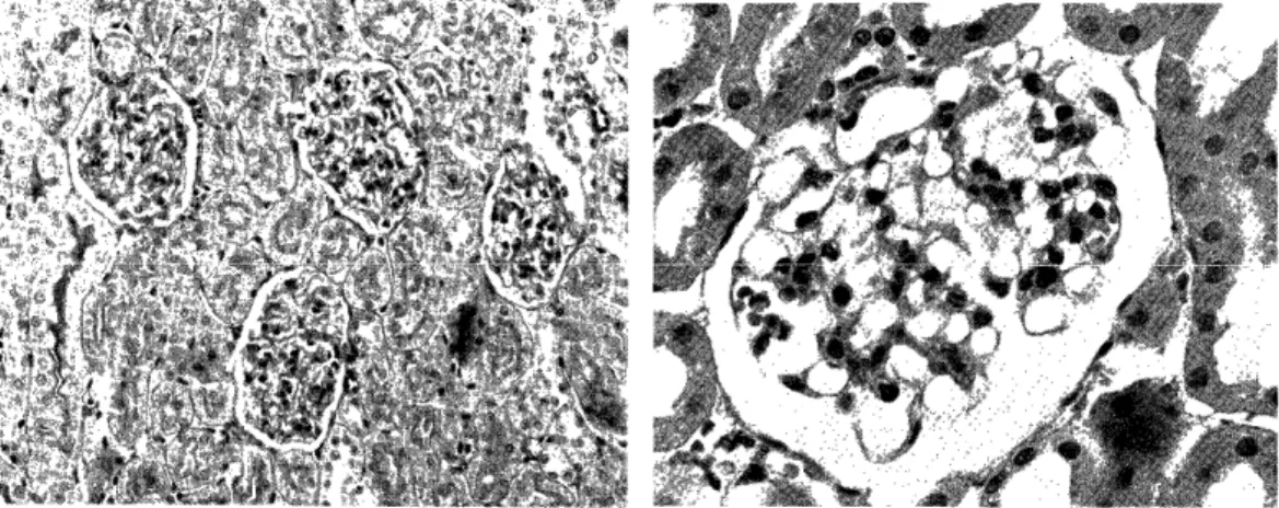
Fig. 7: Kidney from a rat which received
heterologous complexes after AN treat-
ment for 7 days. Swelling of glome-
ruli, hypercellularity and ischemia ap-
pear. These lesions are noted in all
glomeruli. PAS stain, low magnisi-
cation.
Fig. 8: Glomerulus from a rat which received
homologous complexes after AN treat-
ment for 7 days. It shows hypercellular,
relatively bloodless glorrierulus. PAS stain, high magnification.
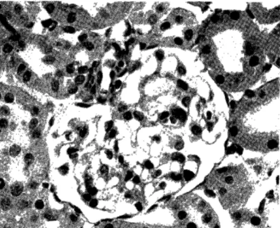
Fig. 9: Photomicrography from same section
illustrated Fig. 7. Hypercellularity,
relatively bloodless, in addition, lobu- lation is observed in this glomerulus.
PAS stain, high magnification.
Fig. 10: Glomerulus from a rat which received
homologous complexes after AN trea-
tment for 6 days. Adhesion between
loops, thickening of basement membrane
is seen. Perfusion fixation, PAS stain,
high magnification.