ヒ トの心臓 は主 として左 側 の要素 か ら成 る
左 上大 静脈 の解 剖 に基づ く考 察
岡 山 大 学 医 学 部 第 二 解 剖 学 教 室(主 任:村 上 宅 郎 教 授)
村 上 宅 郎
(平 成8年7月5日 受 稿)
Key words: Human heart, coronary arteries, sinus coronarius, conductive system, left superior vena cava
緒 言 動 物 の 体 は 上 下,前 後,内 外,左 右 な どの 区 別 が で き る.体 内 の 臓 器 で も 同様 の 区 別,区 分 が で き る. 腸や 心 臓 で も左 右 両 側 の 神 経 支 配 を うけ,ま た 発 生 的 に左 右 の 原 基 の癒 合 を 出 発 点 と し て い るの で,左 と右 の 要 素 か ら構 成 さ れ て い る こ と は 明 らか で あ る.こ の 観 点 か ら,私 達 は ヒ トの 腹 部 内臓 の動 脈 系 に つ い て 類 型 学 的 に 上 下,左 右 の 区 分 け を 試 み た1). 本稿 で は ヒ トの 左 右 上 大 静 脈 の 共 存 例 を報 告 し,心 臓 に お け る左 側 優 位 性 を論 じ る. 材 料 と 方 法 平 成3年 度 の 岡 山 大 学 医 学 部 人 体 解 剖 実 習 で, 89歳 の 男 性 ご遺 体(脳 出 血 で 死 亡)に 左 と 右 の 上 大 静 脈 の 共 存 例 に 遭 遇 した.本 例 を調 査 し,心 臓 の 血 管 を調 べ た. 結 果 1. 右 の 上 大 静 脈 右 側 の 上 大 静 脈 は正 常 に 存 在 し,左 と右 の 腕 頭 静 脈 を うけ て右 心 房 に 注 い で い た.右 側 の 下 大 静脈 も正 常 で,異 常 な 左 側 の 下 大 静 脈 は 認 め な か っ た. 2. 左 の 上 大 静 脈(Fig. 1)
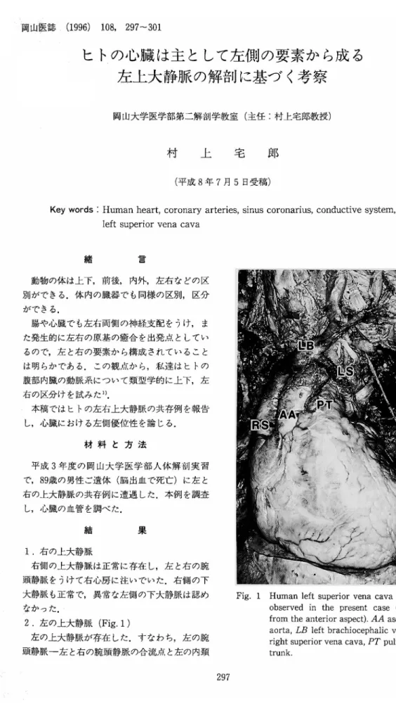
Fig. 1•@ Human left superior vena cava (LS) as observed in the present case (viewed
from the anterior aspect). AA ascending aorta, LB left brachiocephalic vein, RS
right superior vena cava, PT pulmonary trunk.
左 の上大静 脈 が存在 した.す な わち,左 の腕
頭静脈-左
と右 の腕 頭 静脈 の合 流点 と左 の内頚
静 脈 の 起 始 部 との 中 間 点-か ら発 達 した一 本 の 静 脈(左 の 上 大 静 脈)が 起 こ っ て い た.こ の 静 脈 は 左 の 腕 頭 静 脈 の 約1/2の 太 さ を も ち,上 行 大 動 脈 と肺 動 脈 幹 の 前,左 肺 静 脈 の 下 で左 心 房 の う し ろ を通 っ て冠 状 静 脈(洞)を うけ 右 心 房 に 注 い で い た.こ の 静 脈 は,左 肺 静 脈 の 下 で, 後 部 体 壁 を上 行 す る一 本 の 静 脈(半 奇 静 脈)を うけ て い た. 3. 心 臓 の 静 脈 a) 冠 状 静 脈(洞):大 心 静 脈,左 心 室 後 静 脈, 中心 静 脈,小 心 静 脈 を うけ,上 述 の 如 く左 上 大 静 脈 を介 し て右 心 房 に 注 い で い た. b) 前 心 静 脈: 3本 存 在 し,右 心 室 前 面 を上 行 し右 心 房 に 直 接 注 い で い た. c) 細 小 心 静 脈:小 さ い 静 脈 孔 が 右 心 房 中 隔 壁 に2つ,他 の 心 房 壁 に3つ 認 め ら れ た. 4. 心 臓 の動 脈 正 常 に 左 と右 の 冠 状 動 脈 が 存 在 し,左 冠 状 動 脈 が 前 室 間 枝 と回 旋 枝 を,右 冠 状 動 脈 が 後 室 間 枝 を分 枝 して い た. 5. 心 臓 の 神 経 左 右 両 側 に お い て,迷 走 神 経 の 上,下 心 臓 枝 が 確 認 で き,交 感 神 経 節 か ら の 上,中,下 心 臓 神 経 も確 認 で き た.
考
察
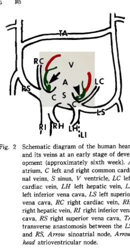
発 生 の 初 期 に 左 右 の 上 大 静 脈(総 主 静 脈)が 出 現 す る が,左 が 退 縮 して 冠 状 静 脈 洞 に 注 ぐ細 い 左 心 房 斜 静 脈 と な る.一 方,こ の 左 上 大 静 脈 が よ く発 達 し て 残 る こ とが あ り, Adachi2)を は じめ 多 くの 報 告 例 が あ る. 本 稿 は左 の 上 大 静 脈 の残 存 例 の1型 を紹 介 し, 冠 状 静 脈(洞)が 左 の 上 大 静 脈 に 注 ぐこ と を確 認 し た.こ の こ と は心 臓 の 主 静 脈 で あ る冠 状 静 脈 洞 と こ の 洞 に 注 ぐ静 脈 群 は左 側 の 静 脈 系 に 属 す る こ と を 明 瞭 に 示 す も の で あ る. 本 稿 は 前 心 静 脈 が 直 接 右 心 房 に 開 口 す る こ と も確 認 し た.私 達 は, Fig. 2とFig. 3に 示 す よ う に,前 心 静 脈 は 総 主 静 脈(上 大 静 脈)の 起 部 が 静 脈 洞 と共 に 心 房 に 取 り込 まれ る際 に,同 時 に 心 房 に 取 り込 まれ た静 脈 で あ る と考 え て い る. す な わ ち,前 心 静 脈 は,左 側 の 冠 状 静 脈 洞 と こ の 洞 に 注 ぐ静 脈 群(大 心 静 脈,左 心 室 後 静 脈, 中 心 静 脈,小 心 静 脈)に 相 当 す る,右 側 の 静 脈 群 で あ る. Fig. 2とFig. 3は 私 達 の 未 発 表 の 人 胎 児 連 続 切 片 標 本 と成 書3)を 参 考 に し て 描 い た.Fig. 2•@ Schematic diagram of the human heart and its veins at an early stage of devel opment (approximately sixth week). A atrium, C left and right common cardi nal veins, S sinus, V ventricle, LC left cardiac vein, LH left hepatic vein, LI left inferior vena cava, LS left superior vena cava, RC right cardiac vein, RH right hepatic vein, RI right inferior vena cava, RS right superior vena cava, TA transverse anastomosis between the LS and RS, Arrow sinoatrial node, Arrow
head atrioventricular node.
細 小 心 静 脈 も,前 心 静 脈 と 同 様 に,心 房 に 取 り込 まれ た 静 脈 群 と考 え られ,冠 状 静 脈 洞近 辺 あ る い は 心 房 中 隔 の 細 小 心 静 脈 は左 側 に,他 は 右 側 に 属 す る と思 わ れ る. 心 筋 は 自動 能 を 有 し,そ の 第 一 次 中 枢(洞 房 結 節)は 右 の 上 大 静 脈 開 口部 に,第 二 次 中枢(房 室 結 節)は 冠 状 静 脈 洞(左 の 上 大 静 脈)の 開 口 部 に 存 在 す る.つ ま り,位 置 的 に み る と, Fig. 4 に 示 す よ うに,第 一 次 中 枢 は右 側 に,第 二 次 中 枢 は左 側 に 属 す る(Fig. 2とFig. 3参 照).第 二 次 中 枢 がHis束,右 脚 ・左 脚 と して 左 右 の 心 室 に 下 るの で,心 室 筋 で は 左 側 絶 対 優 位 と い う こ とが で き る.な お,私 達 は 第 一 次 中枢 は 体 循 環 に,第 二 次 中 枢 は肺 循 環 に 帰 属 す る と考 え て い る.そ して,通 常 拍 数 の 多 い 第 一 次 中 枢 が 機 能 的 に上 位 に あ るの は こ の た め(体 循 環 優 先)で
ある と思 っ てい る.
動 脈系 で は前室 間枝 と回旋枝 を分枝 す る左冠
状動 脈 が右 冠状 動 脈 よ り分 布域 が広 く,明 らか
に左 側優位 であ る.神 経 系 の左右 の優 劣 は解析
で きなか った.し か し,自 律 神経 は 血管 に沿 っ
て分 布 す る とい う一般 論 か ら,神 経支 配 で も左
側優 位 とす るこ とが で きる.ま た狭 心症 の痛 み
が左肩 や左 腕 内側 に放 散 す るこ とは よ く知 られ
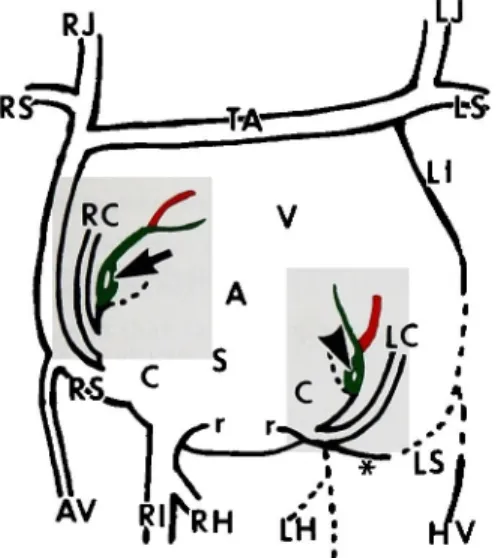
て お り,こ れ は 関 接 的 に 心 臓 の 神 経 支 配 は 左 側 優 位 で あ る こ と を示 し て い る. 以 上 の よ う に,左 右 の観 点 か ら ヒ トの 心 臓 を み る と一 貫 し て左 側 優 位 の 議 論 を展 開 で き る. 最 近,内 臓 の 発 生 を 規 定 す る 因 子[形 質 転 換 成 長 因 子-ア ク チ ビ ン 等 のTGF-β フ ァ ミ リー] の 存 在 が 確 認 さ れ4,5),形 態 形 成 に つ い て新 しい 展 開 が は じ ま っ て い る6∼8).そ して,こ の 因 子 が 私 達 の 類 型 学1)(本 稿 の 左 右 論 を含 む)に ど の よ う に か か わ っ て くる の か 興 味 の あ る と こ ろ で あ る.Fig. 3•@ Further developed form of the human heart and its veins (approximately sev
enth week). The sinus (S) is incorpo rated, together with the original seg ments of the left and right cardinal veins (C), into the atrium (A). Thus, the right superior vena cava (RS), right inferior vena cava (RI) and right cardiac vein
(RC, anterior cardiac veins) drain, as the independent vessels, into the heart. The
left superior vena cava (LS) disappears, but remains as the oblique vein of the
left atrium (*) which drains into the left cardiac vein (LC, sinus coronarius). The
left hepatic vein (LH) also disappears. The TA vessel remains as the left bra chiocephalic vein. The atrioventricular node (arrow) is moved deeply, and locat
ed close to the ventricle. Arrowhead indicates the sinoatrial node. AV
azygos vein, HV hemiazygos vein, LI left superior intercostal vein, LJ and RJ left and right jugular veins, LS and RS left and right subclavian veins, r rem
nants of venous valves.
Fig. 4•@ Schematic diagram of the conductive system in the human heart. The sinoatrial (arrow) and atrioventricular
(arrowhead) nodes are homologous; the sinoatrial node is the right-sided center of the conductive system, the atrio ventricular node being the left-sided center of this system. The bundles of His
(H) and Kent (K) are also homologous. The sinoatrial node with the systemic circulation rhythm acts as the pace
maker, and controls the atrioventricular node with the pulmonary circulation rhythm. L and R letf and right branches of His's atrioventricular bundle. C mus
cle fibers in the terminal crest (Thorel), F muslce fibers in the rims of the oval fossa (Bachmann and Wenckebach). J bundle (muscle fibers) of James, M bun
dle of Maheim.
本 稿 で 紹 介 し た左 上 大 静 脈 の 遺 存 を含 む 血 管 系 の 発 達 や 消 退 な ど一 旦 形 成 さ れ た 形 態 の 退 行 や 消 滅 が 遺 伝 子 が 直 接 関 与 し て お こ る細 胞 群 の ア ポ トー シ ス(Apoptosis)あ る い は 計 画 的 死 (Programmed cell death)に よ る の か,あ る
い は 別 の 機 構(例 え ば 神 経 支 配 あ る い は ホ ル モ ン の コ ン トロー ル な ど を 失 な っ た 結 果 と して の 間 接 的 あ る い は 廃 用 性 細 胞 死)に よ るの か も興 味 の あ る と こ ろ で あ る. な お,体 循 環 系 の 幹 動 脈 群 で は,通 常 左 の 大 動 脈 弓 が 残 存 す る の で,左 側 優 位 で あ る.一 方 同 静 脈 群 で は,上 述 の 如 く通 常 左 側 は 退 縮 す る の で,右 側 優 位 で あ る.つ ま り,正 常 で は,左 側 優 位 の動 脈 系 と右 側 優 位 の 静 脈 系 が 仲(バ ラ ン ス)良 く棲 み わ け て い る こ とに な る.私 達 は Fallotの 四徴 症 等 の 心 臓 の 破 格 は 左 と右 の 原 基 の 優 劣 に 関 係 が あ る と思 っ て い る.す な わ ち, この 四 徴 症 は 右 側 の 原 基 の 関 与(発 達)が 不 十 分 な た め に 起 る と考 え て い る. 結 論 左 上 大 静 脈 の1例 を報 告 し,冠 状 静 脈 洞 は左 側,前 心 静 脈 は 右 側 の 血 管 で あ る と した.さ ら に,刺 激 伝 導 系 の 第 一 次 中枢 は右 側,第 二 次 中 枢 は 左 側 に 属 す る こ と を 述 べ た.左 冠 状 動 脈 が, 右 冠 状 動 脈 よ り,よ く発 達 して い る こ と も確 認 した.以 上 の こ とか ら,心 臓 で は 左 側 優 位 で あ る と結 論 し た. 文 献 1) 村 上 宅 郎,大 塚 愛 二,朴 大 動:ヒ ト腹 腔,左 胃,脾,肝,上 腸 間 膜,下 横 隔 膜 動 脈 群 の 類 型 解 剖 学.岡 山 医 誌(1995) 107, 219-226.
2) Adachi B: V. cava superior; in Das Venensystem der Japaner. Erste Lieferung, Adachi ed, Kenkyu
sha, Tokyo (1933) pp 65-78.
3) Langman J: Cardiovascular
system; in Medical Embryology, Third Asian Edition, Langman ed,
Igagu Shoin, Tokyo (1975) pp 201-257.
4) Asashima M: Mesoderm induction during early amphibian development. Develop Growth and Differ
(1994) 36, 343-355.
5) 浅 島 誠:卵 か ら の 幼 生 へ の 〈形 づ く りの な ぞ 〉;ミ ク ロ ス コ ピ アVol. 12 (No. 4),藤 田 恒 夫 編,考 古 堂 書 店,新 潟(1995) pp 220-232 (4-16).