INVITED PAPER
Special Section on Electronic DisplaysContinuous Liquid Phase Synthesis of Europium and Bismuth Co-Doped Yttrium Vanadate Nanophosphor Using Microwave Heating
Takashi KUNIMOTO†,††a),Member, Yoshiko FUJITA†,andHiroshi OKURA†††,Nonmembers
SUMMARY A continuous flow reactor equipped with a low-loss flow channel and a microwave cavity was developed for synthesizing nanophos- phors. A continuous solution synthesis of YVO4:Eu,Bi nanophosphor was succeeded through the rapid hydrothermal method using this equipment.
Internal quantum efficiency of YVO4:Eu,Bi nanophosphor obtained by 20 minutes microwave heating is about 30% at 320 nm as high as that obtained by 6 hours hydrothermal treatment in autoclave.
key words: nanophosphor, flow reactor, microwave heating, vanadate
1. Introduction
Nanophosphors including quantum dots (QDs) have been extensively investigated during the last two decades due to their potential for display[1]–[5], photovoltaic cells[6]–
[8], bio-labels[9],[10], security[11]and so on. Especially, QDs-utilized light emitting diode (LED) backlights realize a wide color gamut display due to their excitonic narrow emis- sion bands[4]. Another important use of nanophosphors is expected to be an in-vivo bio-sensing application[10]. In any case, a high quantum efficiency (QE) and a size-control are required for nanophosphors in order to utilize in real ap- plications.
Preparation of nanoparticles is generally done by liquid-phase synthses including coprecipitation[12]–[14], colloidal[15], sol-gel[16]–[19], solvothermal[20]–[22], and so on. Nanophosphors are also normally produced via liquid-phase synthses[4], [6], [7], [9], [11],[23]. To ob- tain an efficient nanophosphor, a long time solvothermal synthesis using an autoclave, which leads a batch reaction with low yield and wide distribution of size and physical properties, is commonly used. Microreaction technology has proven beneficial for producing nanoparticles[24]be- cause of homogeneous mixing of source solutions and pH- controllability. This technology has been also applied to producing nanophosphors[25]–[28]. Since microwave syn- thesis has advantages of a very rapid and a homogeneous heating of solution and/or solids, a few minutes synthesis of
Manuscript received February 26, 2016.
Manuscript revised May 31, 2016.
†The authors are with Faculty of Science and Engineering, Tokushima Bunri University, Sanuki-shi, 769–2193 Japan.
††The author is with Center for Advanced Science and Engi- neering, Tokushima Bunri University, Sanuki-ku, 769–2193 Japan.
†††The author is with Merck Ltd., Kanagawa-ken, 243–0303 Japan.
a) E-mail: [email protected] DOI: 10.1587/transele.E99.C.1249
inorganic phosphor materials were reported using a single- mode microwave oven[29]. The single-mode cavity real- izes an efficient coupling of the works and microwaves in the cavity in comparison to the multi-mode one, resulting in the rapid reactive sintering of the thin disk of source ma- terials. Microwave-assisted solvothermal synthesis of inor- ganic materials using a combination of a quartz tube ves- sel and a single mode cavity or a low-loss autoclave and a multi-mode cavity has been also developed[30]and well- established equipments are commercially available in recent years.
Our work addressed the possibility of obtaining well- characterized oxide nanophosphors in a short time using a combination of a microreactor and a microwave heating method. Eu and Bi co-doped YVO4 phosphor is selected as a target material. YVO4:Eu is a well-known efficient red phosphor used in plasma display panels[31], high-pressure mercury lamps[32] and scintillator[33]. YVO4:Eu,Bi nanophosphors were also developed as a wavelength conver- tor for the Si-solar panel[6],[7]. In this study, the suspen- sion including the precursor of YVO4:Eu,Bi nanophosphor was injected into the flow channel placed into the single- mode rectangular microwave cavity. The cavity was de- signed as the electric field of microwaves was uniform in the horizontal plane which was parallel to the flow chan- nel, and then it could be expected that the suspension was heated simultaneously and homogeneously in the channel under microwave irradiation. Physical properties of ob- tained YVO4:Eu,Bi nanophosphors were investigated.
2. Experimental
The YVO4:Eu,Bi nanophosphor particles were prepared by the citric-acid-gel method[23]. An aqueous solution of yt- trium acetate tetrahydrate (20.182 mmol) and europium ac- etate tetrahydrate (4.838 mmol) and an ethylene glycol so- lution of bismuth nitrate pentahydtrate (1.104 mmol) were mixed with an aqueous solution of sodium citrate (7.742 mmol). The molar ratio of Y : Eu : Bi in the starting mixture is 0.772 : 0.185 : 0.042. An aqueous solution of sodium orthovanadate (22.312 mmol) and sodium hydrate (8.000 mmol) was added to the prepared white suspension.
The suspension was aged at 70◦C for 1 hour. After cooling, the suspension was diluted with deionized water; the diluted suspension was then hydrothermally treated using a contin- Copyright c2016 The Institute of Electronics, Information and Communication Engineers
1250
IEICE TRANS. ELECTRON., VOL.E99–C, NO.11 NOVEMBER 2016
Fig. 1 Schematic diagram of developed flow reactor system using mi- crowave heating. Microwave components were manufactured by IDX Ltd.
uous flow reactor equipped with a syringe pump, a low-loss flow channel made of polyether-ether-ketone (PEEK), a sin- gle mode microwave rectangular cavity and a magnetron (2.45 GHz, ≤2 kW) as shown in Fig. 1. The magnetron was operated with an output power up to 1 kW. The resi- dence time of the suspension in the channel was 20 minutes.
The temperature of suspension at the outlet of flow channel was measured using thermocouple. The resulting colloidal solution was washed with deionized water and centrifuged (11000 rpm, 1 hour) several times in order to obtain the gel including YVO4:Eu,Bi nanophosphor particles. Powder phosphors for some analyses were collected from gels using freeze drying for several days.
The obtained YVO4:Eu,Bi nanophosphor particles were analyzed by X-ray diffraction (XRD) using diffrac- tometer (Rint-2000, Rigaku) and photoluminescence (PL) and PL excitation (PLE) spectra recorded using spectroflu- orophotometer (RF-5300PC, Shimadzu). The internal QE (iQE) was also measured with the absolute photolumines- cence quantum yields measurement system (C9920-02G, Hamamatsu photonics). The size distribution of the nano particles were obtained by dynamic light scattering (Zeta- sizerNano, Malvern Inst.). Chemical composition of the obtained powders were analyzed using X-ray fluorescence spectroscopy (Axios, PANalytical). All measurements were carried out at room temperature.
3. Results and Discussion
Figure 2 shows the XRD profiles of as-aged sample (Ref) and the sample obtained through microwave treatment (MW). The broad peaks observed in both samples are originated from the tetragonal zircon type structure of YVO4[34]. The diffraction angles of observed peaks sug- gest that the approximately 25% of Y ions are replaced into the Eu and Bi ions as shown in Fig. 2. The crystalline sizes, D, along the a-axis of the Ref and MW samples are esti- mated to be 7.1 and 7.8 nm, respectively, using following Scherrer formula,
Fig. 2 XRD profiles of as-aged (Ref) and microwave treated (MW) sam- ples. The profile of MW sample is drawn by using offset (0.6 kcps). JCPDS pattern of Y0.75Gd0.25VO4(PDF 85-2318) is also shown as reference.
Fig. 3 Typical particle size distribution of colloidal suspension after mi- crowave treatment.
D= 0.9λ
βcosθ (1)
where λis the wavelength of X-ray (CuKα, 0.154 nm), θ is the Bragg’s angle of diffraction peak, andβis the half width of the peak after subtracting instrumental broadening, in radians. From theseDvalues, the crystalline volume in- creases 30% after microwave irradiation. The absorption of microwave power by flow-channel is negligibly small be- cause the rise in temperature of the channel without solu- tion is only 10◦C or below under microwave irradiation, which is quite insufficient for the hydrothermal synthesis.
Thus the irradiated microwave power was absorbed by aque- ous solution, then the solution was heated, resulting in the slight crystal growth via microwave treatment. As shown in Fig. 3, the mean size of obtained dispersed nanoparticle is estimated to be 35±10 nm, which are about five times larger than crystalline size (∼7 nm), because of the agglom- eration.
Fig. 4 PL spectra of as-aged (Ref) and microwave treated (MW) sam- ples. Excitation wavelengths are (a) 340 nm and (b) 395nm.
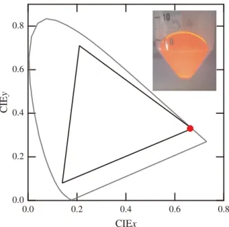
Fig. 5 CIE color coordinate of obtained YVO4:Eu,Bi nanophosphor (closed circle). The triangle area shows the NTSC standard. The inset shows the photograph of transparent gel including nanophosphor under UV illumination.
As shown in Fig. 4 and Fig. 5, the both samples exhibit saturated red emission due to Eu3+ substituted into the Y site of YVO4 crystal as reported elsewhere[6], [7], [23], [28],[31]–[33],[35]–[37]. The sharp lines with saturated red color; (CIEx, CIEy)=(0.663, 0.330) has an advantage in the display application, which is superior to the QDs. How- ever it is difficult to apply the YVO4:Eu,Bi nanophosphor to the LED backlight due to the lack of absorption in the blue
Fig. 6 (a) PLE and (b) iQE spectra of as-aged (Ref) and microwave treated (MW) samples. Monitored wavelength is 619 nm.
spectral region. The shape of PL spectra and the color coor- dinate do not change before and after microwave treatment.
On the contrary PLE spectrum significantly varies after mi- crowave treatment as shown in Fig. 6. The PLE spectrum consists of the sharp peaks due to f-f transitions of Eu3+and two broad bands due to charge transfer transitions related to Bi (∼350 nm) and VO43−group (∼320 nm)[23]. The strong sharp line at about 395 nm, which is attributed to the direct absorption of Eu3+, suggests that the high-concentration Eu ions up to approximately 20 mol% are activated in obtained nanophosphors in contrast with the bulk material which nor- mally shows the maximum iQE at about 5 mol%[35]. Some YVO4:Eu[37]and YVO4:Eu,Bi[28]nanophosphors shows relatively high iQE even at the high Eu concentration (about 20 mol%) as high as in this study. These indicate that the interactions between Eu ions are less efficient or the de- excitation of VO4 groups seriously occurs until the exci- tation reaches a Eu ion. In the bulk material, an efficient energy-transfer pathway from VO4groups might result in a high iQE (beyond 70%) for a small Eu content. Compar- ison to this, Huignard et al. suggested that surface effects and a structural disorder which limit the iQE might lead an apparent high quenching concentration[36],[37]in the nanophosphors. In our study, similar behaviors are thought to be observed, however, the reason is not well understood at the present time.
The intensities of the peaks slightly decrease while the intensities of the bands significantly increase through the microwave treatment. The decrease of PLE intensity of the sharp line at about 395 nm, which is attributed to the di- rect absorption of Eu3+, is due to the decrease of Eu con-
1252
IEICE TRANS. ELECTRON., VOL.E99–C, NO.11 NOVEMBER 2016
Table 1 Cation ratio of obtained powders.
Y Eu Bi V
Ref 0.423 0.108 0.025 0.443
MW(1kW) 0.347 0.085 0.019 0.552
Table 2 (Y,Eu,Bi) ratio in Y site of obtained powders.
Y Eu Bi
Ref 0.760 0.194 0.045
MW(1kW) 0.769 0.188 0.043
centration and decrease of energy migration between Eu ions. Chemical compositions of the obtained powders and the cation (Y,Eu,Bi) ratio in Y site are listed in Table 1 and Table 2, respectively. The Eu concentration of the MW sample slightly decreases in comparison to the Ref sam- ple, which is consistent with the decrease of PLE inten- sity of the sharp line. On the other hand iQE increases via short-time microwave irradiation over the all excitation wavelength region as similar to the long-time hydrothermal treatment[28]. These facts suggest that the pathway of non- radiative relaxation, such as Eu-Eu concentration quench- ing, energy-dissipation of excitons in VO4 groups and OH quenching[36], reduces through the hydrothermal treatment in the solution heated by microwave irradiation. Therefore a continuous solution synthesis of YVO4:Eu,Bi nanophos- phor was succeeded. At present, the iQE of YVO4:Eu,Bi nanophosphor is not sufficient to the practical use. The lim- itation of iQE might be originated from the composition de- viation in the precursor suspension. This problem will be solved by applying the microreaction method.
Finally the synthesis processes in the reactor presented are discussed. Figure 7 shows the microwave power depen- dence of solution temperature and iQE. The (Y,Eu,Bi) ratio is almost kept (0.77:0.19:0.04) with slight deviation before and after microwave heating, and this ratio is almost same as one in the starting material, while (Y+Eu+Bi)/V ratio is significantly varied from 1.25 to 0.82 before and after mi- crowave heating, respectively. In the case of as-aged (Ref) sample, (Y,Eu,Bi)-hydroxide, which does not react with or- thovanadate ions, might still remain in the gel. After mi- crowave heating, by-products such as metavanadate and/or poly-vanadate might be yielded due to the variation of pH under heating[38]. As shown in Fig. 7, the iQE values re- lated to the V-O and Bi-V absorption as mentioned above similarly increase with increasing the solution temperature.
This increase of iQE means that the solution temperature in the flow channel exceeds 100◦C even at the 0.8 kW. The mi- crowave power in the cavity is directly absorbed by the so- lution in the channel due to the low dielectric loss of PEEK, then the so-called “super-heating phenomenon” might oc- cur inside the solution, resulting in the high temperature be- yond the boiling point of aqueous solution. The tempera- ture might be enough to realize the condition for the hy- drothermal reaction. It can be expected that a part of re- mained (Y,Eu,Bi)-hydroxide reacts with orthovanadate ions via microwave heating. To clarify the reaction process as
Fig. 7 The iQE of resultant phosphors and average solution temperature as a function of output microwave power. The points at 0 kW mean the iQE and aging temperature of as-aged sample. The average solution tempera- ture is measured during the synthesis period at the outlet of flow channel.
discussed above, further systematical syntheses and struc- tural studies must be carried out. These are the future prob- lems.
4. Conclusion
A continuous flow reactor equipped with a low-loss flow channel and a microwave cavity was developed for synthe- sizing nanocrystalline phosphors. Continuous solution syn- thesis of nanocrystalline YVO4:Eu,Bi phosphor was suc- ceeded through the rapid hydrothermal method using this equipment. After 20 minutes microwave irradiation, iQE of YVO4:Eu,Bi nanophosphor obtained in this study reaches about 30% at 320 nm as high as that obtained by 6 hours hy- drothermal treatment. At present, the iQE of YVO4:Eu,Bi nanophosphor obtained by our process is not sufficient to the practical use. Further improvement of this continuous flow reactor is now in progress. In near future, advanced syn- thesis using a combination of micro reaction and microwave heating will be examined.
References
[1] H.-S. Chen, C.-K. Hsu, and H.-Y. Hong, “InGaN-CdSe-ZnSe Quan- tum Dots White LEDs,” IEEE Photonics Technol. Lett, vol.18, no.1, pp.193–195, 2006.
[2] E. Jang, S. Jun, H. Jang, J. Lim, B. Kim, and Y. Kim, “White- Light-Emitting Diodes with Quantum Dot Color Converters for Dis- play Backlights,” Adv. Mater., vol.22, no.28, pp.3076–3080, 2010.
[3] H.V. Demir, S. Nizamoglu, T. Erdem, E. Mutlugun, N. Gaponik, and A. Eychm¨uller, “Quantum dot integrated LEDs using photonic and excitonic color conversion,” Nanotoday, vol.6, no.6, pp.632–647, 2011.
[4] S. Coe-Sullivan, W. Liu, P. Allen, and J.S. Steckel, “Quantum Dots
for LED Downconvertsion in Display Applications,” ECS J. Solid State Sci. Tech., vol.2, no.2, pp.R30226–3030, 2013.
[5] Y. Shirasaki, G.J. Supran, M.G. Bawendi, and V. Bulovi´c, “Emer- gence of colloidal quantum-dot light emitting technologies,” Nature Photonics, vol.7, no.1, pp.12–23, 2013.
[6] Y. Iso, S. Takeshita, and T. Isobe, “Effects of YVO4:Bi3+,Eu3+
Nanophosphors Spectral Down Shifter on Properties of Monocrys- talline Silicon Photovoltaic Module,” J. ElectroChem. Soc., vol.159, no.3, pp.J72–J76, 2012.
[7] H. Okura and K. Ohmi, “Advanced Nanophosphors Synthesized by Microreaction Method and Their Application to Wavelength Con- version Layer,” Proc. IDW’14, pp.506–509, 2014.
[8] P. Du, J.-H. Lim, J.-W. Leem, S.-M. Cha and J.S. Yu, “En- hanced Photovoltaic Performance of Dye-Sensitized Solar Cells by Efficient Near- Infrared Sunlight Harvesting using Upconverting Y2O3:Er3+/Yb3+Phosphor Nanoparticles,” Nanoscale Res. Lett., vol.10, no.1, p.321, 2015.
[9] Q.M. Le, T.H. Tran, T.H. Nguyen, T.K. Hoang, T.B. Nguyen, K.T.
Do, K.A. Tran, D.H. Nguyen, T.L. Le, T.Q. Nguyen, M.D. Dang, N.A.T. Nguyen, and V.M. Nguyen, “Development of a fluorescent label tool based on lanthanide nanophosphors for viral biomedical application,” Adv. Nat. Sci.: Nanosci. Nanotechnol., vol.3, no.3, 035003, 2012.
[10] X. Liu, R. Wang, F. Zhang, L. Zhou, D. Shen, C. Yao, and D. Zhao,
“Nd3+Sensitized Up/Down Converting Dual-Mode Nanomaterials for Efficient In-vitro and In-vivo Bioimaging Excited at 800 nm,”
Sci. Rep., vol.3, 3536, 2013.
[11] B.K. Gupta, D. Haranath, S. Saini, V.N. Singh, and V. Shanker,
“Synthesis and characterization of ultra-fine Y2O3:Eu3+nanophos- phors for luminescent security ink applications,” Nanotechnology, vol.21, no.5, 055607, 2010.
[12] B.L. Cushing, V.L. Kolesnichenko, and C.J. O’Connor, “Recent Ad- vances in the Liquid-Phase Syntheses of Inorganic Nanoparticles,”
Chem. Rev., vol.104, no.9, pp.3893–3946, 2004.
[13] X. Wang and Y. Li, “Monodisperse nanocrystals: general syn- thesis, assembly, and their applications,” Chem. Commun., no.28, pp.2901–2910, 2007.
[14] C.N.R. Rao, S.R.C. Vivekchand, K. Biswasa, and A. Govindaraja,
“Synthesis of inorganic nanomaterials,” Dalton Trans., no.34, pp.3728–3749, 2007.
[15] S.G. Kwon and T. Hyeon, “Colloidal Chemical Synthesis and For- mation Kinetics of Uniformly Sized Nanocrystals of Metals, Oxides, and Chalcogenides,” Acc. Chem. Res., vol.41, no.12, pp.1696–1709, 2008.
[16] J. Livage, M. Henry, and C. Sanchez, “Sol-gel chemistry of transition metal oxides,” Prog. Solid State Chem., vol.18, no.4, pp.259–341, 1988.
[17] L.L. Hench and J.K. West, “The sol-gel process,” Chem. Rev., vol.90, no.1, pp.33–72, 1990.
[18] P.H. Mutin and A. Vioux, “Nonhydrolytic Processing of Ox- ide-Based Materials: Simple Routes to Control Homogeneity, Morphology, and Nanostructure,” Chem. Mater., vol.21, no.4, pp.582–596, 2009.
[19] J. Park, J. Joo, S.G. Kwon, Y. Jang, and T. Hyeon, “Synthesis of monodisperse spherical nanocrystals,” Angew. Chem., Int. Ed., vol.46, no.25, pp.4630–4660, 2007.
[20] K. Byrappa and T. Adschiri, “Hydrothermal technology for nan- otechnology,” Prog. Cryst. Growth Charact. Mater., vol.53, no.2, pp.117–166, 2007.
[21] M. Rajamathi and R. Seshadri, “Oxide and chalcogenide nanopar- ticles from hydrothermal/solvothermal reactions,” Curr. Opin. Solid State Mater. Sci., vol.6, no.4, pp.337–345, 2002.
[22] K. Riwotzki, M. Haase, “Wet-Chemical Synthesis of Doped Col- loidal Nanoparticles: YVO4:Ln (Ln=Eu, Sm, Dy),” J. Phys. Chem.
B, vol.102, no.50, p10129, 1998.
[23] S. Takeshita, T. Isobe, T. Sawayama, and S. Niikura, “Effects of the homogeneous Bi3+doping process on photoluminescence proper-
ties of YVO4:Bi3+,Eu3+nanophosphor,” J. Lumin., vol.129, no.9, pp.1067–1072, 2009.
[24] H.Z. Wang, H. Nakamura, M. Uehara, M. Miyazaki, and H.
Maeda, “Preparation of titania particles utilizing the insoluble phase interface in a microchannel reactor,” Chem. Commun., 2002, pp.1462–1463, 2002.
[25] H. Nakamura, Y. Yamaguchi, M. Miyazaki, H. Maeda, M. Uehara, and P. Mulavaney, “Preparation of CdSe nanocrystals in a mi- cro-flow-reactor,” Chem. Commun., 2002, pp.2844–2845, 2002.
[26] H. Wang, H. Nakamura, M. Uehara, Y. Yamaguchi, M. Miyazaki, and H. Maeda, “Highly Luminescent CdSe/ZnS Nanocrystals Syn- thesized Using a Single-Molecular ZnS Source in a Microfluidic Re- actor,” Adv. Func. Mater., vol.15, no.4, pp.603–608, 2005.
[27] H. Okura, T. Murakawa, Y. Miyamoto, and K. Ohmi, “Novel Solu- tion Synthesis of White LED Phosphors by Microreaction Method,”
ECS Trans., vol.33, no.33, pp.95–100, 2011.
[28] K. Yamashina, H. Okura, R. Sakata, R. Komiyama, H. Miyashita, S.-S. Lee, and K. Ohmi, “Advanced Microreactor system with Glass Mixer Cell for Synthesizing Nanophosphors ” Proc. IDW’13, pp.797–798, 2013.
[29] T. Hiragi and T. Kunimoto, “Synthesis of ZnO-based phosphor ma- terials using microwave heating,” J. Ceram. Process. Res., vol.12, pp.s9–12, 2011.
[30] I. Bilecka and M. Niederberger, “Microwave chemistry for inorganic nanomaterials synthesis,” Nanoscacle, vol.2, no.8, pp.1358–1374, 2010.
[31] T. Kojima and T. Hisamune, “Phosphors for plasma display” in Phosphor Handbook, 2nd ed. W.M. Yen, S. Shionoya, and H.
Yamamoto, pp.731–768, CRC, Boca Raton, 2007.
[32] F.C. Palila and A.K. Levine, “YVO4: a highly efficient phos- phor for high pressure mercury lamps,” Appl. Opt., vol.5, no.9, pp.1467–1468, 1996.
[33] G. Panayiotakis, D. Cavouras, I. Kandarksis, and C. Nomicos, “A study of X-ray luminescence and spectral compatibility of europi- um-activated yttrium-vanadate (YVO4: Eu) screens for medical imaging applications,” Appl. Phys. A, vol.62, no.5, pp.483–486, 1996.
[34] J. Isasi, M.L. Veiga, Y. Laureiro, R. Saez-Puche, and C. Pico,
“Synthesis, structural determination, and magnetic behaviour of YxGd1−xVO4 phases (x=0.25, 0.50, 0.75),” J. Alloys Compds., vol.177, no.1, pp.143–147, 1991.
[35] R.C. Ropp, “Spectra of Some Rare Earth Vanadates,” J. Elec- trochem. Soc., vol.115, no.9, pp.940–945, 1968.
[36] A. Huignard, V. Buissette, A.-C. Franville, T. Gacoin, and J.-P.
Boilot, “Emission Processes in YVO4:Eu Nanoparticles,” J. Phys.
Chem. B, vol.107, no.28, pp.6754–6759, 2003.
[37] A. Huignard, V. Buissette, G. Laurent, T. Gacoin, and J.-P. Boilot,
“Synthesis and Characterizations of YVO4:Eu Colloids,” Chem.
Mater., vol.14, no.5, pp.2264–2269, 2002.
[38] N. McCann, M. Wagner, and H. Hasse, “A thermodynamic model for vanadate in aqueous solution - equilibria and reaction enthalpies,”
Dalton Trans., vol.42, no.7, pp.2622–2688, 2013.
1254
IEICE TRANS. ELECTRON., VOL.E99–C, NO.11 NOVEMBER 2016
Takashi Kunimoto studied physics at Tokyo University of Science, where he also received the B.Sc., M.Sc. and Ph.D. degrees in Physics in 1992, 1994 and 1999, respectively. He is currently a professor in Tokushima Bunri Uni- versity, Japan. He is currently working on the synthesis and characterization of phosphor ma- terials. His area of research includes ultrafast lasers, electron spin resonance and spectroscopy of condensed matters from the X-ray to far in- frared region. Prof. Kunimoto is a member of the Society for Information Display, the Japan Society of Applied Physics, the Physical Society of Japan, the Laser Society of Japan and the Japan Society of Infrared Science and Technology.
Yoshiko Fujita studied material science and technology and earned a master’s degree in Nano material and Bio Engineering from Toku- shima Bunri University, Japan, in 2012 and 2014, respectively. She is currently working on the processing of phosphor materials and in- vestigating the properties of phosphors for plant cultivation, display and lighting. Ms. Fujita is a member of the Japan Society of Applied Physics.
Hiroshi Okura graduated the electrochem- ical laboratory in Kyoto University with the M.Eng. degree in 1998 and he has been work- ing at Merck Ltd. as a phosphor and nanoma- terial researcher now. Regarding the Ph.D. de- gree, he got it from Tottori University in 2012 while working in Merck. Dr. Okura is a mem- ber of the Japan Society of Applied Physics, the Electrochemical Society of Japan, the Chemical Society of Japan, the Ceramic Society of Japan and the Society of Chemical Engineers, Japan.